Review
. 2022 Apr 20;27(9):2658.
doi: 10.3390/molecules27092658.

G Protein-coupled Receptor (GPCR) Reconstitution and Labeling for Solution Nuclear Magnetic Resonance (NMR) Studies of the Structural Basis of Transmembrane Signaling

Affiliations
Review

G Protein-coupled Receptor (GPCR) Reconstitution and Labeling for Solution Nuclear Magnetic Resonance (NMR) Studies of the Structural Basis of Transmembrane Signaling

Haoyi Ge et al. Molecules. .

Abstract

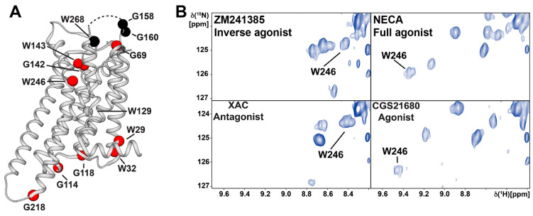
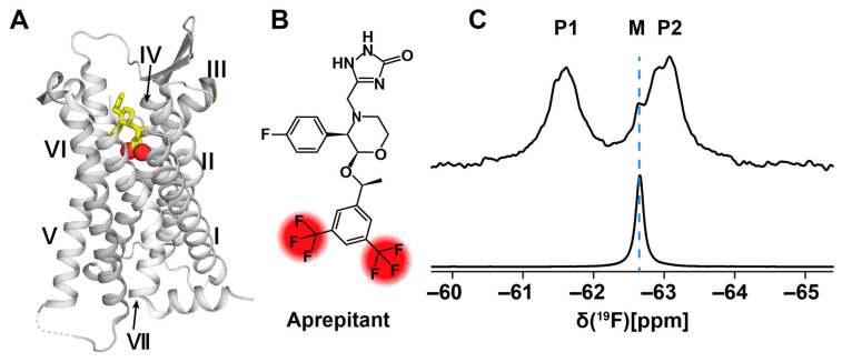
G protein-coupled receptors (GPCRs) are a large membrane protein family found in higher organisms, including the human body. GPCRs mediate cellular responses to diverse extracellular stimuli and thus control key physiological functions, which makes them important targets for drug design. Signaling by GPCRs is related to the structure and dynamics of these proteins, which are modulated by extrinsic ligands as well as by intracellular binding partners such as G proteins and arrestins. Here, we review some basics of using nuclear magnetic resonance (NMR) spectroscopy in solution for the characterization of GPCR conformations and intermolecular interactions that relate to transmembrane signaling.

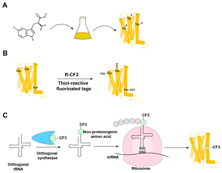
Keywords: 19F-NMR; G protein-coupled receptors; amino-acid-specific NMR labeling; in-membrane chemical modification; membrane mimetics; sequence-specific NMR labeling; stable-isotope labeling.


Conflict of interest statement

The authors declare no conflict of interest.

Figures

Figure 1
Figure 1
Overview of the intermolecular interactions involved in cellular signaling by GPCRs. The orthosteric binding pocket (indicated as a yellow ball) is usually targeted by endogenous ligands (indicated as small colored balls), which interact with the orthosteric binding pocket; they alter the activation state of the receptors according to their efficacy as full agonists, partial agonists, antagonists or inverse agonists. Allosteric ligands (indicated as triangles) typically bind to sites that are spatially distinct from the orthosteric binding pocket and modulate the affinity and/or efficacy of the ligand bound to the orthosteric site. Representative intracellular interaction partners of GPCRs are heterotrimeric G proteins (α, β, γ) and arrestins. Biased GPCR ligands preferentially activate either the G protein or the arrestin signaling pathways. (Adapted from Figure 2 in reference [12]).
Figure 2
Figure 2
Cartoon representation of GPCRs embedded in five different membrane mimics in use for reconstitution and solubilization for NMR experiments in solution. (A) GPCR (blue) stabilized in detergent micelles (gray). (B) GPCR (blue) embedded in a nanodisc composed of a lipid bilayer (pink) and membrane scaffold proteins (dark blue). (C) GPCR (blue) embedded in a saposin A nanoparticle composed of a lipid bilayer (pink) and saposin A molecules (dark blue). (D) GPCR (blue) embedded in amphipathic polymers or amphipols (gray). (E) GPCR (blue) embedded in bicelles composed of long-chain lipids (pink) and short-chain lipids or detergent molecules (gray). (Adapted from Figures 2, 4 and 5 of reference [21]; the size ranges indicated below the individual cartoons are taken from reference [23]).
Figure 3
Figure 3
Overview of the methods in use for incorporation of 19F-NMR labels into GPCRs. (A) Biosynthetic incorporation by adding fluorinated amino acids, such as 5F-Trp, to the expression system; all Trp residues in the protein are then labeled with 19F. (B) Post-translational chemical modification by reacting the GPCR with thiol-reactive fluorinated tags; all reagent-accessible Cys residues are then labeled with the fluorinated tag. (C) Genetic labeling using an extrinsic orthogonal tRNA/aminoacyl-tRNA synthetase pair to incorporate non-proteinogenic 19F-labeled amino acids at positions defined by a TAG amber codon.
Figure 4
Figure 4
Sequence-specific assignment of the 19F-NMR signals of TET moieties attached to the three native Cys residues near the intracellular surface of the transmembrane domain (TMD) of the GLP-1R. (A) Crystal structure of the GLP-1R[TMD] (PDB: 5VEX; generated using the PyMOL Molecular Graphics System, Schrödinger, LLC, New York, USA) shown as a green cartoon. Three native Cys residues, C174, C329 and C341, shown as black spheres, are exposed on the intracellular surface and are thus accessible for TET labeling with the IMCM method [56]. (B) 1D 19F-NMR spectra. The GLP-1R[TMD] spectrum contains three signals corresponding to the three Cys sites shown in (A). Individual assignments of the 19F-NMR signals were obtained using site-specific mutagenesis to replace cysteines with serine residues. The peak assignments indicated at the top by the one-letter amino acid code and the residue number are based on the disappearance of the signals of the replaced cysteines. (Adapted from Figure 1 in reference [57]).
Figure 5
Figure 5
Spectral region containing the Trp2466.48 indole 15N–1H signal of 2D [15N, 1H]-TROSY correlation spectra of [u-15N, ~70% 2H]-A2AAR in complexes with ligands of different efficacies. (A) Locations of glycines and tryptophans in the crystal structure of A2AAR in complex with the inverse agonist ZM241385 (PDB: 3PWH), for which NMR signals were assigned. Assigned residues which show a response in their NMR signals to bound ligands with different efficacies are depicted by red spheres. Residues that showed no response are depicted by black spheres. The disordered portion of the ECL2 that was not observed in the crystal structure is shown as a dashed line. (B) The indole 15N–1H NMR signal of the toggle switch tryptophan W2466.48 is highly responsive to variable drug efficacy, as observed by comparing the spectra for A2AAR in complexes with the inverse agonist ZM241385, the antagonist XAC, the selective agonist CGS21680 and the full agonist NECA. (Adapted from Figure 4D in reference [88]).
Figure 6
Figure 6
19F-NMR observation of the ligand aprepitant bound to NK1R. (A) Crystal structure of the NK1R complex with aprepitant (PDB: 6J20). NK1R is shown in ribbon presentation with grey color. Aprepitant is shown in yellow stick presentation, with red spheres representing the trifluoromethyl groups. The transmembrane helices are identified with roman numerals. (B) Chemical structure of the drug aprepitant. The two –CF3 groups are highlighted in red. (C) 1D 19F-NMR spectra of aprepitant in complex with NK1R (upper trace) and “free” in DDM/CHS micelles (lower trace). M is the 19F-NMR signal of micelle-associated “free” aprepitant; P1 and P2 are the two –CF3 signals of NK1R-bound aprepitant. (Adapted from Figure 4, A and E, in reference [99]).

Similar articles

Cited by

References

    1. Rask-Andersen M., Almén M.S., Schiöth H.B. Trends in the exploitation of novel drug targets. Nat. Rev. Drug Discov. 2011;10:579–590. doi: 10.1038/nrd3478. - DOI - PubMed
    1. Oprea T.I., Bologa C.G., Brunak S., Campbell A., Gan G.N., Gaulton A., Gomez S.M., Guha R., Hersey A., Holmes J., et al. Unexplored therapeutic opportunities in the human genome. Nat. Rev. Drug Discov. 2018;17:317–332. doi: 10.1038/nrd.2018.14. - DOI - PMC - PubMed
    1. Hauser A.S., Attwood M.M., Rask-Andersen M., Schioth H.B., Gloriam D.E. Trends in GPCR drug discovery: New agents, targets and indications. Nat. Rev. Drug Discov. 2017;16:829–842. doi: 10.1038/nrd.2017.178. - DOI - PMC - PubMed
    1. Weis W.I., Kobilka B.K. The molecular basis of G protein–coupled receptor activation. Annu. Rev. Biochem. 2018;87:897–919. doi: 10.1146/annurev-biochem-060614-033910. - DOI - PMC - PubMed
    1. Kenakin T. Inverse, protean, and ligand-selective agonism: Matters of receptor conformation. FASEB J. 2001;15:598–611. doi: 10.1096/fj.00-0438rev. - DOI - PubMed

LinkOut - more resources