. 2022 Apr 24;27(9):2740.
doi: 10.3390/molecules27092740.

Novel Chitosan-Based Schiff Base Compounds: Chemical Characterization and Antimicrobial Activity

Affiliations

Novel Chitosan-Based Schiff Base Compounds: Chemical Characterization and Antimicrobial Activity

Riccardo Fontana et al. Molecules. .

Abstract

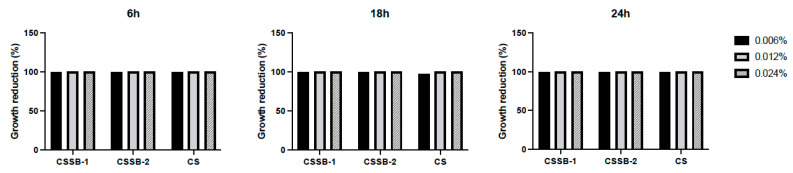
Chitosan (CS) and its derivatives are receiving considerable attention for their great biocompatibility and broad-spectrum activities in many fields. In this work, we aimed to characterize the antimicrobial activity of novel chitosan Schiff bases (CSSB). CS was synthesized by double deacetylation of chitin (Cn) after its extraction from the armors of crustaceans Astacus leptodactylus, and CSSB-1 and CSSB-2 were synthesized by interaction of CS with 4-(2-chloroethyl) benzaldehyde (aldehyde-1) and 4-(bromoethyl) benzaldehyde (aldehyde-2), respectively, at room temperature. The synthesized compounds were characterized by elemental analysis, gel permeation chromatography (GPC), infrared spectroscopy (FTIR), thermogravimetry (TG), and differential scanning calorimetry (DSC). The antimicrobial activity against Gram-positive (Staphylococcus aureus) and Gram-negative (Pseudomonas aeruginosa) bacteria and against yeasts (Candida albicans) was significantly increased due to their higher solubility as compared to unmodified CS opening perspectives for the use of these compounds for antimicrobial prevention in different fields as, for example, food industry, cosmetics, or restoration.

Keywords: antibacterial activity; antifungal activity; chitosan; chitosan-based Schiff base.


Conflict of interest statement

The authors declare no conflict of interest.

Figures

Figure 1
Figure 1
Characterization of the compounds. TG and DSC data of CS (a), CSSB−1 (b), and CSSB−2 (c) samples.
Figure 2
Figure 2
Antimicrobial activity of CS, CSSB-1, and CSSB-2 compounds on S. Aureus. Fresh cultures of S. aureus were incubated in liquid cultures with three different concentrations (0.006%, 0.012%, and 0.024%) of CS, CSSB-1, and CSSB-2 for 6, 18, and 24 h or without the compounds (untreated cells). After incubation, the antimicrobial activity was evaluated by measuring the optical density at 600 nm of each liquid culture (A) and by determining the number of colony-forming units/mL (CFU/mL) (B) after plating the cultures onto agar plates and incubation to allow colony formation. *, p = 0.01; **, p = 0.001; ****, p < 0.0001.
Figure 3
Figure 3
Antimicrobial activity of CS, CSSB-1, and CSSB-2 compounds on P. aeurginosa. Fresh cultures of P. aeruginosa were incubated with three different concentrations (0.006%, 0.012%, and 0.024%) of CS, CSSB-1, and CSSB-2 for 6, 18, and 24 h or without the compounds (untreated cells). After incubation, the antimicrobial activity was evaluated by measuring the optical density at 600 nm of each liquid culture (A) and by determining the number of colony-forming units/mL (CFU/mL) (B) after plating the cultures onto agar plates and incubation to allow colony formation. *, p = 0.01; **, p = 0.001; ***, p = 0.0001; ****, p < 0.0001.
Figure 4
Figure 4
Antimicrobial activity of CS, CSSB-1, and CSSB-2 compounds on C. albicans. Fresh cultures of C. albicans were incubated with three different concentrations (0.006%, 0.012%, and 0.024%) of CS, CSSB-1, and CSSB-2 for 6, 18, and 24 h or without the compounds (untreated cells). After incubation, the antimicrobial activity was evaluated by measuring the optical density at 600 nm of each liquid culture (A) and by determining the number of colony-forming units/mL (CFU/mL) (B) after plating the cultures onto agar plates and incubation to allow colony formation. ***, p = 0.0001; ****, p < 0.0001.
Figure 5
Figure 5
Growth reduction in S. Aureus after treatment with CS, CSSB-1, and CSSB-2 compounds. Fresh cultures of S. aureus were incubated in liquid cultures with three different concentrations (0.006%, 0.012%, and 0.024%) of CS, CSSB-1, and CSSB-2 for 6, 18, and 24 h or without the compounds (untreated cells). After incubation, each culture was plated onto agar plates and incubated to allow colony formation (CFU/mL, as illustrated in Figure 2). The results are represented as percentage (%) of cell growth reduction after each treatment as compared to control, untreated cells.
Figure 6
Figure 6
Growth reduction in P. aeruginosa after treatment with CS, CSSB-1, and CSSB-2 compounds. Fresh cultures of P. aeruginosa were incubated in liquid cultures with three different concentrations (0.006%, 0.012%, and 0.024%) of CS, CSSB-1, and CSSB-2 for 6, 18, and 24 h or without the compounds (untreated cells). After incubation, each culture was plated onto agar plates and incubated to allow colony formation (CFU/mL, as illustrated in Figure 3). The results are represented as percentage (%) of cell growth reduction after each treatment as compared to control, untreated cells.
Figure 7
Figure 7
Growth reduction in C. albicans after treatment with CS, CSSB-1, and CSSB-2 compounds. Fresh cultures of C. albicans were incubated in liquid cultures with three different concentrations (0.006%, 0.012%, and 0.024%) of CS, CSSB-1, and CSSB-2 for 6, 18, and 24 h or without the compounds (untreated cells). After incubation, each culture was plated onto agar plates and incubated to allow colony formation (CFU/mL, as illustrated in Figure 4). The results are represented as percentage (%) of cell growth reduction after each treatment as compared to control, untreated cells.

References

    1. La Fauci V., Costa G.B., Genovese C., Palamara M.A.R., Alessi V., Squeri R. Drug-resistant bacteria on hands of healthcare workers and in the patient area: An environmental survey in Southern Italy’s hospital. Rev. Esp. Quimioter. Publ. Soc. Esp. Quimioter. 2019;32:303–310. - PMC - PubMed
    1. Kong M., Chen X.G., Xing K., Park H.J. Antimicrobial properties of chitosan and mode of action: A state of the art review. Int. J. Food Microbiol. 2010;144:51–63. doi: 10.1016/j.ijfoodmicro.2010.09.012. - DOI - PubMed
    1. Allan C.R., Hadwiger L.A. The fungicidal effect of chitosan on fungi of varying cell wall composition. Exp. Mycol. 1979;3:285–287. doi: 10.1016/S0147-5975(79)80054-7. - DOI
    1. Hassan M.A., Omer A.M., Abbas E., Baset W.M.A., Tamer T.M. Preparation, physicochemical characterization and antimicrobial activities of novel two phenolic chitosan Schiff base derivatives. Sci. Rep. 2018;8:11416. doi: 10.1038/s41598-018-29650-w. - DOI - PMC - PubMed
    1. Aranaz I., Alcántara A.R., Civera M.C., Arias C., Elorza B., Caballero A.H., Acosta N. Chitosan: An Overview of Its Properties and Applications. Polymers. 2021;13:3256. doi: 10.3390/polym13193256. - DOI - PMC - PubMed

MeSH terms

LinkOut - more resources