. 2022 Apr 27;27(9):2791.
doi: 10.3390/molecules27092791.

Synthesis and Investigation of the G-Quadruplex Binding Properties of Kynurenic Acid Derivatives with a Dihydroimidazoquinoline-3,5-dione Core

Affiliations

Synthesis and Investigation of the G-Quadruplex Binding Properties of Kynurenic Acid Derivatives with a Dihydroimidazoquinoline-3,5-dione Core

Stefania Mazzini et al. Molecules. .

Abstract

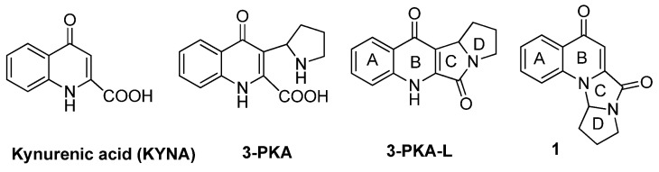
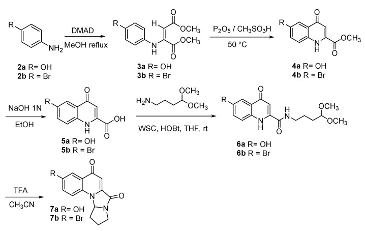
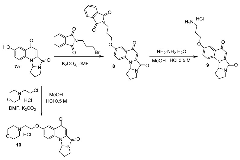
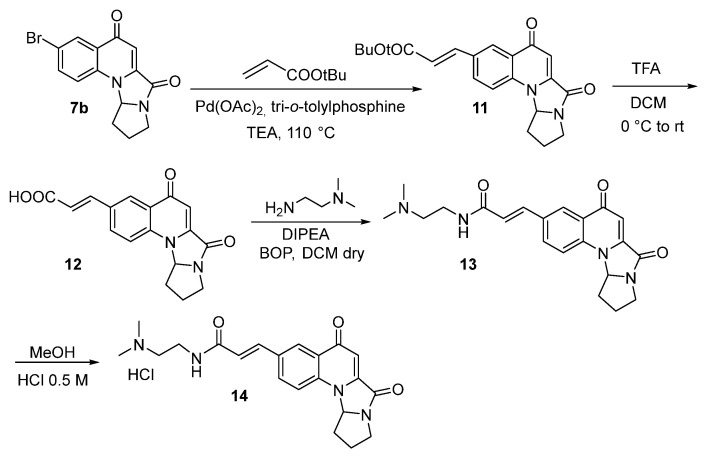
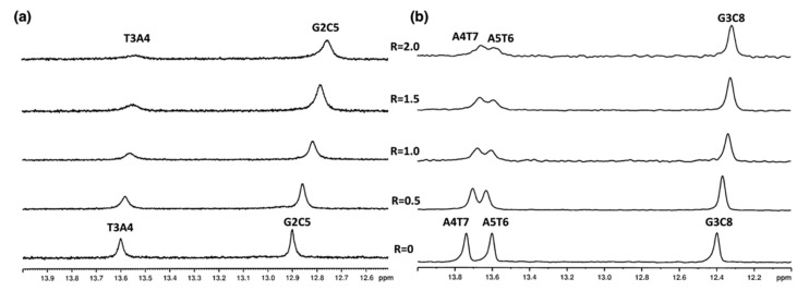
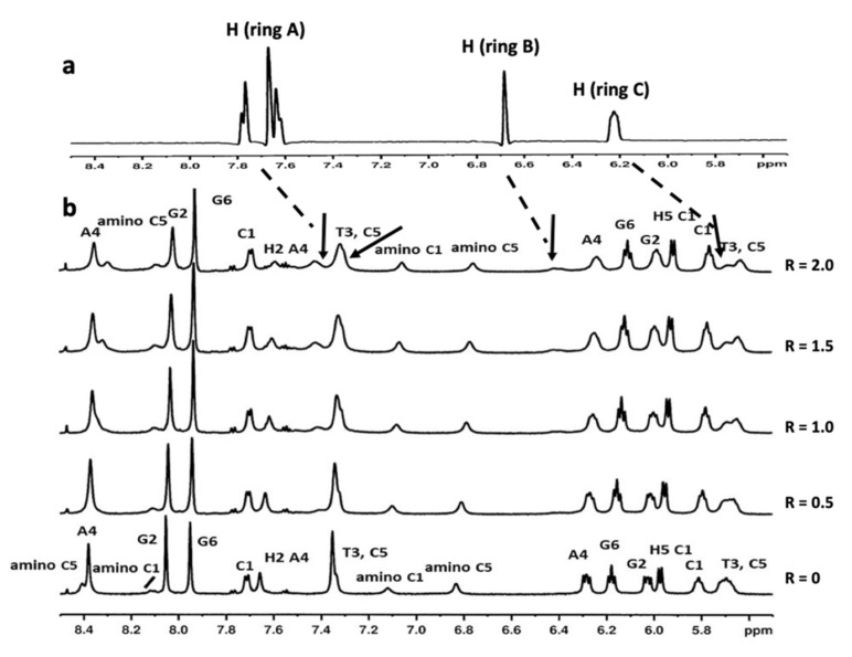
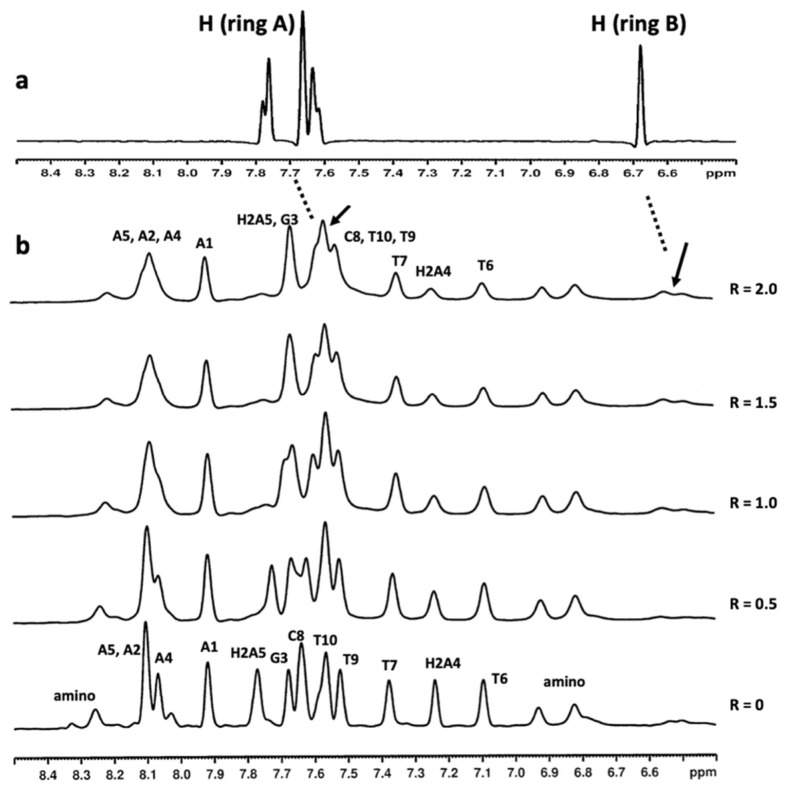
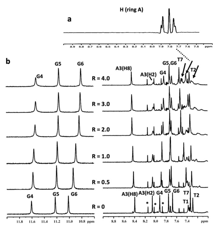
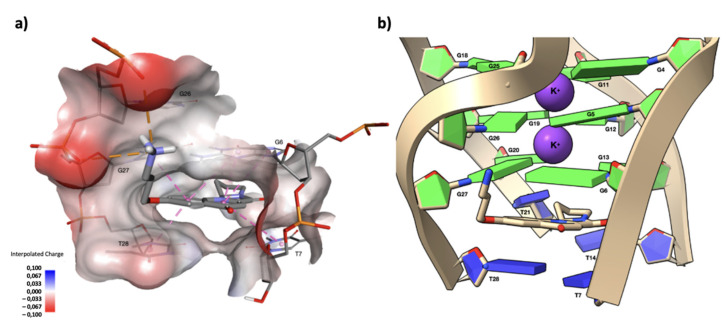
G-quadruplexes are secondary structures originating from nucleic acid regions rich in guanines, which are well known for their involvement in gene transcription and regulation and DNA damage repair. In recent studies from our group, kynurenic acid (KYNA) derivative 1 was synthesized and found to share the structural features typical of G-quadruplex binders. Herein, structural modifications were conducted on this scaffold in order to assist the binding with a G-quadruplex, by introducing charged hydrophilic groups. The antiproliferative activity of the new analogues was evaluated on an IGROV-1 human ovarian cancer cell line, and the most active compound, compound 9, was analyzed with NMR spectrometry in order to investigate its binding mode with DNA. The results indicated that a weak, non-specific interaction was set with duplex nucleotides; on the other hand, titration in the presence of a G-quadruplex from human telomere d(TTAGGGT)4 showed a stable, although not strong, interaction at the 3'-end of the nucleotidic sequence, efficiently assisted by salt bridges between the quaternary nitrogen and the external phosphate groups. Overall, this work can be considered a platform for the development of a new class of potential G-quadruplex stabilizing molecules, confirming the crucial role of a planar system and the ability of charged nitrogen-containing groups to facilitate the binding to G-quadruplex grooves and loops.

Keywords: DNA; G-quadruplex; KYNA; cytotoxicity; kynurenic acid; stabilization.


Conflict of interest statement

The authors declare no conflict of interest.

Figures

Figure 1
Figure 1
Structures of KYNA, its metabolites 3-PKA and 3-PKA-L and the analogue 1.
Scheme 1
Scheme 1
Synthesis of compounds 7a,b.
Scheme 2
Scheme 2
Synthesis of compounds 9 and 10.
Scheme 3
Scheme 3
Synthesis of compound 14.
Figure 2
Figure 2
Imino proton region of the 1H NMR titration spectra of (a) 5′-d(CGTACG)2-3′ and (b) 5′d-(AAGAATTCTT)2-3′ with compound 9 at 15 °C in H2O/D2O (9:1), 100 mM NaCl and 10 mM sodium phosphate buffer (pH 7.0), at different ratios R = [9]/[DNA].
Figure 3
Figure 3
(a) Selected 1H proton region of compound 9 at 15 °C in H2O/D2O (9:1), 100 mM NaCl and 10 mM sodium phosphate buffer (pH 7.0); (b) aromatic and anomeric proton region of the 1H NMR titration spectra of 5′-d(CGTACG)2-3′ with compound 9 at 15 °C in H2O/D2O (9:1), 100 mM NaCl and 10 mM sodium phosphate buffer (pH 7.0), at different ratios R = [9]/[DNA]. The arrows indicate the broad proton signals of compound 9.
Figure 4
Figure 4
(a) Selected 1H proton region of compound 9 at 15 °C in H2O/D2O (9:1), 100 mM NaCl and 10 mM sodium phosphate buffer (pH 7.0); (b) aromatic proton region of the 1H NMR titration spectra of 5′d-(AAGAATTCTT)2-3′ with compound 9 at 15 °C in H2O/D2O (9:1), 100 mM NaCl and 10 mM sodium phosphate buffer (pH 7.0), at different ratios R = [9]/[DNA]. The arrows indicate the broad proton signals of compound 9.
Figure 5
Figure 5
(a) Selected region of 1H NMR spectrum of compound 9 at 25 °C in H2O/D2O (9:1), 150 mM KCl, 25 mM K+ phosphate buffer and EDTA 1 mM (pH 6.7); (b) imino and aromatic proton region of the 1H NMR titration spectra of d(TTAGGGT)4 with compound 9 at 25 °C in H2O/D2O (9:1), 150 mM KCl, 25 mM K+ phosphate buffer and EDTA 1 mM (pH 6.7), at different ratios R = [9]/[DNA]. The arrows indicate the broad proton signals of compound 9. The peaks labelled with * correspond to the single-strand oligonucleotide.
Figure 6
Figure 6
(a) Drawing that shows the first interaction shell of compound 9 complexed with the d(TTAGGGT)4 G-quadruplex, as obtained from the molecular docking experiment. In this position, compound 9 is capable of forming multiple π–π stacking interactions between the aromatic rings of the polycycle and those of the G6 and T7 units. The side chain of compound 9 orients along the groove, with the quaternary nitrogen forming two salt bridges with G5OP2 and G6OP2, contributing to the stabilization of the complex. In the figure, G5, G6 and T7 are labelled as G26, G27 and T28, respectively, due to the loss of symmetry in the complex. The non-bonded interactions are displayed as dashed lines, the π–π stacking interactions in pink, and the two salt bridge interactions in orange. A surface near the current ligand is created around the target and colored by the interpolated atomic charge of the d(TTAGGGT)4 G-quadruplex atoms. Nucleotides and the ligand are represented in sticks and colored according to their atom types. (b) A close-up view of the capping at the ligands interacting with the 5′-end tetrad. Nucleotide bases are represented as slabs and fill sugars, with the guanine residues colored in green and thymine residues colored in blue. Potassium ions are represented as purple van der Waals spheres, while the ligand is drawn in stick and colored according to its atom types. The drawing was created using the Discovery Studio® Visualizer (BIOVIA, Dassault Systèmes Discovery Studio Modeling Environment, Release 2017) and UCSF ChimeraX, developed by the Resource for Biocomputing, Visualization, and Informatics at the University of California, San Francisco, with support from the National Institutes of Health R01-GM129325 and the Office of Cyber Infrastructure and Computational Biology, National Institute of Allergy and Infectious Diseases.

References

    1. Walczak K., Wnorowski A., Turski W.A., Plech T. Kynurenic Acid and Cancer: Facts and Controversies. Cell. Mol. Life Sci. 2020;77:1531–1550. doi: 10.1007/s00018-019-03332-w. - DOI - PMC - PubMed
    1. Moroni F., Cozzi A., Sili M., Mannaioni G. Kynurenic Acid: A Metabolite with Multiple Actions and Multiple Targets in Brain and Periphery. J. Neural Transm. 2012;119:133–139. doi: 10.1007/s00702-011-0763-x. - DOI - PubMed
    1. Schwarcz R., Bruno J.P., Muchowski P.J., Wu H.-Q. Kynurenines in the Mammalian Brain: When Physiology Meets Pathology. Nat. Rev. Neurosci. 2012;13:465–477. doi: 10.1038/nrn3257. - DOI - PMC - PubMed
    1. Wirthgen E., Hoeflich A., Rebl A., Günther J. Kynurenic Acid: The Janus-Faced Role of an Immunomodulatory Tryptophan Metabolite and Its Link to Pathological Conditions. Front. Immunol. 2018;8:1957. doi: 10.3389/fimmu.2017.01957. - DOI - PMC - PubMed
    1. Małaczewska J., Siwicki A.K., Wójcik R.M., Turski W.A., Kaczorek E. The Effect of Kynurenic Acid on the Synthesis of Selected Cytokines by Murine Splenocytes—In Vitro and Ex Vivo Studies. Cent. Eur. J. Immunol. 2016;41:39–46. doi: 10.5114/ceji.2016.58815. - DOI - PMC - PubMed