Improvement of Biological Effects of Root-Filling Materials for Primary Teeth by Incorporating Sodium Iodide
- PMID: 35566277
- PMCID: PMC9105270
- DOI: 10.3390/molecules27092927
Improvement of Biological Effects of Root-Filling Materials for Primary Teeth by Incorporating Sodium Iodide
Abstract
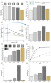
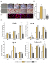
Therapeutic iodoform (CHI3) is commonly used as a root-filling material for primary teeth; however, the side effects of iodoform-containing materials, including early root resorption, have been reported. To overcome this problem, a water-soluble iodide (NaI)-incorporated root-filling material was developed. Calcium hydroxide, silicone oil, and NaI were incorporated in different weight proportions (30:30:X), and the resulting material was denoted DX (D5~D30), indicating the NaI content. As a control, iodoform instead of NaI was incorporated at a ratio of 30:30:30, and the material was denoted I30. The physicochemical (flow, film thickness, radiopacity, viscosity, water absorption, solubility, and ion releases) and biological (cytotoxicity, TRAP, ARS, and analysis of osteoclastic markers) properties were determined. The amount of iodine, sodium, and calcium ion releases and the pH were higher in D30 than I30, and the highest level of unknown extracted molecules was detected in I30. In the cell viability test, all groups except 100% D30 showed no cytotoxicity. In the 50% nontoxic extract, D30 showed decreased osteoclast formation compared with I30. In summary, NaI-incorporated materials showed adequate physicochemical properties and low osteoclast formation compared to their iodoform-counterpart. Thus, NaI-incorporated materials may be used as a substitute for iodoform-counterparts in root-filling materials after further (pre)clinical investigation.
Keywords: iodoform; primary teeth; root canal treatment; root resorption; root-filling material; sodium iodide.
Conflict of interest statement
The authors declare no conflict of interest.
Figures


Similar articles
-
Histological Evaluation of Sodium Iodide-Based Root Canal Filling Materials in Canine Teeth.Materials (Basel). 2024 Dec 12;17(24):6082. doi: 10.3390/ma17246082. Materials (Basel). 2024. PMID: 39769682 Free PMC article.
-
Physicochemical, Pre-Clinical, and Biological Evaluation of Viscosity Optimized Sodium Iodide-Incorporated Paste.Pharmaceutics. 2023 Mar 27;15(4):1072. doi: 10.3390/pharmaceutics15041072. Pharmaceutics. 2023. PMID: 37111558 Free PMC article.
-
Calcium hydroxide/iodoform nanoparticles as an intracanal filling medication: synthesis, characterization, and in vitro study using a bovine primary tooth model.Odontology. 2021 Jul;109(3):687-695. doi: 10.1007/s10266-021-00591-7. Epub 2021 Jan 26. Odontology. 2021. PMID: 33495859
-
Root canal filling materials for primary teeth: a review of the literature.ASDC J Dent Child. 1992 May-Jun;59(3):225-7. ASDC J Dent Child. 1992. PMID: 1629445 Review.
-
Effectiveness of iodoform-based filling materials in root canal treatment of deciduous teeth: a systematic review and meta-analysis.Biomater Investig Dent. 2022 May 19;9(1):52-74. doi: 10.1080/26415275.2022.2060232. eCollection 2022. Biomater Investig Dent. 2022. PMID: 35615468 Free PMC article. Review.
Cited by
-
Histological Evaluation of Sodium Iodide-Based Root Canal Filling Materials in Canine Teeth.Materials (Basel). 2024 Dec 12;17(24):6082. doi: 10.3390/ma17246082. Materials (Basel). 2024. PMID: 39769682 Free PMC article.
-
Optimization of Sodium Iodide-Based Root Filling Material for Clinical Applications: Enhancing Physicochemical Properties.Pharmaceutics. 2024 Aug 2;16(8):1031. doi: 10.3390/pharmaceutics16081031. Pharmaceutics. 2024. PMID: 39204376 Free PMC article.
-
Physicochemical, Pre-Clinical, and Biological Evaluation of Viscosity Optimized Sodium Iodide-Incorporated Paste.Pharmaceutics. 2023 Mar 27;15(4):1072. doi: 10.3390/pharmaceutics15041072. Pharmaceutics. 2023. PMID: 37111558 Free PMC article.
References
-
- Waterhouse P.J., Whitwortth J.M. Pediatric endodontics: Endodontic treatment for the primary and young permanent dentition. Cohen’s Pathw. Pulp. 2011;24:e6-10
-
- Tchaou W.S., Turng B.F., Minah G.E., Coll J.A. Inhibition of pure cultures of oral bacteria by root canal filling materials. Pediatr. Dent. 1996;18:444–449. - PubMed
-
- Chonat A., Rajamani T., Rena E. Obturating Materials in Primary Teeth—A Review. J. Dent. Sci. 2018;6:20–25.
-
- Estrela C., Sydney G.B., Bammann L.L., Felippe Júnior O. Mechanism of action of calcium and hydroxyl ions of calcium hydroxide on tissue and bacteria. Braz. Dent. J. 1995;6:85–90. - PubMed
MeSH terms
Substances
LinkOut - more resources
Full Text Sources
Research Materials

