

In Vivo Confocal Microscopy in Different Types of Dry Eye and Meibomian Gland Dysfunction
- PMID: 35566475
- PMCID: PMC9099706
- DOI: 10.3390/jcm11092349
In Vivo Confocal Microscopy in Different Types of Dry Eye and Meibomian Gland Dysfunction
Abstract
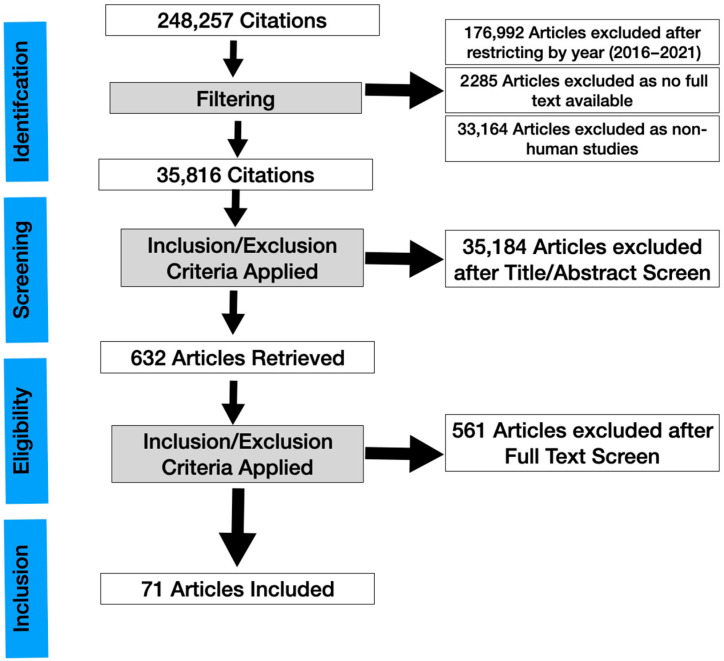
In vivo confocal microscopy (IVCM) imaging is increasingly popular in ocular surface disease diagnosis and management. We conducted a systematic review to update the use of IVCM in the diagnosis and treatment of dry eye and meibomian gland dysfunction (MGD). A literature review was conducted on IVCM studies in MGD, dry eye disease, systemic disease causing dry eye, dry eye in glaucoma patients, contact lens-associated ocular conditions, graft-versus-host disease, and Sjogren's syndrome-related dry eye. The articles were identified through PubMed and a total number of 63 eligible publications were analyzed in detail. All primary research studies on confocal microscopy on dry eye and related conditions from 2017 onwards were included. The reports were reviewed for their contribution to the existing literature as well as potential biases and drawbacks. Despite limitations such as small field of view, lack of population-based norms, and lack of standardization of image acquisition, interpretation, and quantification, IVCM is useful as a complementary technique for clinical diagnosis in various ocular surface disorders related to dry eye. With advances in hardware and software in the near future, it has the potential for further practical impact.
Keywords: diagnostic device; dry eye; in vivo confocal microscopy (IVCM); inflammation; ocular surface; review; tear disorder.
Conflict of interest statement
The authors declare no conflict of interest.
Figures
References
-
- Khamar P., Nair A.P., Shetty R., Vaidya T., Subramani M., Ponnalagu M., Dhamodaran K., D’Souza S., Ghosh A., Pahuja N., et al. Dysregulated Tear Fluid Nociception-Associated Factors, Corneal Dendritic Cell Density, and Vitamin D Levels in Evaporative Dry Eye. Investig. Opthalmol. Vis. Sci. 2019;60:2532–2542. doi: 10.1167/iovs.19-26914. - DOI - PubMed
Publication types
LinkOut - more resources
Full Text Sources
