Review
. 2022 Apr 22;11(9):2360.
doi: 10.3390/jcm11092360.

New Advanced Imaging Parameters and Biomarkers-A Step Forward in the Diagnosis and Prognosis of TTR Cardiomyopathy

Affiliations
Review

New Advanced Imaging Parameters and Biomarkers-A Step Forward in the Diagnosis and Prognosis of TTR Cardiomyopathy

Roxana Cristina Rimbas et al. J Clin Med. .

Abstract

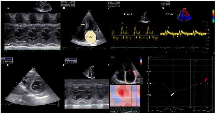
Transthyretin amyloid cardiomyopathy (ATTR-CM) is an infiltrative disorder characterized by extracellular myocardial deposits of amyloid fibrils, with poor outcome, leading to heart failure and death, with significant treatment expenditure. In the era of a novel therapeutic arsenal of disease-modifying agents that target a myriad of pathophysiological mechanisms, timely and accurate diagnosis of ATTR-CM is crucial. Recent advances in therapeutic strategies shown to be most beneficial in the early stages of the disease have determined a paradigm shift in the screening, diagnostic algorithm, and risk classification of patients with ATTR-CM. The aim of this review is to explore the utility of novel specific non-invasive imaging parameters and biomarkers from screening to diagnosis, prognosis, risk stratification, and monitoring of the response to therapy. We will summarize the knowledge of the most recent advances in diagnostic, prognostic, and treatment tailoring parameters for early recognition, prediction of outcome, and better selection of therapeutic candidates in ATTR-CM. Moreover, we will provide input from different potential pathways involved in the pathophysiology of ATTR-CM, on top of the amyloid deposition, such as inflammation, endothelial dysfunction, reduced nitric oxide bioavailability, oxidative stress, and myocardial fibrosis, and their diagnostic, prognostic, and therapeutic implications.

Keywords: PET; SPECT; TTR amyloidosis; cardiac magnetic resonance; cardiac scintigraphy; new biomarkers; prognosis; speckle-tracking echocardiography.


Conflict of interest statement

The authors declare no conflict of interest.

Figures

Figure 3
Figure 3
Apical sparing pattern in ATTR cardiac amyloidosis. (A) Longitudinal deformation bull’s eye plot of the left ventricle by speckle−tracking−echocardiography in a 46-year-old patient with ATTR-CM with significantly decreased global LS of −10%, with longitudinal deformation impaired at the basal, midventricular segments, and relatively preserved at the apex, in an “apical sparing” pattern. (B) The corresponding myocardial work index (MWI) bull’s eye plot, confirming a similar apical sparing pattern, with preserved MWI in the apical segments; (C) Extracellular volume (ECV) bull’s eye plot evaluated by cardiac magnetic resonance, using a 16-segment model, displaying a progressive decrease in the ECV from base to apex, explaining the substrate of apical sparing deformation pattern.
Figure 4
Figure 4
Reversed Look–Locker inversion time nulling pattern in cardiac amyloidosis. (A) Sequential Look–Locker inversion time (TI) mid-ventricular short-axis images are shown at increasing inversion times from left to right. Blood pool nulls after myocardium, which is an abnormal (reversed) nulling pattern. (B) Midventricular short-axis, 4-chamber, and 2-chamber LGE images demonstrate a dark blood pool and difficulty nulling the myocardium.
Figure 1
Figure 1
Anatomopathological main findings suggestive of cardiac amyloidosis. (Upper panel). Salivary glands biopsy, showing Congo red positivity (red arrows) (A), with apple green birefringence on polarization yellow arrows (B); (Mid panel). Abdominal fat pad, showing important Congo red positivity (black arrow) (C), with apple green birefringence on polarization (black arrow) (D); (Lower panel). Rectal biopsy, with important and diffuse infiltration with amyloid fibrils, Congo red positivity (black arrows) (E), with apple green birefringence on polarization (yellow arrows) (F).
Figure 2
Figure 2
Transthoracic echocardiographic images in advanced ATTR-CM. (A) M−mode parasternal long-axis view of the LV displaying concentric severe LV hypertrophy (LV mass = 157 g/m2, RWT = 0.8), with mild pericardial effusion (white arrow); (B) apical 4-chamber view demonstrating a sparkling texture of the IVS and severely dilated LA (64 mL/m2); (C) Pseudo normal transmitral filling pattern (E/A = 1.8); (D) TDI tracing of the mitral annulus showing that all tissue velocities are <5 cm/s, with increased E/E’ ratio. (E) parasternal short axis view showing biventricular hypertrophy—RV free wall = 8 mm; (F) RV severe systolic dysfunction (TAPSE = 13 mm) (G) LA speckle-tracking showing significantly decreased pump function (white arrow) and even more severe reservoir function (red arrow), with the absence of conduit function. RWT, relative wall thickness, E = early filling velocity; A = late atrial filling velocity; S’, systolic TDI velocity; E’, early diastolic TDI velocity; A’, late diastolic TDI velocity; LA, left atrium; LV, left ventricular; RV, right ventricular; TAPSE, tricuspid annular plane systolic excursion; STE, speckle tracking echocardiography.
Figure 5
Figure 5
Typical cardiac magnetic resonance findings in a patient with ATTR cardiac amyloidosis. Short axis views at basal (bSAX), midventricular (mSAX), and apical (aSAX) levels showing concentric left ventricle hypertrophy in SSFP cine images (the first column), with diffuse elevation in native T1 mapping (the second column), transmural hyperenhancement, more pronounced at the basal and midventricular levels, with a dark blood pool in LGE images (the third column) and diffuse reduction in post-contrast T1 mapping (forth column). SSFP: Steady-state free precession; LGE: late gadolinium enhancement.
Figure 6
Figure 6
Cardiac scintigraphy with 99mTc-PYP in ATTR cardiac amyloidosis. Upper panel illustrating with 3D chest impression Perugini grading scale of myocardial uptake. Lower panel illustrating the presence of a heart silhouette on chest planar imaging (middle row), and the radionuclide distribution at the level of the myocardial walls on SPECT imaging (bottom row) in varying degrees, from left to right. (A) Perugini score 0—no cardiac uptake. (B) Perugini score 1—low cardiac uptake below the bone uptake. (C) Perugini score 2—moderate cardiac uptake similar to the bone tissue. (D) Perugini score 3—high cardiac uptake greater than rib uptake, with biventricular involvement.
Figure 7
Figure 7
Mechanisms of cardiac impairment in ATTR amyloidosis. Mutations in TTR, a tetrameric protein, lead to its dissociation into dimers, subsequently into monomers that rapidly misfold and aggregate into oligomers, and eventually into amyloid fibrils. Amyloid fibrils infiltrate the myocardial walls, triggering the inflammatory response and fibroblast activation. Amyloid deposits increase oxidative stress, which exacerbates inflammation and induces endothelial dysfunction. These responses lead to myocardial fibrosis and extracellular volume expansion, with myocardial thickening and stiffening and diastolic and systolic dysfunction. TTR: transthyretin; RBP4: retinol binding protein 4; IL: interleukin; TNFα: tumor necrosis factor α; TGFβ:transforming growth factor β; IFN β: interferon β; RO: reactive oxygen species; RNS: reactive nitrogen species; NO: nitric oxide; cGMP: cyclic guanosine monophosphate; sST2: soluble suppression of tumorigenesis 2; Gal-3: galectin-3; MMPs: matrix metallopeptidases; TIMPs: tissue inhibitors of metalloproteinases; vWF: von Willebrand factor; ADAMTS-13: a disintegrin-like and metalloprotease with thrombospondin type 1 repeats-13; VCAM-1: vascular cell adhesion molecule-1; HGF: hepatocyte growth factor (images for tetrameric proteins and their dissociation products were modified from Coelho T. et al. Neurol Ther 5, 1–25 (2016) with permission under CC BY 4.0).

References

    1. Ahmad M., Syed B., Yvonne S.E., Prem S. Molecular Imaging of Cardiac Amyloidosis. J. Nucl. Med. 2020;61:965–970. - PMC - PubMed
    1. Nitsche C., Scully P.R., Patel K.P., Kammerlander A.A., Koschutnik M., Dona C., Wollenweber T., Ahmed N., Thornton G.D., Kelion A.D., et al. Prevalence and Outcomes of Concomitant Aortic Stenosis and Cardiac Amyloidosis. J. Am. Coll. Cardiol. 2021;77:128–139. doi: 10.1016/j.jacc.2020.11.006. - DOI - PMC - PubMed
    1. Kittleson M.M., Maurer M.S., Ambardekar A.V., Bullock-Palmer R.P., Chang P.P., Eisen H.J., Nair A.P., Nativi-Nicolau J., Ruberg F.L. Cardiac Amyloidosis: Evolving Diagnosis and Management. A Scientific Statement Fron the American Heart Association. Circulation. 2020;142:e7–e22. doi: 10.1161/CIR.0000000000000792. - DOI - PubMed
    1. Wechalekar A.D., Gillmore J.D., Hawkins P.N. Systemic amyloidosis. Lancet. 2016;387:2641–2654. doi: 10.1016/S0140-6736(15)01274-X. - DOI - PubMed
    1. Ruberg F.L., Berk J.L. Transthyretin (TTR) cardiac amyloidosis. Circulation. 2012;126:1286–1300. doi: 10.1161/CIRCULATIONAHA.111.078915. - DOI - PMC - PubMed