Review
. 2022 Apr 24;11(9):2382.
doi: 10.3390/jcm11092382.

Secondary Osteoporosis and Metabolic Bone Diseases

Affiliations
Review

Secondary Osteoporosis and Metabolic Bone Diseases

Mahmoud M Sobh et al. J Clin Med. .

Abstract

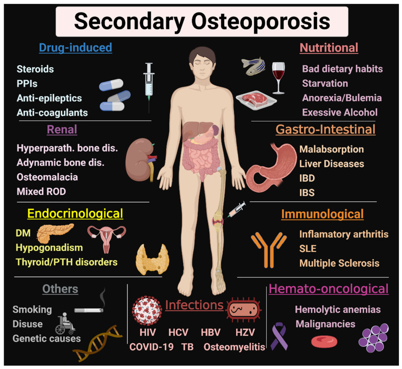
Fragility fracture is a worldwide problem and a main cause of disability and impaired quality of life. It is primarily caused by osteoporosis, characterized by impaired bone quantity and or quality. Proper diagnosis of osteoporosis is essential for prevention of fragility fractures. Osteoporosis can be primary in postmenopausal women because of estrogen deficiency. Secondary forms of osteoporosis are not uncommon in both men and women. Most systemic illnesses and organ dysfunction can lead to osteoporosis. The kidney plays a crucial role in maintaining physiological bone homeostasis by controlling minerals, electrolytes, acid-base, vitamin D and parathyroid function. Chronic kidney disease with its uremic milieu disturbs this balance, leading to renal osteodystrophy. Diabetes mellitus represents the most common secondary cause of osteoporosis. Thyroid and parathyroid disorders can dysregulate the osteoblast/osteoclast functions. Gastrointestinal disorders, malnutrition and malabsorption can result in mineral and vitamin D deficiencies and bone loss. Patients with chronic liver disease have a higher risk of fracture due to hepatic osteodystrophy. Proinflammatory cytokines in infectious, autoimmune, and hematological disorders can stimulate osteoclastogenesis, leading to osteoporosis. Moreover, drug-induced osteoporosis is not uncommon. In this review, we focus on causes, pathogenesis, and management of secondary osteoporosis.

Keywords: bone loss; bone mineral density; causes; fracture; management.


Conflict of interest statement

All the authors declared no conflict of interest in this work.

Figures

Figure 1
Figure 1
Causes of secondary osteoporosis. Various causes of secondary osteoporosis are illustrated in this figure. They include ROD, DM, thyroid and parathyroid disorders, malabsorption, IBD, IBS, nutritional causes, drug-induced, infections, anemia, malignancies, inflammatory arthritis, SLE, smoking, and genetic causes. PPIs: proton pump inhibitors, ROD: renal osteodystrophy, DM: diabetes mellitus, PTH: parathyroid, IBD: inflammatory bowel disease, IBS: irritable bowel syndrome, SLE: systemic lupus erythematosus. HIV: human immunodeficiency virus, HCV: hepatitis C virus, HBV: hepatitis B virus, HZV: herpes zoster virus, TB: tuberculosis. This Figure was created with BioRender.com (accessed on 1 February 2022).
Figure 2
Figure 2
Pragmatic diagnostic approach for newly diagnosed patients with osteoporosis. A systematic approach for the analysis and detection of a secondary cause of osteoporosis is recommended for all patients with a new diagnosis of osteoporosis. A full history and physical examination followed by a routine laboratory investigation for the most common and simple underlying causes of osteoporosis are required for most cases. Some additional investigation may be considered after routine lab for the suspected cases. CKD, chronic kidney disease, CRP: C-reactive protein; ESR: erythrocyte sedimentation rate; IBD: inflammatory bowel diseases; HCV: hepatitis C virus; HBV: hepatitis B virus; HIV: human immunodeficiency virus; TB: tuberculosis; FSH: follicle stimulating hormone; LH: luteinizing hormone.
Figure 3
Figure 3
Approach for prevention and management of secondary osteoporosis. Correction of the underlying causes of secondary osteoporosis is the cornerstone of prevention and treatment. All patients can benefit from non-pharmacological intervention, DEXA scan and assessment of fracture risk. Anti-osteoporotic medications (antiresorptives and osteoanabolics) can be used in selected cases with high fracture risk. DEXA: dual-energy X-ray absorptiometry, FRAX: fracture-risk algorithm, SERM: Selective estrogen receptor modulators.
Figure 4
Figure 4
Mechanism of action of common antiosteoporotic medications. Antiosteoporotic medications can be divided into two main categories: 1. Antiresorptives “on the right side” act mainly by inhibiting osteoclasts. Bisphosphonates act by inhibiting osteoclast differentiation from osteoclast precursors. The monoclonal antibody “denausumab” inhibits osteoclast differentiation by binding to RANKL, preventing its interaction with RANK. SERMs increase OPG production, thus inhibiting osteoclastogenesis. 2. Osteoanabolics “on the left side” stimulate bone formation via activation of PTH (teriparatide) or PTH-related peptide (abaloparatide) receptors. Romosuzumab is an anti-sclerostin monoclonal antibody. Thus, it stimulates osteoblast differentiation and function. MSC: mesenchymal stem cells, HSC: hematopoietic stem cells, SERMs: selective estrogen receptor modulators, OPG: osteoprotegerin, RANK: Receptor activator of nuclear factor κ B, RANKL: receptor activator of nuclear factor kappa-Β ligand. this figure was created with BioRender.com (accessed on 1 February 2022).

References

    1. Tannenbaum C., Clark J., Schwartzman K., Wallenstein S., Lapinski R., Meier D., Luckey M. Yield of Laboratory Testing to Identify Secondary Contributors to Osteoporosis in Otherwise Healthy Women. J. Clin. Endocrinol. Metab. 2002;87:4431–4437. doi: 10.1210/jc.2002-020275. - DOI - PubMed
    1. Kanis J.A., Hans D., Cooper C., Baim S., Bilezikian J.P., Binkley N., Cauley J.A., Compston J.E., Dawson-Hughes B., El-Hajj Fuleihan G., et al. Interpretation and use of FRAX in clinical practice. Osteoporos. Int. 2011;22:2395. doi: 10.1007/s00198-011-1713-z. - DOI - PubMed
    1. Najar M.S., Mir M.M., Muzamil M. Prevalence of osteoporosis in patients with chronic kidney disease (stages 3–5) in comparison with age-and sex-matched controls: A study from Kashmir Valley Tertiary Care Center. Saudi J. Kidney Dis. Transpl. 2017;28:538. - PubMed
    1. Aggarwal H., Jain D., Yadav S., Kaverappa V. Bone mineral density in patients with predialysis chronic kidney disease. Ren. Fail. 2013;35:1105–1111. doi: 10.3109/0886022X.2013.815102. - DOI - PubMed
    1. Fidan N., Inci A., Coban M., Ulman C., Kursat S. Bone mineral density and biochemical markers of bone metabolism in predialysis patients with chronic kidney disease. J. Investig. Med. 2016;64:861–866. doi: 10.1136/jim-2015-000043. - DOI - PubMed

LinkOut - more resources