

Intracardiac Echocardiogram: Feasibility, Efficacy, and Safety for Guidance of Transcatheter Multiple Atrial Septal Defects Closure
- PMID: 35566520
- PMCID: PMC9100238
- DOI: 10.3390/jcm11092394
Intracardiac Echocardiogram: Feasibility, Efficacy, and Safety for Guidance of Transcatheter Multiple Atrial Septal Defects Closure
Abstract
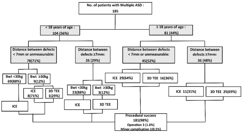
We aimed to determine the feasibility, efficacy, success, and safety of intracardiac echocardiography (ICE) in transcatheter multiple atrial septal defect (ASD) closure. Of 185 patients with multiple ASDs who underwent transcatheter closure, 140 (76%) patients who weighed <30kg with a narrow distance between defects or in whom single device closure was anticipated were guided by ICE and 45 patients were guided by three-dimensional (3D) transesophageal echocardiography (TEE) with or without ICE. Patients in the ICE group were relatively younger and weighed less than those in the 3D TEE group (p < 0.0001). The ratio of the distance between defects >7 mm was high, and more cases required ≥2 devices in the 3D TEE group than those in the ICE group (p < 0.0001). All patients in the 3D TEE group and seven patients (5%) in the ICE group were operated on under general anesthesia (p < 0.0001). The fluoroscopic time was shorter in the ICE group (13.98 ± 6.24 min vs. 24.86 ± 16.47 min, p = 0.0005). No difference in the complete closure rate and complications was observed. ICE-guided transcatheter and 3D TEE were feasible, safe, and effective in successful multiple ASD device closures, especially for young children and patients at high risk under general anesthesia.
Keywords: intracardiac echocardiography; multiple atrial septal defects; real-time three-dimensional transesophageal echocardiography; transcatheter closure.
Conflict of interest statement
The authors declare no conflict of interest.
Figures
References
-
- Du Z.D., Hijazi Z.M., Kleinman C.S., Silverman N.H., Larntz K., Amplatzer I. Comparison between transcatheter and surgical closure of secundum atrial septal defect in children and adults: Results of a multicenter nonrandomized trial. J. Am. Coll. Cardiol. 2002;39:1836–1844. doi: 10.1016/S0735-1097(02)01862-4. - DOI - PubMed
-
- Stout K.K., Daniels C.J., Aboulhosn J.A., Bozkurt B., Broberg C.S., Colman J.M., Crumb S.R., Dearani J.A., Fuller S., Gurvitz M., et al. 2018 AHA/ACC Guideline for the Management of Adults With Congenital Heart Disease: A Report of the American College of Cardiology/American Heart Association Task Force on Clinical Practice Guidelines. J. Am. Coll. Cardiol. 2019;73:e81–e192. doi: 10.1016/j.jacc.2018.08.1029. - DOI - PubMed
-
- Farhaj Z., Hongxin L., Wenbin G., Zhang W.L., Liang F., Zhang H.Z., Yuan G.D., Zou C.W. Device closure of diverse layout of multi-hole secundum atrial septal defect: Different techniques and long-term follow-up. J. Cardiothorac. Surg. 2019;14:130. doi: 10.1186/s13019-019-0952-5. - DOI - PMC - PubMed
-
- Hascoet S., Warin-Fresse K., Baruteau A.E., Hadeed K., Karsenty C., Petit J., Guerin P., Fraisse A., Acar P. Cardiac imaging of congenital heart diseases during interventional procedures continues to evolve: Pros and cons of the main techniques. Arch. Cardiovasc. Dis. 2016;109:128–142. doi: 10.1016/j.acvd.2015.11.011. - DOI - PubMed
LinkOut - more resources
Full Text Sources
