. 2022 Apr 26;11(9):2441.
doi: 10.3390/jcm11092441.

Impact of Whole Body Vibration and Zoledronic Acid on Femoral Structure after Ovariectomy: Morphological Evaluation

Affiliations

Impact of Whole Body Vibration and Zoledronic Acid on Femoral Structure after Ovariectomy: Morphological Evaluation

Nazar M Kostyshyn et al. J Clin Med. .

Abstract

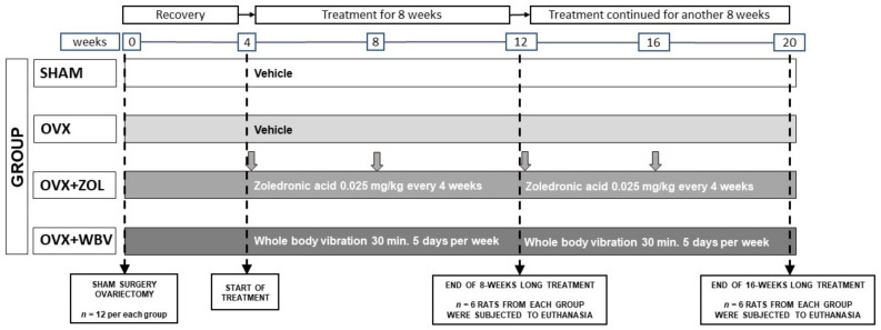
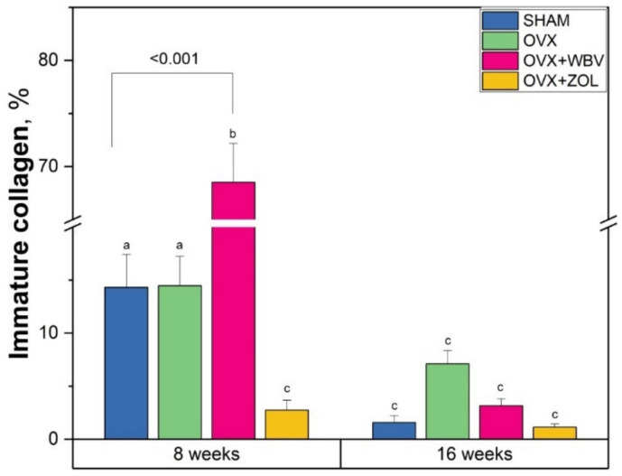
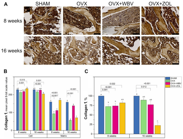
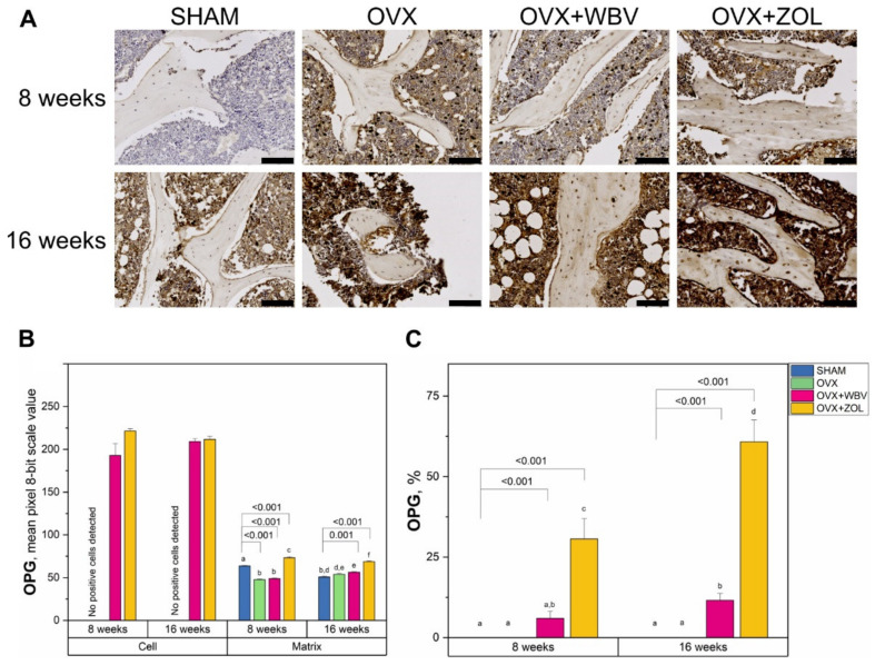
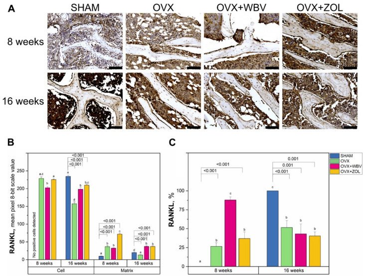
Our study aimed to evaluate the effect of whole body vibration (WBV) treatment as an non-pharmacological method of treatment for early osteopenia in ovariectomized female rats. In total, 48 female Wistar rats were assigned to two groups: sham-operated control (SHAM, n = 12) and ovariectomized (n = 36). Four weeks after ovariectomy, the animals were divided into three experimental groups (n = 12 each): ovariectomized (OVX), ovariectomized subjected to whole body vibration with acceleration level of 0.3 g (OVX + WBV), or ovariectomized subjected to i.m. injection of Zoledronic acid at a dose of 0.025 mg/kg (OVX + ZOL). After the 8th and 16th week of treatment n = 6 rats from each group were euthanized and isolated femora were subjected to histological examination of trabecular bone and analysis of the expression of collagen 1 (Col1), osteoprotegerin (OPG), and receptor activator of nuclear factor kappa-Β ligand (RANKL) involved in bone turnover. The obtained results indicated that widespread vibration therapy can provide negative outcomes such as deterioration of trabecular bone histomorphometry.

Keywords: osteoporosis; ovariectomy; rat model; whole body vibration.


Conflict of interest statement

The authors declare no conflict of interest.

Figures

Figure 1
Figure 1
Scheme of the experimental design. Four weeks after the ovariectomy, rats from the OVX + WBV group were subjected to whole body vibration treatment (30 min., 5 days per week) while rats from the OVX + ZOL group were injected with Zoledronic acid (i.m. 0.025 mg/kg every 4 weeks, vertical gray arrows show the time of injections). Rats from the SHAM, OVX and OVX + WBV groups were injected with the vehicle (0.9% NaCl) at the same volume. After the 8th and the 16th week of treatment, six animals from each group were euthanized.
Figure 2
Figure 2
Immature collagen content in femora trabeculae of controls (SHAM surgery), ovariectomized rats (OVX), ovariectomized rats with whole body vibration (OVX + WBV), or Zoledronic acid (OVX + ZOL). Data are presented as mean ± SE. Statistically significant differences between groups are indicated by a, b and c letters as shown by two-way ANOVA with Tukey’s post hoc test (p < 0.05). Brackets show statistically significant results of Dunnett’s test post-hoc performed to evaluate the differences between the experimental groups and the corresponding sham operated controls (SHAM).
Figure 3
Figure 3
(A) Representative images of the immunohistochemical reactions for collagen 1 (Col1) in the trabecular bone of femora from controls (SHAM surgery), ovariectomized rats (OVX), ovariectomized rats with whole body vibration (OVX + WBV), or Zoledronic acid (OVX + ZOL). All the scale bars represent 100 μm. (B) Bar graphs show the intensity of expression of Col1 in cells (osteocytes) and matrix measured by the comparison of the pixel brightness value in the microscopic images converted to 8-bit grayscale—the higher the pixel value, the higher the intensity of the immunohistochemical reaction. (C) The percentage of cells with a positive response. Data are presented as mean ± SE with corresponding statistical analysis. Statistically significant differences between groups (at p < 0.05) are indicated by a, b, c, d, e, and f letters. Dunnett post-hoc significant p-values are placed over brackets.
Figure 4
Figure 4
(A) Representative images of the immunohistochemical reactions for osteoprotegerin (OPG) in the trabecular bone of femora from controls (SHAM surgery), ovariectomized rats (OVX), ovariectomized rats with whole body vibration (OVX + WBV), or Zoledronic acid (OVX + ZOL). All the scale bars represent 100 μm. (B) Bar graphs show the intensity of expression of OPG in cells (osteocytes) and matrix measured by the comparison of the pixel brightness value in the microscopic images converted to 8-bit grayscale—the higher the pixel value, the higher the intensity of the immunohistochemical reaction. (C) The percentage of cells with a positive response. Data are presented as mean ± SE with corresponding statistical analysis. Statistically significant differences between groups (at p < 0.05) are indicated by a, b, c, d, e, and f letters. Dunnett post-hoc significant p-values are placed over brackets.
Figure 5
Figure 5
(A) Representative images of the immunohistochemical reactions for receptor activator of nuclear factor kappa-Β ligand (RANKL) in the trabecular bone of femora from controls (SHAM surgery), ovariectomized rats (OVX), ovariectomized rats with whole body vibration (OVX + WBV), or Zoledronic acid (OVX + ZOL). All the scale bars represent 100 μm. (B) Bar graphs show the intensity of expression of RANKL in cells (osteocytes) and matrix measured by the comparison of the pixel brightness value in the microscopic images converted to 8-bit grayscale—the higher the pixel value, the higher the intensity of the immunohistochemical reaction. (C) The percentage of cells with a positive response. Data are presented as mean ± SE with corresponding statistical analysis. Statistically significant differences between groups (at p < 0.05) are indicated by a, b, c, and d letters. Dunnett post-hoc significant p-values are placed over brackets.

Similar articles

Cited by

References

    1. Akkawi I., Zmerly H. Osteoporosis: Current concepts. Joints. 2018;6:122–127. doi: 10.1055/s-0038-1660790. - DOI - PMC - PubMed
    1. Cooper C., Melton L.J., 3rd Epidemiology of osteoporosis. Trends Endocrinol. Metab. 1992;3:224–229. doi: 10.1016/1043-2760(92)90032-V. - DOI - PubMed
    1. Khosla S., Monroe D.G. Regulation of Bone Metabolism by Sex Steroids. Cold Spring Harb. Perspect. Med. 2018;8:a031211. doi: 10.1101/cshperspect.a031211. - DOI - PMC - PubMed
    1. Singh-Ospina N., Maraka S., Rodriguez-Gutierrez R., Davidge-Pitts C., Nippoldt T.B., Prokop L.J., Murad M.H. Effect of sex steroids on the bone health of transgender individuals: A systematic review and meta-analysis. J. Clin. Endocrinol. Metab. 2017;102:3904–3913. doi: 10.1210/jc.2017-01642. - DOI - PubMed
    1. Sözen T., Özışık L., Başaran N.Ç. An overview and management of osteoporosis. Eur. J. Rheumatol. 2017;4:46–56. doi: 10.5152/eurjrheum.2016.048. - DOI - PMC - PubMed

LinkOut - more resources