

Electrospun Medical Sutures for Wound Healing: A Review
- PMID: 35566807
- PMCID: PMC9105379
- DOI: 10.3390/polym14091637
Electrospun Medical Sutures for Wound Healing: A Review
Abstract
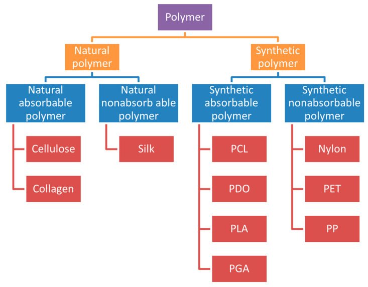
With the increasing demand for wound healing around the world, the level of medical equipment is also increasing, but sutures are still the preferred medical equipment for medical personnel to solve wound closures. Compared with the traditional sutures, the nanofiber sutures produced by combining the preparation technology of drug-eluting sutures have greatly improved both mechanical properties and biological properties. Electrospinning technology has attracted more attention as one of the most convenient and simple methods for preparing functional nanofibers and the related sutures. This review firstly discusses the structural classification of sutures and the performance analysis affecting the manufacture and use of sutures, followed by the discussion and classification of electrospinning technology, and then summarizes the relevant research on absorbable and non-absorbable sutures. Finally, several common polymers and biologically active substances used in creating sutures are concluded, the related applications of sutures are discussed, and the future prospects of electrospinning sutures are suggested.
Keywords: drug delivery; electrospinning; medical polymers; nanofibers; sutures; wound healing.
Conflict of interest statement
The authors declare no conflict of interest.
Figures
Similar articles
-
Electrospun Medicated Nanofibers for Wound Healing: Review.Membranes (Basel). 2021 Oct 9;11(10):770. doi: 10.3390/membranes11100770. Membranes (Basel). 2021. PMID: 34677536 Free PMC article. Review.
-
State-of-the-Art Review of Advanced Electrospun Nanofiber Composites for Enhanced Wound Healing.AAPS PharmSciTech. 2023 Nov 29;24(8):246. doi: 10.1208/s12249-023-02702-9. AAPS PharmSciTech. 2023. PMID: 38030812 Review.
-
Electrospun nanofibers promote wound healing: theories, techniques, and perspectives.J Mater Chem B. 2021 Apr 14;9(14):3106-3130. doi: 10.1039/d1tb00067e. Epub 2021 Mar 24. J Mater Chem B. 2021. PMID: 33885618
-
Electrospun nanofibrous membrane for biomedical application.SN Appl Sci. 2022;4(6):172. doi: 10.1007/s42452-022-05056-2. Epub 2022 May 13. SN Appl Sci. 2022. PMID: 35582285 Free PMC article. Review.
-
Applications of Electrospun Drug-Eluting Nanofibers in Wound Healing: Current and Future Perspectives.Polymers (Basel). 2022 Jul 20;14(14):2931. doi: 10.3390/polym14142931. Polymers (Basel). 2022. PMID: 35890706 Free PMC article. Review.
Cited by
-
Effect of Electrospun PLGA/Collagen Scaffolds on Cell Adhesion, Viability, and Collagen Release: Potential Applications in Tissue Engineering.Polymers (Basel). 2023 Feb 21;15(5):1079. doi: 10.3390/polym15051079. Polymers (Basel). 2023. PMID: 36904322 Free PMC article.
-
Electrospun Zein/Polyoxyethylene Core-Sheath Ultrathin Fibers and Their Antibacterial Food Packaging Applications.Biomolecules. 2022 Aug 12;12(8):1110. doi: 10.3390/biom12081110. Biomolecules. 2022. PMID: 36009003 Free PMC article.
-
Study on Filtration Performance of PVDF/PUL Composite Air Filtration Membrane Based on Far-Field Electrospinning.Polymers (Basel). 2022 Aug 12;14(16):3294. doi: 10.3390/polym14163294. Polymers (Basel). 2022. PMID: 36015550 Free PMC article.
-
Assessment of the Mechanical Properties of Different Suture Materials for Oral Surgery: An In Vitro Tensile Strength Study.Cureus. 2024 Aug 1;16(8):e65952. doi: 10.7759/cureus.65952. eCollection 2024 Aug. Cureus. 2024. PMID: 39221394 Free PMC article.
-
A Review on Electrospun Poly(amino acid) Nanofibers and Their Applications of Hemostasis and Wound Healing.Biomolecules. 2022 Jun 7;12(6):794. doi: 10.3390/biom12060794. Biomolecules. 2022. PMID: 35740919 Free PMC article. Review.
References
Publication types
LinkOut - more resources
Full Text Sources
