. 2022 Apr 20;14(9):1677.
doi: 10.3390/polym14091677.

Biocompatible Nanoparticles Based on Amphiphilic Random Polypeptides and Glycopolymers as Drug Delivery Systems

Affiliations

Biocompatible Nanoparticles Based on Amphiphilic Random Polypeptides and Glycopolymers as Drug Delivery Systems

Natalia Zashikhina et al. Polymers (Basel). .

Abstract

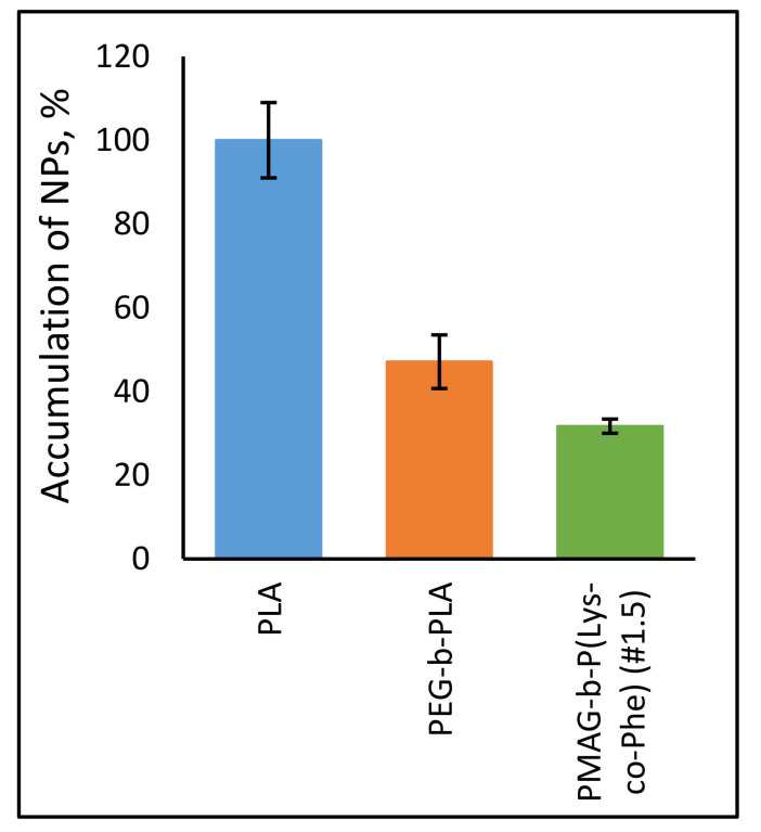
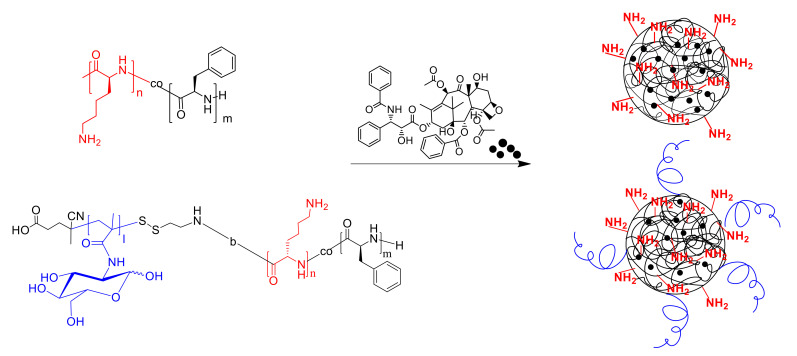
In this research, the development and investigation of novel nanoobjects based on biodegradable random polypeptides and synthetic non-degradable glycopolymer poly(2-deoxy-2-methacrylamido-d-glucose) were proposed as drug delivery systems. Two different approaches have been applied for preparation of such nanomaterials. The first one includes the synthesis of block-random copolymers consisting of polypeptide and glycopolymer and capable of self-assembly into polymer particles. The synthesis of copolymers was performed using sequential reversible addition-fragmentation chain transfer (RAFT) and ring-opening polymerization (ROP) techniques. Amphiphilic poly(2-deoxy-2-methacrylamido-d-glucose)-b-poly(l-lysine-co-l-phenylalanine) (PMAG-b-P(Lys-co-Phe)) copolymers were then used for preparation of self-assembled nanoparticles. Another approach for the formation of polypeptide-glycopolymer particles was based on the post-modification of preformed polypeptide particles with an oxidized glycopolymer. The conjugation of the polysaccharide on the surface of the particles was achieved by the interaction of the aldehyde groups of the oxidized glycopolymer with the amino groups of the polymer on particle surface, followed by the reduction of the formed Schiff base with sodium borohydride. A comparative study of polymer nanoparticles developed with its cationic analogues based on random P(Lys-co-d-Phe), as well as an anionic one-P(Lys-co-d-Phe) covered with heparin--was carried out. In vitro antitumor activity of novel paclitaxel-loaded PMAG-b-P(Lys-co-Phe)-based particles towards A549 (human lung carcinoma) and MCF-7 (human breast adenocarcinoma) cells was comparable to the commercially available Paclitaxel-LANS.

Keywords: amphiphilic copolymers; cellular uptake of particles; drug delivery systems; polymer particles; polypeptides; random and block-random copolymers; synthetic glycopolymers.


Conflict of interest statement

The authors declare no conflict of interest.

Figures

Figure 1
Figure 1
Scheme of synthesis of amphiphilic PMAG-b-P(Lys-co-Phe).
Figure 2
Figure 2
1H NMR spectrum of PMAG-b-P(Lys(Z)-co-Phe) (sample #1.2).
Figure 3
Figure 3
Scheme of synthesis of oxidized PMAG.
Figure 4
Figure 4
Schematic representation of: (1) PMAG-b-P(Lys-co-Phe) self-assembly; (2) P(Lys-co-Phe) self-assembly; (3) covering of P(Lys-co-d-Phe)-based NPs with heparin; (4) covalent modification of P(Lys-co-dl-Phe)-based NPs with ox-PMAG.
Figure 4
Figure 4
Schematic representation of: (1) PMAG-b-P(Lys-co-Phe) self-assembly; (2) P(Lys-co-Phe) self-assembly; (3) covering of P(Lys-co-d-Phe)-based NPs with heparin; (4) covalent modification of P(Lys-co-dl-Phe)-based NPs with ox-PMAG.
Figure 5
Figure 5
Dependence of (P(Lys-co-dl-Phe)-PMAG) particle ζ-potential on ox-PMAG concentration of different content of aldehyde groups (a) and dependence of P(Lys-co-dl-Phe) and P(Lys-co-d-Phe) particle size and ζ-potential on ox-PMAG (obtained at [NaIO4]/[MAG] = 0.3) (b) and heparin concentration, respectively (c) (0.01 M PBS, рН 7.4, CNPs = 0.1 mg/mL).
Figure 6
Figure 6
Hydrodynamic diameters of different nanoparticles (C = 0.1 mg/mL) in the buffer solution (0.01 M PBS, pH 7.4) and culture medium (DMEM + 10% FCS (v/v). The compositions of different polymer samples are provided in Table 2.
Figure 7
Figure 7
Changes in hydrodynamic diameters of NPs over time (DLS): (a) P(Lys-co-Phe) and (b) PMAG-b-P(Lys-co-Phe) NPs. Conditions of biodegradation: 0.01 M PBS, pH 7.4, T = 37 °C, CNPs = 1.0 mg/mL; Cpapain = 0.5 mg/mL. The compositions of different polymer samples are provided in Table 2.
Figure 8
Figure 8
Fluorescent images of A549 cells treated with Cy3-labeled NPs: (1) PMAG-b-P(Lys-co-Phe), sample #1.5, (2) P(Lys-co-d-Phe), sample #2.1 and (3) (P(Lys-co-d-Phe)-HEP, sample #3.1 for 4 h (CNPs = 60 µg/mL). Images from left to right show Cy3-labeled nanoparticles in cells (red), cell nuclei stained by DAPI (blue), and overlays of two images (×20). Scale bar: 50 μm.
Figure 9
Figure 9
Dependence of NPs accumulation on their concentration (t = 24 h) (a) and kinetics of cell uptake of Cy3-labeled NPs (b): P(Lys-co-d-Phe) (sample #2.1), (P(Lys-co-d-Phe)-HEP (sample #3.1) and PMAG-b-P(Lys-co-Phe) (sample #1.5) NPs (CNPs = 25 μg/mL, CCy3 = 50 μg/mg of NPs, A549 cells). The compositions of different polymer samples are provided in Table 2.
Figure 10
Figure 10
Dependence of NPs accumulation in A549 cells and J774 A.1 macrophages on their composition and surface properties (CNPs = 25 μg/mL, CCy3 = 50 μg/mg of NPs, t = 24 h).
Figure 11
Figure 11
Uptake of the Cy5-labeled NPs by J774 A.1 macrophages. (CNPs = 50 μg/mL, CCy5 = 3 μg/mg of NPs, t = 6 h).
Figure 12
Figure 12
Schematic representation of paclitaxel-loaded NPs formulations.

Similar articles

Cited by

References

    1. Raj S., Khurana S., Choudhari R., Kesari K.K., Kamal M.A., Garg N., Ruokolainen J., Das B.C., Kumar D. Specific targeting cancer cells with nanoparticles and drug delivery in cancer therapy. Semin. Cancer Biol. 2021;69:166–177. doi: 10.1016/j.semcancer.2019.11.002. - DOI - PubMed
    1. Ding S., Khan A.I., Cai X., Song Y., Lyu Z., Du D., Dutta P., Lin Y. Overcoming blood–brain barrier transport: Advances in nanoparticle-based drug delivery strategies. Mater. Today. 2020;37:112–125. doi: 10.1016/j.mattod.2020.02.001. - DOI - PMC - PubMed
    1. Luo Y., Yang H., Zhou Y.F., Hu B. Dual and multi-targeted nanoparticles for site-specific brain drug delivery. J. Control. Release. 2020;317:195–215. doi: 10.1016/j.jconrel.2019.11.037. - DOI - PubMed
    1. Jeevanandam J., Pal K., Danquah M.K. Virus-like nanoparticles as a novel delivery tool in gene therapy. Biochimie. 2019;157:38–47. doi: 10.1016/j.biochi.2018.11.001. - DOI - PubMed
    1. Petros R.A., DeSimone J.M. Strategies in the design of nanoparticles for therapeutic applications. Nat. Rev. Drug Discov. 2010;9:615–627. doi: 10.1038/nrd2591. - DOI - PubMed

LinkOut - more resources