. 2022 May 2;14(9):1865.
doi: 10.3390/polym14091865.

Aquaponics-Derived Tilapia Skin Collagen for Biomaterials Development

Affiliations

Aquaponics-Derived Tilapia Skin Collagen for Biomaterials Development

Nunzia Gallo et al. Polymers (Basel). .

Abstract

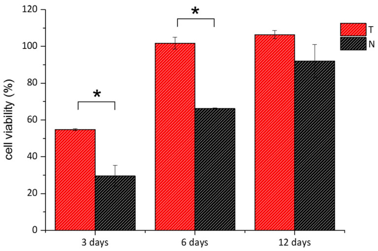
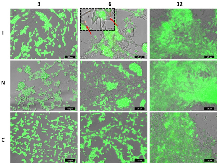
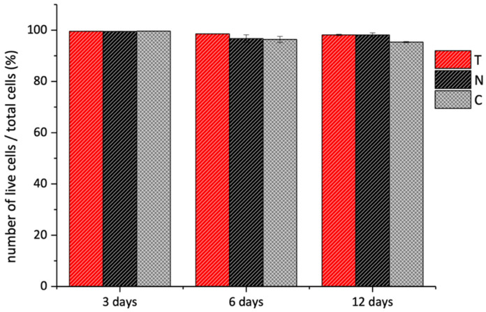
Collagen is one of the most widely used biomaterials in health-related sectors. The industrial production of collagen mostly relies on its extraction from mammals, but several issues limited its use. In the last two decades, marine organisms attracted interest as safe, abundant, and alternative source for collagen extraction. In particular, the possibility to valorize the huge quantity of fish industry waste and byproducts as collagen source reinforced perception of fish collagen as eco-friendlier and particularly attractive in terms of profitability and cost-effectiveness. Especially fish byproducts from eco-sustainable aquaponics production allow for fish biomass with additional added value and controlled properties over time. Among fish species, Oreochromis niloticus is one of the most widely bred fish in large-scale aquaculture and aquaponics systems. In this work, type I collagen was extracted from aquaponics-raised Tilapia skin and characterized from a chemical, physical, mechanical, and biological point of view in comparison with a commercially available analog. Performed analysis confirmed that the proprietary process optimized for type I collagen extraction allowed to isolate pure native collagen and to preserve its native conformational structure. Preliminary cellular studies performed with mouse fibroblasts indicated its optimal biocompatibility. All data confirmed the eligibility of the extracted Tilapia-derived native type I collagen as a biomaterial for healthcare applications.

Keywords: aquaponic; biomaterials; skin; tilapia; type I collagen.


Conflict of interest statement

The authors declare no conflict of interest.

Figures

Figure 1
Figure 1
On the left: Comparison of electrophoretic pattern of T and N; proteins were separated by SDS-PAGE and Coomassie stained; high molecular weight protein markers (MW) ranging from 10 to 250 kDa were used to estimate the molecular weight of the proteins, type I collagen from horse tendon (C) was used as an example of integrity and purity. On the right: comparison of the molecular weight of α1 and α2 chains of T (red) and N (black) with literature data about α1 and α2 of ASC (green) and PSC (grey) isolated from Tilapia skin [23,38,44,45,46,49,50,51,52,53,54,55,57].
Figure 2
Figure 2
Comparison of the amino acid relative abundance of T (red) and N (black) with the literature data from ASC (green) and PSC (grey) isolated from Tilapia skin [22,23,24,44,45,50,51,52,54,57,58,59].
Figure 3
Figure 3
FT-IR spectra of T (red) and N (black) samples (A).
Figure 4
Figure 4
On the left the 2D WAXS patterns on N and T samples. The black and the red arrows show the directions along which the equatorial and the meridional diffraction signals, respectively. On the right the 1D diffraction profiles of both samples. The equatorial peak (q2) is marked with the black line. The magenta lines show the peak positions of Ultralene sachet.
Figure 5
Figure 5
Representative DSC thermograms of T (red) and N (black) samples showing the endothermic phenomena of type I collagen denaturation.
Figure 6
Figure 6
Correlation of the denaturation temperature with the hydroxyproline content (left) and the molecular weight (right), and comparison of T (red) and N (black) data with literature data on ASC (green) and PSC (grey) [12,13,22,23,24,44,45,51,52,54,56,57,58,59,66].
Figure 7
Figure 7
Instant contact angle on T (left) and N (right) substrates.
Figure 8
Figure 8
Representative stress-strain curves of T film (red) compared to N film (black).
Figure 9
Figure 9
MTT viability assay of 3T3 cells grown over T (red bars) and N (black bars) was performed after 3, 6, and 12 days. Data were reported as the percentage viability of the samples over the control cells (i.e., cells grown on standard plates). (*) indicates statistical significance with p < 0.01.
Figure 10
Figure 10
Live/Dead assay of 3T3 cells grown over T and N was performed after 3, 6, and 12 days. The images correspond to the overlapped fluorescent channels (green for calcein signal and red for ethidium homodimer) and the bright field. The inset in the upper-central panel shows some dead cells among the live ones. Note: in the panels in which cells are localized on different focal lanes (elongated on the bottom plane and clustered on the upper plane), the green fluorescent channel has been slightly reduced to allow the detection of the dead cells. Please, refer to the images of Figures S2–S4 for the original signal intensity for each fluorescent channel. Scale bar is 125 µm.
Figure 11
Figure 11
Live/Dead assay of 3T3 cells grown over T (red bars) and N (black bars) substrates performed after 3, 6, and 12 days. The data are presented as the average percentage of live cells over the number of total cells. The analysis was performed through Image J Software over three different fluorescent images for each sample and measuring the green (i.e., live cells) and red (i.e., dead cells) pixels of each image.

References

    1. Gelse K., Pöschl E., Aigner T. Collagens—Structure, function, and biosynthesis. Adv. Drug Deliv. Rev. 2003;55:1531–1546. doi: 10.1016/j.addr.2003.08.002. - DOI - PubMed
    1. Salvatore L., Gallo N., Natali M.L., Terzi A., Sannino A., Madaghiele M. Mimicking the Hierarchical Organization of Natural Collagen: Toward the Development of Ideal Scaffolding Material for Tissue Regeneration. Front. Bioeng. Biotechnol. 2021;9:644595. doi: 10.3389/fbioe.2021.644595. - DOI - PMC - PubMed
    1. Gallo N., Natali M.L., Sannino A., Salvatore L. An overview of the use of equine collagen as emerging material for biomedical applications. J. Funct. Biomater. 2020;11:79. doi: 10.3390/jfb11040079. - DOI - PMC - PubMed
    1. Bianchini P., Parma B. Immunological safety evaluation of a horse collagen haemostatic pad. Arzneim. Forsch./Drug Res. 2001;51:414–419. doi: 10.1055/s-0031-1300056. - DOI - PubMed
    1. Lynn A., Yannas I., Bonfield W. Antigenicity and immunogenicity of collagen. J. Biomed. Mater. Res. Part B Appl. Biomater. 2004;71:343–354. doi: 10.1002/jbm.b.30096. - DOI - PubMed

LinkOut - more resources