Review
. 2022 May 7;14(9):1913.
doi: 10.3390/polym14091913.

Recent Advances in Fluorescence Recovery after Photobleaching for Decoupling Transport and Kinetics of Biomacromolecules in Cellular Physiology

Affiliations
Review

Recent Advances in Fluorescence Recovery after Photobleaching for Decoupling Transport and Kinetics of Biomacromolecules in Cellular Physiology

Ning Cai et al. Polymers (Basel). .

Abstract

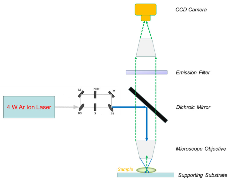
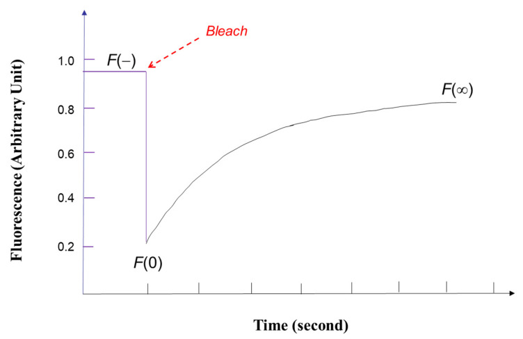
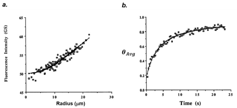
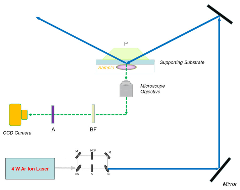
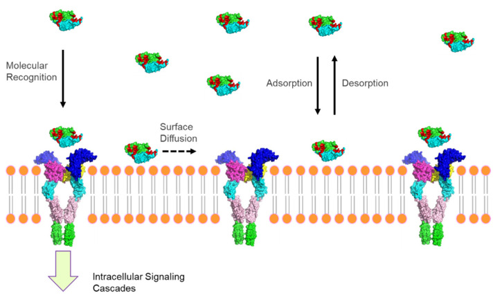
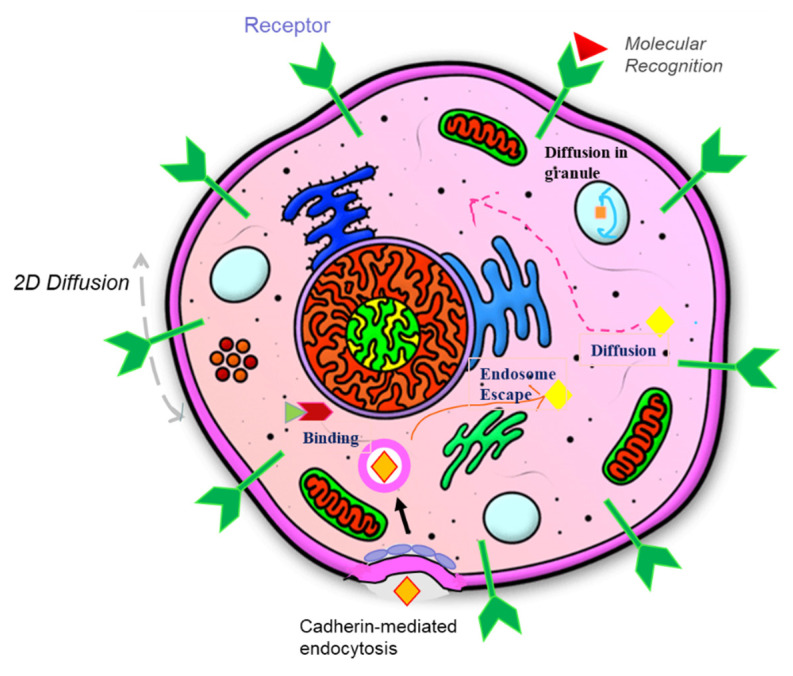
Among the new molecular tools available to scientists and engineers, some of the most useful include fluorescently tagged biomolecules. Tools, such as green fluorescence protein (GFP), have been applied to perform semi-quantitative studies on biological signal transduction and cellular structural dynamics involved in the physiology of healthy and disease states. Such studies focus on drug pharmacokinetics, receptor-mediated endocytosis, nuclear mechanobiology, viral infections, and cancer metastasis. In 1976, fluorescence recovery after photobleaching (FRAP), which involves the monitoring of fluorescence emission recovery within a photobleached spot, was developed. FRAP allowed investigators to probe two-dimensional (2D) diffusion of fluorescently-labelled biomolecules. Since then, FRAP has been refined through the advancements of optics, charged-coupled-device (CCD) cameras, confocal microscopes, and molecular probes. FRAP is now a highly quantitative tool used for transport and kinetic studies in the cytosol, organelles, and membrane of a cell. In this work, the authors intend to provide a review of recent advances in FRAP. The authors include epifluorescence spot FRAP, total internal reflection (TIR)/FRAP, and confocal microscope-based FRAP. The underlying mathematical models are also described. Finally, our understanding of coupled transport and kinetics as determined by FRAP will be discussed and the potential for future advances suggested.

Keywords: bio-interfaces; biomolecules; biophysical techniques; fluorescence recovery after photobleaching; polymers; reaction; transport.


Conflict of interest statement

The authors declare no conflict of interest.

Figures

Figure 1
Figure 1
The general experimental setup of epifluorescence spot FRAP based on a high-power argon laser, upright microscope, and CCD camera. Beam Splitter: BS; Dichroic Mirror: DM; Mirror: M; Shutter: S; Neutral Density Filler: NDF.
Figure 2
Figure 2
The fluorescence intensity measured by a CCD camera or PMT before and after photobleaching during FRAP experiments.
Figure 3
Figure 3
Photobleaching recovery for (a) fluorescence intensity (ITACTA) against r plotted from t = 0, immediately after photobleaching is fitted with Equation (22) for determining the value of R0 and ITB. (b) Deff is determined by fitting the dimensionless average fluorescence intensity data θAvg against time by fitting to Equation (31) and β is determined by measuring D in nonbinding system. Reprinted with permission from [95]. Copyright 1991, Elsevier.
Figure 4
Figure 4
The overall optical train for the TIR/FRAP setup attached to an inverted microscope. The basic equipment is a 4 W argon ion laser, a low-light level-cooled CCD camera, an inverted microscope, and a computer for image capture and analysis. Beam Splitter: BS; Mirror: M; Dichroic Mirror: DM; Aperture: A; Prism: P; Shutter: S; Band Pass Filter: BF; Neutral Density Filler: NDF.
Figure 5
Figure 5
Total internal reflection (TIR) microscopy taps on the production of an evanescent wave, which specifically illuminates biomacromolecules (circles) close to the solid substrate (e.g., fused silica). Immediately after photobleaching, the fluorescent tag on the biomacromolecules were deactivated by the intense laser beam (purple circles) and lost the fluorescence signal to be detected by CCD camera or photomultiplier tube. Afterwards, the fluorescence signal recovers by the exchange of the bleached molecules inside the region of interest with those unbleached biomacromolecules (red circles) through adsorption/desorption kinetics and surface diffusion.
Figure 6
Figure 6
A conventional TIR/FRAP recovery curve (F(t) vs. time) of fluorescently tagged DNA in PBS on amino-coated glass which was fitted with two adsorption state model (extended version of Equation (49)). Reprinted/adapted with permission from [96]. Copyright 1997, Vincent Chan.
Figure 7
Figure 7
The overall optical train for the Confocal FRAP setup. Beam Splitter: BS; Aperture: A; Prism: P; Shutter: S; Neutral Density Filler: NDF.
Figure 8
Figure 8
Transport and reaction kinetics involved in drug delivery in circulation, from blood vessel to lymphatics, which are detectable with FRAP techniques.
Figure 9
Figure 9
Interfacial phenomena involved in the molecular recognition of biomacromolecule (e.g., ligand) by transmembrane proteins (receptors) on plasma membrane.
Figure 10
Figure 10
Transport and binding kinetics including ligand-receptor binding, cadherin-mediated endocytosis, diffusion in cytoplasm, diffusion in membrane-bound organelles, escape from endosomes, etc., for maintaining the homeostasis of cellular functions.

References

    1. Manzoni C., Kia D.A., Vandrovcova J., Hardy J., Wood N.W., Lewis P.A., Ferrari R. Genome, transcriptome and proteome: The rise of omics data and their integration in biomedical sciences. Brief. Bioinform. 2018;19:286–302. doi: 10.1093/bib/bbw114. - DOI - PMC - PubMed
    1. Hartl D., de Luca V., Kostikova A., Laramie J., Kennedy S., Ferrero E., Siegel R., Fink M., Ahmed S., Millholland J., et al. Translational precision medicine: An industry perspective. J. Transl. Med. 2021;19:245. doi: 10.1186/s12967-021-02910-6. - DOI - PMC - PubMed
    1. Ganapathiraju M.K., Orii N. Research prioritization through prediction of future impact on biomedical science: A position paper on inference-analytics. GigaScience. 2013;2:11. doi: 10.1186/2047-217X-2-11. - DOI - PMC - PubMed
    1. Nair A., Chauhan P., Saha B., Kubatzky K.F. Conceptual evolution of cell signaling. Int. J. Mol. Sci. 2019;20:3292. doi: 10.3390/ijms20133292. - DOI - PMC - PubMed
    1. Tarbell J.M., Simon S.I., Curry F.-R.E. Mechanosensing at the vascular interface. Annu. Rev. Biomed. Eng. 2014;16:505–532. doi: 10.1146/annurev-bioeng-071813-104908. - DOI - PMC - PubMed

LinkOut - more resources