. 2022 Sep 10;31(18):3144-3160.
doi: 10.1093/hmg/ddac108.

Mice lacking MBNL1 and MBNL2 exhibit sudden cardiac death and molecular signatures recapitulating myotonic dystrophy

Affiliations

Mice lacking MBNL1 and MBNL2 exhibit sudden cardiac death and molecular signatures recapitulating myotonic dystrophy

Kuang-Yung Lee et al. Hum Mol Genet. .

Abstract

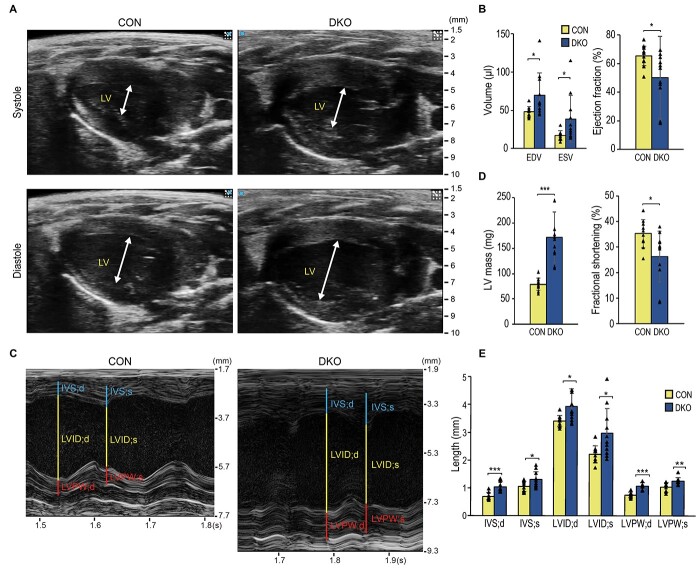
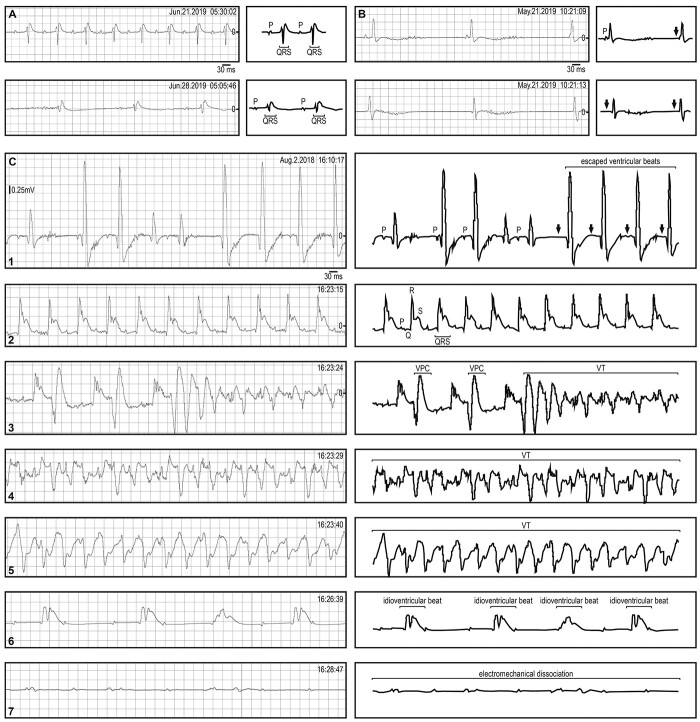
Myotonic dystrophy (DM) is caused by expansions of C(C)TG repeats in the non-coding regions of the DMPK and CNBP genes, and DM patients often suffer from sudden cardiac death due to lethal conduction block or arrhythmia. Specific molecular changes that underlie DM cardiac pathology have been linked to repeat-associated depletion of Muscleblind-like (MBNL) 1 and 2 proteins and upregulation of CUGBP, Elav-like family member 1 (CELF1). Hypothesis solely targeting MBNL1 or CELF1 pathways that could address all the consequences of repeat expansion in heart remained inconclusive, particularly when the direct cause of mortality and results of transcriptome analyses remained undetermined in Mbnl compound knockout (KO) mice with cardiac phenotypes. Here, we develop Myh6-Cre double KO (DKO) (Mbnl1-/-; Mbnl2cond/cond; Myh6-Cre+/-) mice to eliminate Mbnl1/2 in cardiomyocytes and observe spontaneous lethal cardiac events under no anesthesia. RNA sequencing recapitulates DM heart spliceopathy and shows gene expression changes that were previously undescribed in DM heart studies. Notably, immunoblotting reveals a nearly 6-fold increase of Calsequestrin 1 and 50% reduction of epidermal growth factor proteins. Our findings demonstrate that complete ablation of MBNL1/2 in cardiomyocytes is essential for generating sudden death due to lethal cardiac rhythms and reveal potential mechanisms for DM heart pathogenesis.


Figures

Graphical Abstract
Graphical Abstract
Figure 1
Figure 1
General features of Myh-Cre DKO mice. (A) No body size differences between control and DKO mice. Scale bar: 10 mm. (B) Kaplan–Meier survival analysis of control versus DKO mice showing a reduced lifespan in the DKOs (n = 11 per genotype. Log-rank test, P < 0.001). The numbers of mice at risk at different time points (0 ~ 50 weeks) were shown below. (C) A representative picture showing a normal control and an enlarged DKO heart. Scale bar: 1 mm. (D) Body weight analysis. (E) Heart weight analysis. (F) Heart-to-body weight ratio showed a significant increase in DKO mice. (From (D) to (F): Control, n = 7; DKO, n = 3. Data represent the mean ± SD. *, P < 0.05; ***, P < 0.001; 2-tailed Student’s t-test).
Figure 2
Figure 2
The evaluation of fibrosis in Myh6-Cre DKO hearts. (A) Hematoxylin and eosin (H&E) staining of control and DKO hearts. (B) Masson’s trichrome staining and (C) Sirius red staining using sections from left ventricles of control and DKOs. Scale bars represent 1 mm in (A) and 100 μM in (B&C) (n = 3 per genotype). (RA: right atrium; RV: right ventricle; LA: left atrium; LV: left ventricle; BV: blood vessel).
Figure 3
Figure 3
Echocardiography for analyzing Myh6-Cre DKO mice. (A) Representative B-mode 2-dimentional images of control and DKO mouse hearts during systolic and diastolic phases. The white arrows indicate internal chamber of the left ventricles. (B) The DKO mice showed increased EDV and ESV and the EF was significantly reduced in the DKOs. (C) The M-mode images of control and DKO mice during systolic and diastolic phases. (D) Increased LV mass and reduced FS were observed in the DKO mice. (E) Significant differences were detectable in the lengths of IVS, LVID and LVPW during systole and diastole. (Control, n = 10; DKO n = 11. Data represent the mean ± SD. *, P < 0.05; **, P < 0.01; ***, P < 0.001; 2-tailed Student’s t-test).
Figure 4
Figure 4
Basic analysis of ambulatory ECG in Myh6-Cre DKO mice. (A) Representative ECG recordings of control (top) and DKO (bottom), in the early stage. (B) Representative ECG recordings of control (top) and DKO (bottom) during the late stage. (C) Statistical analysis of RR, PR, QRS, QT and QTc intervals during early stage and late stage (n = 3 per genotype. Data represent the mean ± SD. *, P < 0.05; **, P < 0.01; ***, P < 0.001; 2-tailed Student’s t-test).
Figure 5
Figure 5
Examples of abnormal cardiac rhythm detected by ambulatory ECG in Myh6-Cre DKO mice. (A) The initial ECG recordings from a DKO mouse showing only PR prolongation and wide QRS complex (top). The pattern changed to sinus arrhythmia with bradycardia (bottom) a week later, 2 days before death. (B) The ECG recordings from a DKO mouse showing sinus bradycardia with isorhythmic atrial-ventricular dissociation (from top to bottom) and the mouse succumbed 12 h later. (C) Serial ambulatory ECG recordings from a Myh6-Cre DKO mouse before death. The initial ECG showing first-degree AV block, sinus bradycardia, intermittent sinus arrest with escaped ventricular beats (1st panel). Thirteen minutes later, an episode of sustained intraventricular conduction delay appeared (2nd panel). Sinus bradycardia with VPCs and VT occurred within seconds (3rd panel). Life-threatening persistent VT (4th panel). Continuous run of VT (5th panel). Complete AV block with escaped idioventricular beats (6th panel). The agonal rhythm and probably with electromechanical dissociation that finally ended the DKO mouse’s life (7th panel).
Figure 6
Figure 6
RNA-seq results of splicing alterations in Myh6-Cre DKO mice. (A) The 20 most affected genes in AS identified by RNA-seq. The results of three replicates of control and DKO hearts were colored based on the percent spliced-in (ψ, psi) values from 0 to 1. The ranking was from left to right including two AS events in Ktn1 gene (No. 2, exon 38 and No. 20, exon 31). (B) Validation of selected 10 targets among top 20 most affected genes that linked to DM cardiac phenotypes by RT-PCR. The numbers on the left side of each gel image indicate isoforms with or without a certain exon. (C) The ψ values for AS in the control and DKO hearts (n = 3 per genotype. Data represent the mean ± SD. **, P < 0.01; ***, P < 0.001; 2-tialed Student’s t-test).
Figure 7
Figure 7
The validation of RNA-seq expression results. (A) The selected upregulated and downregulated genes identified by RNA-seq were listed with values of log2 fold change (FC). (B) The qPCR for RNA-seq validation was shown by the CT (cycle of threshold) values (n = 3 per genotype, *, P < 0.05; **, P < 0.01; ***, P < 0.001; 2-tailed Student’s t-test). (C) The IPA using mRNA expression results in RNA-seq. The blue arrowhead indicated the location of CASQ1. The color codes represented upregulation (red) and downregulation (green) and the FCs were reflected by different saturations. (D) Immunoblots of selected targets showing expression changes in RNA-seq. The GAPDH and TUBULIN served as the loading controls. The signal intensities of the tested target proteins have been normalized with loading controls. Then control and DKO mouse groups were compared and the ratios of DKO/control were shown in the bar graph. (n = 3 per genotype, *, P < 0.05; ***, P < 0.001; 2-tailed Student’s t-test).
Figure 8
Figure 8
Hypothesis of the effect of CASQ1 upregulation in the DKO mice and the potential therapeutic strategy. (A) In the normal heart, Calsequestrin 2 (CASQ2) is the primary CASQ protein that keeps large SR calcium (Ca2+) capacity through inhibiting Ryanodine receptor 2 (RYR2). Upon stimulation, polymeric CASQ2 undergoes depolymerization, relieves the inhibition on RYR2 and releases Ca2+ from SR. This allows an increase in cytoplasmic [Ca2+] and induces transient amplitude for cardiac contraction. (B) While Calsequestrin 1 (CASQ1) is upregulated in the DKOs, it is hypothesized that the SR Ca2+ capacity increases but RYR2 is further inhibited. The decreased cytosolic [Ca2+] causes reduced excitation-contraction (E-C) coupling and cardiac contractility. (C) To rescue, phospholamban inhibitor (PLBi) may counteract the inhibitory effect of phospholamban (PLB) on SERCA2, which accounts for SR Ca2+ restoration. Consequently, it is hypothesized that the amount of SR Ca2+ capacity rises, inhibition on RYR2 is lifted, low cytoplasmic [Ca2+] is corrected and the cardiac contractility reverses.

Similar articles

Cited by

References

    1. Tome, S. and Gourdon, G. (2020) DM1 phenotype variability and triplet repeat instability: challenges in the development of new therapies. Int. J. Mol. Sci., 21, 457. - PMC - PubMed
    1. Ashizawa, T. and Sarkar, P.S. (2011) Myotonic dystrophy types 1 and 2. Handb. Clin. Neurol., 101, 193–237. - PubMed
    1. Thornton, C.A. (2014) Myotonic dystrophy. Neurol. Clin., 32, 705–719viii. - PMC - PubMed
    1. Mankodi, A., Lin, X., Blaxall, B.C., Swanson, M.S. and Thornton, C.A. (2005) Nuclear RNA foci in the heart in myotonic dystrophy. Circ. Res., 97, 1152–1155. - PubMed
    1. Kuyumcu-Martinez, N.M., Wang, G.S. and Cooper, T.A. (2007) Increased steady-state levels of CUGBP1 in myotonic dystrophy 1 are due to PKC-mediated hyperphosphorylation. Mol. Cell, 28, 68–78. - PMC - PubMed

Publication types

MeSH terms