. 2022 Jul 4;221(3):iyac081.
doi: 10.1093/genetics/iyac081.

SUMOylation of Dorsal attenuates Toll/NF-κB signaling

Affiliations

SUMOylation of Dorsal attenuates Toll/NF-κB signaling

Sushmitha Hegde et al. Genetics. .

Abstract

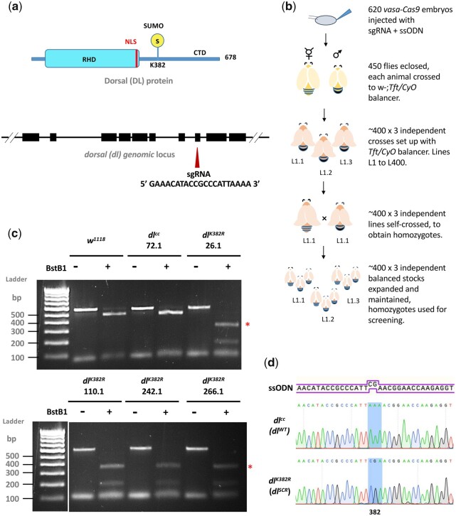
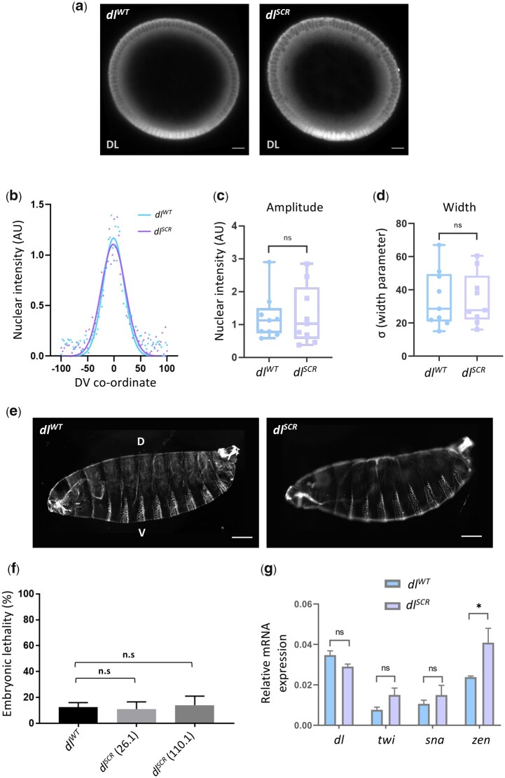
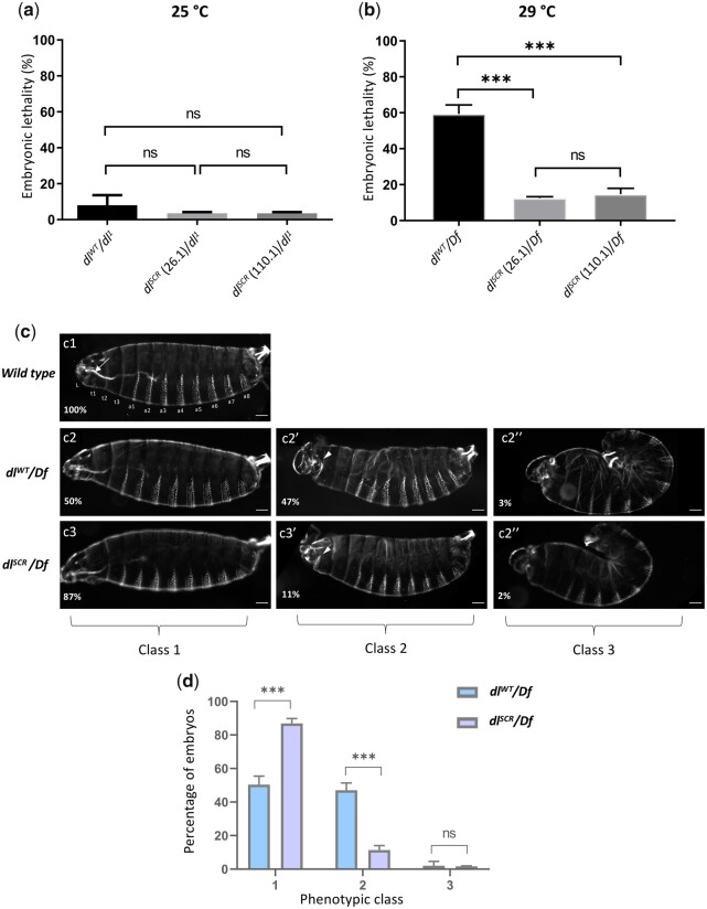
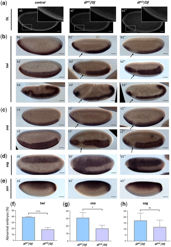
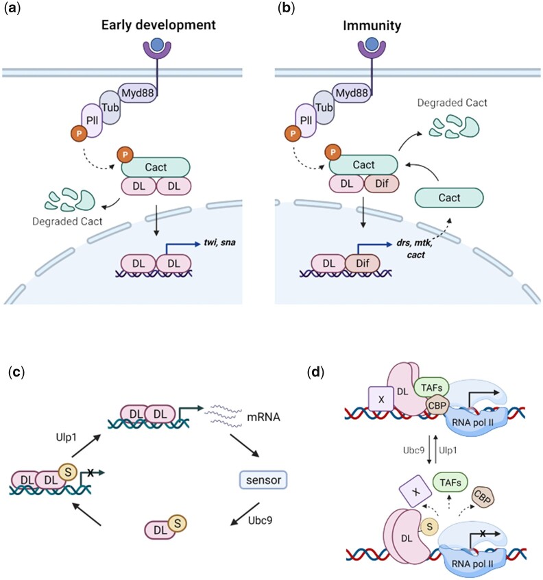
In Drosophila, Toll/NF-κB signaling plays key roles in both animal development and in host defense. The activation, intensity, and kinetics of Toll signaling are regulated by posttranslational modifications such as phosphorylation, SUMOylation, or ubiquitination that target multiple proteins in the Toll/NF-κB cascade. Here, we have generated a CRISPR-Cas9 edited Dorsal (DL) variant that is SUMO conjugation resistant. Intriguingly, embryos laid by dlSCR mothers overcome dl haploinsufficiency and complete the developmental program. This ability appears to be a result of higher transcriptional activation by DLSCR. In contrast, SUMOylation dampens DL transcriptional activation, ultimately conferring robustness to the dorso-ventral program. In the larval immune response, dlSCR animals show an increase in crystal cell numbers, stronger activation of humoral defense genes, and high cactus levels. A mathematical model that evaluates the contribution of the small fraction of SUMOylated DL (1-5%) suggests that it acts to block transcriptional activation, which is driven primarily by DL that is not SUMO conjugated. Our findings define SUMO conjugation as an important regulator of the Toll signaling cascade, in both development and host defense. Our results broadly suggest that SUMO attenuates DL at the level of transcriptional activation. Furthermore, we hypothesize that SUMO conjugation of DL may be part of a Ubc9-dependent mechanism that restrains Toll/NF-κB signaling.

Keywords: Drosophila; SUMO; haploinsufficiency; innate immunity; transcription.


Figures

Fig. 1.
Fig. 1.
Creating the dlK382R mutant using the CRISPR-Cas9 system. A schematic representation of the DL protein (in blue) and gene locus (in black) is presented in (a). DL is SUMOylated at K382, part of the consensus motif IKTE. A 20-bp sgRNA was designed to create a double-strand break in the vicinity of dlK382, in exon 8. The detailed crossing scheme for the generation of the dlK382R allele after injection of the gRNA plasmid and ssODN is outlined in (b). Homozygous flies obtained were screened by genomic PCR and digestion with the BstBI enzyme, which recognizes the engineered site of mutation, TTCGAA (c). Four independent lines—26.1, 110.1, 242.1, and 266.1 showed a distinct digest of the PCR product (indicated by red asterisks), while line 72.1 served as a control. d) The presence of the mutation was confirmed through sequencing (codon CGA is highlighted).
Fig. 2.
Fig. 2.
SUMO conjugation is dispensable for embryonic development. Transverse sections of representative cellular blastoderm embryos stained for DL (a). Localization in the nuclei was observed in embryos oriented dorsal-side up and ventral-side at the bottom. b) Representative nuclear intensity profiles of dlWT and dlSCR embryos, fitted to a Gaussian. The amplitude (c) and width (d) of the gradient centered at the ventral midline are plotted. n = 9, Student’s t-test, (ns) P > 0.05. Cuticle preparations (e) indicate regular arrangement of denticle bands and normal DV patterning. The percentage of unhatched embryos is plotted as embryonic lethality for control and 2 of the mutant lines, 26.1 and 110.1 (f). Genotype of mated mothers is listed on the X-axis. N = 3, ordinary 1-way ANOVA, (ns) P > 0.05. g) qRT-PCR analysis of dl transcripts and DL target genes twi, sna, and zen for embryos from mated females of the genotypes dlWT and dlSCR. N = 3, 2-way ANOVA, (ns) P > 0.05, (*) P < 0.05.
Fig. 3.
Fig. 3.
dlSCR is haplo-sufficient. Progeny of mothers of the indicated genotype, with 1 functional copy of DL, were scored for viability, 48 hr after egg lay, at 25°C (a) and 29°C (b). N = 3, mean ± SEM, ordinary 1-way ANOVA, (ns) P > 0.05, (***) P < 0.001. Cuticle preparations of progeny of the maternal genotypes dlWT/Df and dlSCR/Df, visualized under a dark-field microscope yielded 3 major ranges of phenotypes, classified as class 1 (intact head region/mouth hook; ventral denticle bands and filzkorper normal), class 2 (defective head structure; denticle bands and filzkorper intact), and class 3 (twisted embryos; defective head structures and filzkorper) (c). Cuticles are oriented dorsal-side up and anterior-side on the left. >100 embryos were scored in each replicate, and the percentage of each phenotypic class is plotted for dlWT/Df and and dlSCR/Df (d). N = 3, mean ± SEM, 2-way ANOVA, (ns) P > 0.05, (***) P < 0.001.
Fig. 4.
Fig. 4.
DL activity is altered in the SUMO-deficient mutant. The DL gradient was visualized with a DL antibody in control, dlWT/Df and dlSCR/Df embryos (a). Insets represent a zoomed-in view of the presumptive cephalic furrow in the ventral region. In situ hybridization images of stage 5 embryos probed with digoxigenin-AP-labeled antisense RNA probes against twi (b), sna (c), sog (d), and zen (e) are shown (b3–b3″ are stage 7 embryos, an exception). Embryos are oriented with the anterior side to the left and ventral side down (b1–b1″; b3–b3″; c1–c1″; e1–e1″), or tilted toward the reader (b2–b2″; c2–c2″; d1–d1″), for control (b1–e1), dlWT/Df (b1′–e1′), and dlSCR/Df (b1″–e1″). Arrows indicate a narrowing or an absence of the twi (a) and sna (b) pattern at the region of the presumptive cephalic furrow. d1′–d1″) A fusion of the sog gradient near the ventral cephalic region. Embryos showing a deviation from the normal pattern (narrowing/absence/fusion) for twi, sna, and sog were plotted as a percentage of total stained embryos, for the control, dlWT/Df, and and dlSCR/Df (f–h). Approximately 50 embryos were scored in each technical replicate, across 3 technical replicates. Data represented as mean ± SEM, unpaired t-test, (ns) P > 0.05, (***) P < 0.001, (*) P < 0.05.
Fig. 5.
Fig. 5.
DLSCR displays higher transcriptional activity. Maternal genotypes of the embryos used for the 3′ RNA-seq analysis and their pairwise comparison to obtain DEGs is presented in (a). Genes that were identified as direct targets of DL from published literature and DEGs across all the conditions are represented as a Venn diagram in (b). The subset of DEGs with known binding sites for DL is represented as a heatmap, for dlWT, dlSCR, dlWT/Df and dlSCR/Df embryos at 29°C in (c). LogCPM values are plotted. (d–f) Relative mRNA expression levels of dl, sna and zen transcripts, respectively, measured by qRT-PCR analysis, for 0–2 hr embryos laid by mothers of the indicated genotypes at 29°C. N = 3, mean ± SEM, ordinary 1-way ANOVA, (ns) P > 0.05, (**) P < 0.01, (*) P < 0.05
Fig. 6.
Fig. 6.
DLSCR is a robust immune effector in the larva. Total circulating hemocytes for dlWT and dlSCR are plotted as a bar graph in (a). N = 3, Mean ± SEM, unpaired t-test, (ns) P > 0.05. Crystal cells in the third-instar larva were observed under a bright-field microscope, for the genotypes indicated in (b). The last 3 posterior segments were imaged with the dorsal side facing the viewer. The number of crystal cells in the posterior segments was counted per animal for each genotype and is represented in (c). N = 3, mean ± SEM, ordinary 1-way ANOVA, (***) P < 0.0001. Transcript levels of Toll-responsive AMPs—drs and mtk (d) analyzed by qRT-PCR are plotted for the control and dlSCR. Data were collected at 0, 2, and 4 hr after septic injury with the Gram-positive pathogen S. saprophyticus, in the third instar larvae. N = 3, mean ± SEM, 2-way ANOVA, (*) P < 0.05, (**) P < 0.01, (****) P < 0.0001. Data are representative of at least 8 larvae per replicate, across 3 independent biological replicates.
Fig. 7.
Fig. 7.
DLSCR is responsive to Toll signaling in the larval fat body. DL is visualized via antibody staining (green), in the uninfected state (a) and infected state (c). Nuclei are labeled with DAPI (blue). Merged images (a1″) and (a2″) indicate uniform distribution in fat body cells. DL levels in the cytoplasm and nucleus were quantified and plotted as a nuclear/cytoplasmic (N/C) ratio for control and mutant (b). The N/C ratio was calculated for >40 cells in at least 5 fat bodies, across 3 independent replicates. Individual values are represented on a scatter plot, bar denotes mean ± SEM, statistical significance inferred by unpaired t-test, (ns) P > 0.05, (***) P < 0.001. DL predominantly partitions to the nucleus 60 min after infection with S. saprophyticus, evident in merged images (c1″) and (c2″). N/C ratio was quantified and plotted for dlSCR and dlWT (d), as in (b). Protein levels of DL and Cact in the unchallenged and immune-challenged fat body were determined by a western blot, shown in (e) and (f), respectively. Protein levels were normalized to the loading control (tubulin) and quantified, represented as relative expression levels below the respective blots for dlWT (yellow bar) and dlSCR (orange bar) (g and h). N = 3, bar chart represents mean ± SEM, statistical significance calculated by unpaired t-test, (ns) P > 0.05, (***) P < 0.001.
Fig. 8.
Fig. 8.
Mathematical model to understand roles for SUMOylated DL. Processes included in the mathematical model (a) UnSUMOylated (DLU) and SUMOylated DL (DLS) reversibly dimerize to form homo- and heterodimers (equilibrium constant KD) which are inhibited by Cact in the cytoplasm through reversible binding (equilibrium constant Ki). Dimers partition into nucleus (Kt) where they bind to the promoter P (binding constant Kp). Bound promoter catalyzes first-order reporter expression with rate constants denoted by k. Parameters for reactions involving the 2 homodimers and the heterodimer are represented by superscripts u, s, and us, respectively. b) Simulating the effect of SUMOylation of DL on transcription of DL target genes. Ratio of reporter expression levels (RSCR/RWT) is obtained by solving Supplementary Equations (23)–(28) with parameters listed in Supplementary SI-2. The rate of transcriptional activation by the DLU homodimer (ku) bound to the promoter is kept constant, and the ratio of reporter expression is calculated when the rate (ks) for promoter-bound DLS dimers is varied 2 orders of magnitude from this level. The process is repeated for different values of relative promoter binding affinity (Kps/Kpu= 0.01, 0.1, 1, 10, and 100). The reporter expression levels for dlSCR are greater than the corresponding WT levels when the relative transcription activity is lower (ks/ku< 1) for DLS and there is tighter binding of SUMOylated dimers to the promoter (Kps/Kpu>1). Other parameter ratios (KDs/KDu, Kts/Ktu, and Kis/Kiu) are kept at 1.
Fig. 9.
Fig. 9.
DL SUMOylation attenuates Toll signaling, acting via a feedback circuit. We suggest the following model for dampening of the Toll signal upon DL SUMOylation. When Toll signaling is initiated, DL migrates to the nucleus, and activates target genes, in both the developmental and immune contexts (a and b, respectively). UnSUMOylated DL activates transcription of DL-target genes. Once optimum levels of target transcripts are reached, or under conditions of stress, SUMOylation of DL is triggered, through as yet unknown mechanisms, curtailing excessive transcription (c). Transcriptional activity of DL may be regulated via conserved interactions with CBP and/or TAFs in association with a protein “X,” most likely GATA-factor Srp in immunity or bHLH proteins like daughterless/achaete-scute, in early development (d). SUMOylation of DL may perturb these interactions, attenuating transcription.

Similar articles

Cited by

References

    1. Akimaru H, Hou DX, Ishii S.. Drosophila CBP is required for dorsal-dependent twist gene expression. Nat Genet. 1997;17(2):211–214. 10.1038/ng1097-211 - DOI - PubMed
    1. Al Asafen H, Bandodkar PU, Carrell-Noel S, Schloop AE, Friedman J, Reeves GT.. Robustness of the Dorsal morphogen gradient with respect to morphogen dosage. PLoS Comput Biol. 2020;16(4):e1007750. 10.1371/journal.pcbi.1007750 - DOI - PMC - PubMed
    1. Ambrosi P, Chahda JS, Koslen HR, Chiel HJ, Mizutani CM.. Modeling of the dorsal gradient across species reveals interaction between embryo morphology and Toll signaling pathway during evolution. PLoS Comput Biol. 2014;10(8):e1003807. 10.1371/journal.pcbi.1003807 - DOI - PMC - PubMed
    1. Anderson KV, Nüsslein-Volhard C.. Information for the dorsal–ventral pattern of the Drosophila embryo is stored as maternal mRNA. Nature. 1984;311(5983):223–227. 10.1038/311223a0 - DOI - PubMed
    1. Anderson KV. Toll signaling pathways in the innate immune response. Curr Opin Immunol. 2000;12(1):13–19. 10.1016/s0952-7915(99)00045-x - DOI - PubMed

Publication types

LinkOut - more resources