. 2022 May 14;43(19):1864-1877.
doi: 10.1093/eurheartj/ehab920. Epub 2022 Feb 9.

Interferon regulatory factor-5-dependent CD11c+ macrophages contribute to the formation of rupture-prone atherosclerotic plaques

Affiliations

Interferon regulatory factor-5-dependent CD11c+ macrophages contribute to the formation of rupture-prone atherosclerotic plaques

Andreas Edsfeldt et al. Eur Heart J. .

Abstract

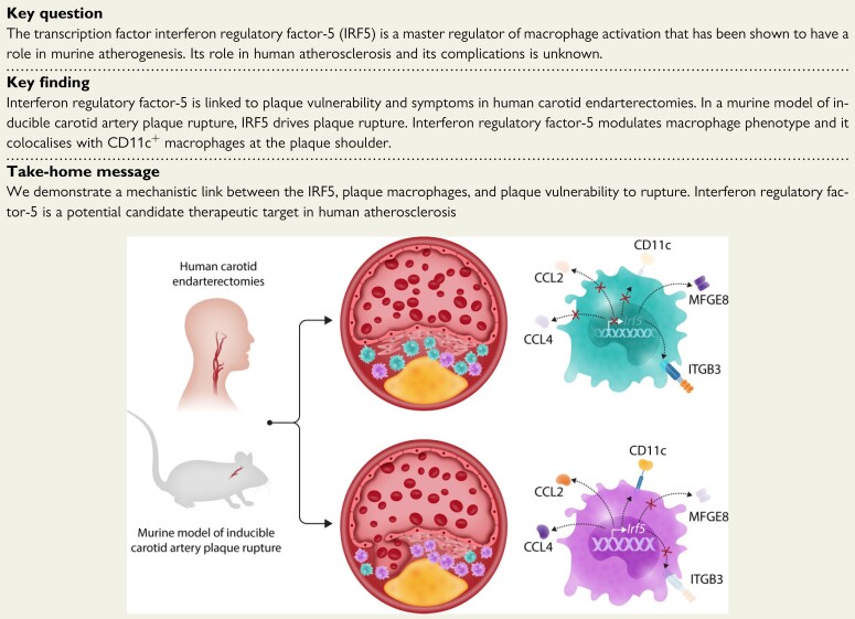
Aims: Inflammation is a key factor in atherosclerosis. The transcription factor interferon regulatory factor-5 (IRF5) drives macrophages towards a pro-inflammatory state. We investigated the role of IRF5 in human atherosclerosis and plaque stability.

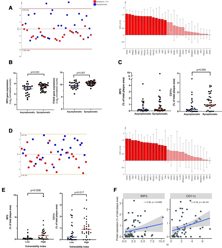
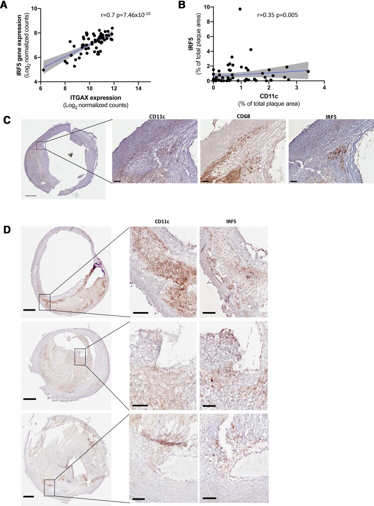
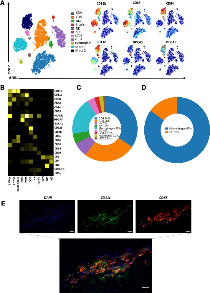
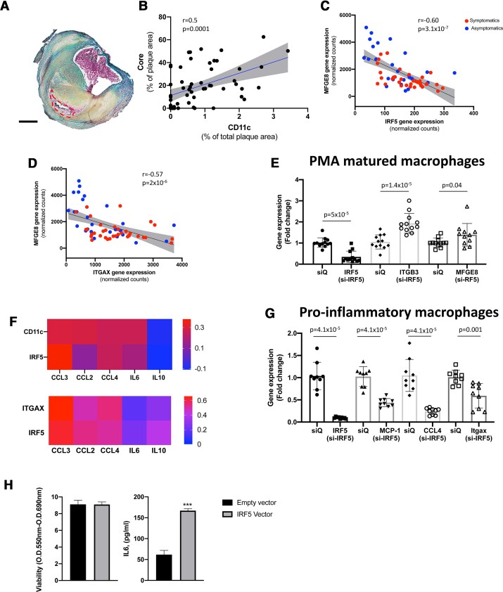
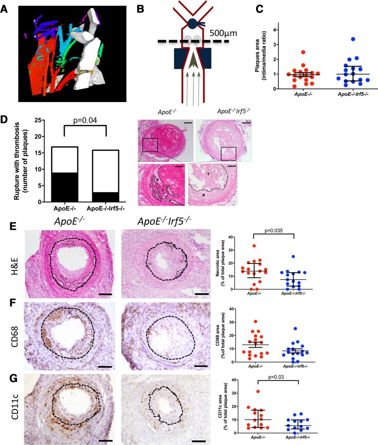
Methods and results: Bulk RNA sequencing from the Carotid Plaque Imaging Project biobank were used to mine associations between major macrophage associated genes and transcription factors and human symptomatic carotid disease. Immunohistochemistry, proximity extension assays, and Helios cytometry by time of flight (CyTOF) were used for validation. The effect of IRF5 deficiency on carotid plaque phenotype and rupture in ApoE-/- mice was studied in an inducible model of plaque rupture. Interferon regulatory factor-5 and ITGAX/CD11c were identified as the macrophage associated genes with the strongest associations with symptomatic carotid disease. Expression of IRF5 and ITGAX/CD11c correlated with the vulnerability index, pro-inflammatory plaque cytokine levels, necrotic core area, and with each other. Macrophages were the predominant CD11c-expressing immune cells in the plaque by CyTOF and immunohistochemistry. Interferon regulatory factor-5 immunopositive areas were predominantly found within CD11c+ areas with a predilection for the shoulder region, the area of the human plaque most prone to rupture. Accordingly, an inducible plaque rupture model of ApoE-/-Irf5-/- mice had significantly lower frequencies of carotid plaque ruptures, smaller necrotic cores, and less CD11c+ macrophages than their IRF5-competent counterparts.

Conclusion: Using complementary evidence from data from human carotid endarterectomies and a murine model of inducible rupture of carotid artery plaque in IRF5-deficient mice, we demonstrate a mechanistic link between the pro-inflammatory transcription factor IRF5, macrophage phenotype, plaque inflammation, and its vulnerability to rupture.

Keywords: Atherosclerosis; IRF5; Macrophages; Plaque rupture.


Figures

None
Macrophages perform both pro- and anti-atherogenic functions depending on their transcriptional status and activation. Using human carotid endarterectomies and a murine model of inducible rupture of carotid plaque, we show that the transcription factor interferon regulatory factor-5 (IRF5) promotes macrophage activation with the production of chemokines CCL2 and CCL4, and the expression of the integrin CD11c ,while it disables the production of proteins required for the uptake of apoptotic cells within the plaque (efferocytosis) such as MFEG8 and the integrin ITGB3. In doing so, IRF5 promotes inflammation and increases the necrotic core size enhancing plaque vulnerability to rupture. MFGE8, Milk fat globule—epidermal growth factor—factor VIII; CCL2, C–C motif chemokine ligand 2; CCL4, C–C motif chemokine ligand 4; ITGB3, Integrin subunit beta 3.
Figure 1
Figure 1
ITGAX (CD11c) gene expression and CD11c plaque area are associated with a vulnerable plaque phenotype and symptomatic carotid plaques in humans. (A) Using an OPLS-DA analysis, IRF5 and ITGAX (CD11c) were identified as the myeloid cell surface marker and transcription factor and with the largest impact on separating symptomatic (<31 days prior to surgery) plaques from asymptomatic plaques. RNAseq data from 47 human carotid plaques (24 symptomatic and 23 asymptomatic). Blue indicates asymptomatic and red indicates symptomatic. (B) Human plaque gene expression levels of IRF5 and ITGAX (CD11c) were also significantly increased in plaques associated with symptoms within 31 days prior to surgery compared with asymptomatic plaques. Lines indicate mean levels, each dot represents an individual value (n = 60, 34 symptomatic and 26 asymptomatic). (C) CD11c+ but not IRF5+ plaque area, as assessed by immunohistochemistry, was increased in symptomatic carotid plaques. Lines indicate median levels, each dot represents an individual value (n = 62, 35 symptomatic <31 days prior to surgery and 27 asymptomatic). Data are presented as % of total plaque area. (D) OPLS-DA analysis identified ITGAX as the myeloid cell gene with the largest impact on separating plaques with a vulnerability index above median from plaques with a vulnerability index below median (n = 47, 24 symptomatic and 23 asymptomatic). Blue indicates less than median vulnerability index and red indicates greater than median vulnerability index. (E) IRF5 and CD11c plaque areas are increased in plaques with a high (above median, n = 31) vulnerability index compared with plaques with a low vulnerability index (below median, n = 31). Mann–Whitney U tests were used. (F) Plaque area stained positive for CD11c and IRF5 correlated with the calculated vulnerability index (n = 62). Spearman test was used for the correlation analysis. CD11c, cluster of differentiation 11c; IRF5, interferon regulatory factor 5.
Figure 2
Figure 2
CD11c associated with IRF5 expression in human carotid plaques. (A) ITGAX (CD11c) and IRF5 gene expression correlate strongly in human carotid plaques. Gene expression is presented as log2 transformed normalized counts. Spearman rank correlation test was used, n = 60. (B) Plaque area of CD11c and IRF5 (% of total plaque area) also correlated in human carotid plaques. Sections from the most stenotic region of the plaque were stained. Spearman rank correlation test was used, n = 62. (C, D) CD11c positive cells were identified in the same plaque regions (commonly close to the core and in the rupture prone shoulder regions) as IRF5 positive cells. CD11c (cytosol and cell membranes) and IRF5 (nuclear) positive cells are seen in brown/black. CD11c, cluster of differentiation 11c; IRF5, interferon regulatory factor 5. Scale bars 1 mm in overview images (left panels) and 200 μm in magnified areas (middle and right panels).
Figure 3
Figure 3
The majority of CD11c+ cells in human carotid plaques are macrophages. Representative CYTOF analysis of a carotid plaque with (A) tSNE and viSNE plots of macrophage and dendritic cell markers expression in human atheroma cells. (B) Heatmap visualizing the expression of markers on different CD45+ cell types. (C) Distribution of main immune cell population in human carotid plaques and (D) distribution of CD11c+ cells between macrophages and dendritic cells in human carotid plaques (n = 4). (E) Immunofluorescent double staining showing that the majority of CD11c+ cells in the human atherosclerotic plaque are CD68+. CD11c, green. DAPI, blue. CD68, red. Scale bars 50 μm. CD68, cluster of differentiation 68; CD11c, cluster of differentiation 11c; DAPI, 4′,6-diamidino-2-phenylindole.
Figure 4.
Figure 4.
CD11c is associated with necrotic core area and reduced gene expression levels of Milk Fat Globule—Epidermal growth factor—factor VIII (MFGE8). (A) Movat pentachrome was used to identify necrotic core areas (acellular non-fibrotic tissue). Core area is marked by red-dotted line. Scale bar 1 mm (n = 57). (B) Human plaque necrotic core area correlate strongly with CD11c plaque area (n = 57). Data are presented as % of total plaque area. Spearman rank correlation test was used. (C, D) MFGE8 gene expression levels correlate inversely to plaque gene expression levels of IRF5 and ITGAX (CD11c). Data are presented as normalized counts. Spearman’s rank correlation coefficient was used (n = 60). Red indicates symptomatic patients and blue indicates asymptomatic patients. (E) Gene expression levels of MFGE8 and ITGB3 were up-regulated in THP-1 PMA matured macrophages upon IRF5 silencing, n = 11–12. Mann–Whitney U tests were used to identify significant differences. Boxes represent median levels and bars represent interquartile range. (F) Heatmap showing correlation coefficients for CD11c and IRF5 positive plaque area, plaque tissue gene expression of ITGAX and IRF5 to plaque protein levels and plaque gene expression levels of interleukin-6 (IL6), Chemokine (C–C motif) ligand 3 (also known as macrophage inflammatory protein-1α), Chemokine ligand 4 (CCL4/macrophage inflammatory protein-1β), monocyte chemoattractant protein-1 (MCP-1/CCL2), and interleukin-10 (IL10). CD11c and IRF5 plaque area were presented as % of total plaque area. IL6, CCL3, CCL4, and MCP-1 were presented as arbitrary units/gram wet weight plaque. Interleukin-10 was presented as picogram/gram wet weight plaque. Spearman’s rho was used for correlations analyses. (G) Gene expression levels of CCL2 (MCP-1), CCL-4, ITGAX (CD11c), and IRF5 were reduced by IRF5 silencing in M1 matured THP-1 cells, n = 9. Mann–Whitney U tests were used to identify significant differences. Boxes represent median levels and bars represent interquartile range. (H) IRF5 overexpression by adenovirus transfection of human plaque cells caused increasing release of IL6 into cell culture supernatants. Four replicates per condition were studied. Unpaired t-test was used to identify significant differences. siQ, silencing control.
Figure 5
Figure 5
Interferon regulatory factor-5 deficiency reduced the frequency of plaque rupture and reduced both necrotic core and CDC11c plaque area in an inducible plaque rupture model. (A) Schematic figure of the common location of plaques ruptures identified in the area between the cast and ligation. (B) Computed tomography (CT) with contrast agent demonstrated that the blood flow was diminished, but not interrupted, after partial ligation proximal to the bifurcation of the carotid artery. Red indicates a high blood flow and blue and green indicate a lower blood flow. (C) No difference in total plaque area, intima/media ratio based on haematoxylin and eosin, was seen when comparing ApoE  −/− and ApoE  −/−  Irf5  −/− carotid lesions. (D) Plaque rupture frequencies are reduced in ApoE  −/−  Irf5  −/− mice compared with ApoE  −/− mice. Representative images of haematoxylin and eosin stained cross-sections of the right carotid artery 4 days after cast placement in the intra-cast region of (left) and ApoE  −/−  Irf5  −/− mice (right). Scale bars, 100 μm. The left insert demonstrates a rupture, characterized by the loss of continuity of the fibrous cap with invasion of thrombus into the plaque tissue and at the right insert a non-ruptured plaque in ApoE  −/−  Irf5  −/−. P indicates plaque tissue and T indicates thrombus. Scale bars insert, 50 μm. Scale bar, 100 μm. (EG) Representative images of cross-sections of carotid arteries after the cast with respective staining and quantification. (E) The sections were stained with haematoxylin and eosin for plaque and necrotic core size (necrotic cores are indicated with a dotted line). A decrease in necrotic core size was observed in the interferon regulatory factor-5-deficient group. (F) CD68 was used to identify macrophages and (G) CD11c. Unless mentioned otherwise, all quantifications are shown as a percentage of the cross-sectional plaque size. All values are expressed as median and interquartile range. Scale bar, 100 μm; n = 16–17.

Comment in

References

    1. Burke AP, Farb A, Malcom GT, Liang Y-H, Smialek J, Virmani R. Coronary risk factors and plaque morphology in men with coronary disease who died suddenly. N Engl J Med 1997;336:1276–1282. - PubMed
    1. Hansson GK. Inflammation, atherosclerosis, and coronary artery disease. N Engl J Med 2005;352:1685–1695. - PubMed
    1. Naghavi M, Libby P, Falk E, Casscells SW, Litovsky S, Rumberger J, et al. From vulnerable plaque to vulnerable patient: a call for new definitions and risk assessment strategies: part I. Circulation 2003;108:1664–1672. - PubMed
    1. Bentzon JF, Otsuka F, Virmani R, Falk E. Mechanisms of plaque formation and rupture. Circ Res 2014;114:1852–1866. - PubMed
    1. Tabas I, Bornfeldt KE. Macrophage phenotype and function in different stages of atherosclerosis. Circ Res 2016;118:653–667. - PMC - PubMed

Publication types