

Responses of promyelocytic leukemia HL60 cells as an inflammatory cell lineage model to silica microparticles used to coat blood collection tubes
- PMID: 35567654
- PMCID: PMC9107555
- DOI: 10.1186/s40729-022-00424-4
Responses of promyelocytic leukemia HL60 cells as an inflammatory cell lineage model to silica microparticles used to coat blood collection tubes
Abstract
Background: The preparation of platelet-rich fibrin (PRF) requires glass blood collection tubes, and thus, the shortage or unavailability of such tubes has driven clinicians to search for suitable substitutes, such as silica-coated plastic tubes. However, we have previously demonstrated the cytotoxicity of silica microparticles (MPs) used in plastic tubes to cultured human periosteal cells. To further establish the effects of silica MPs on inflammation, we examined silica MP-induced changes in a human promyelocytic cell model in vitro.
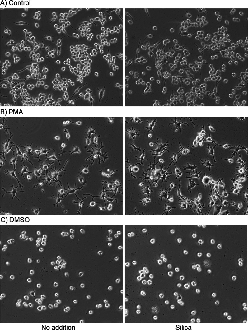
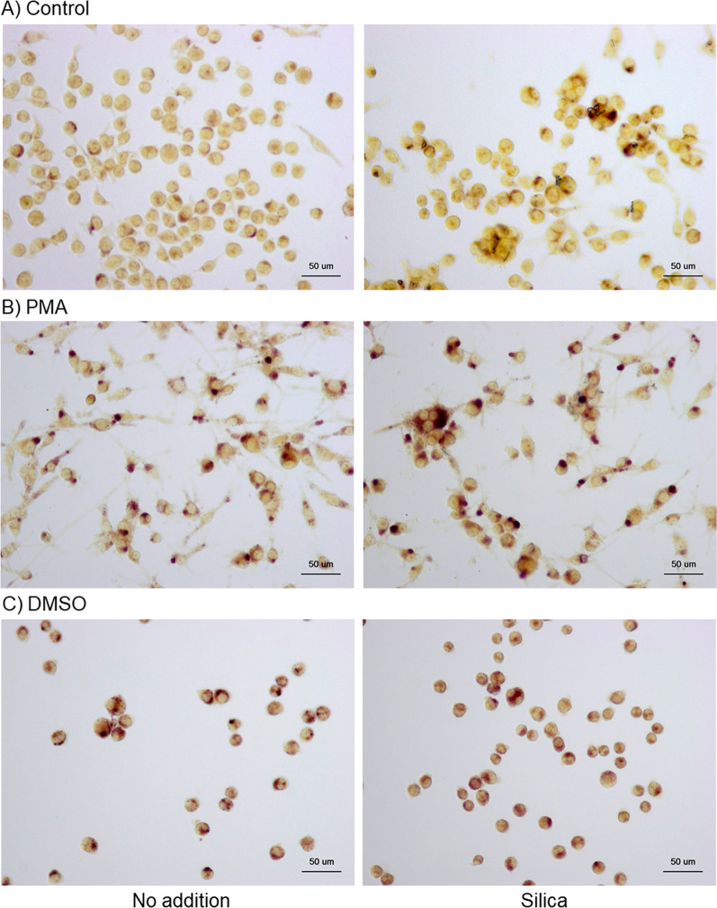
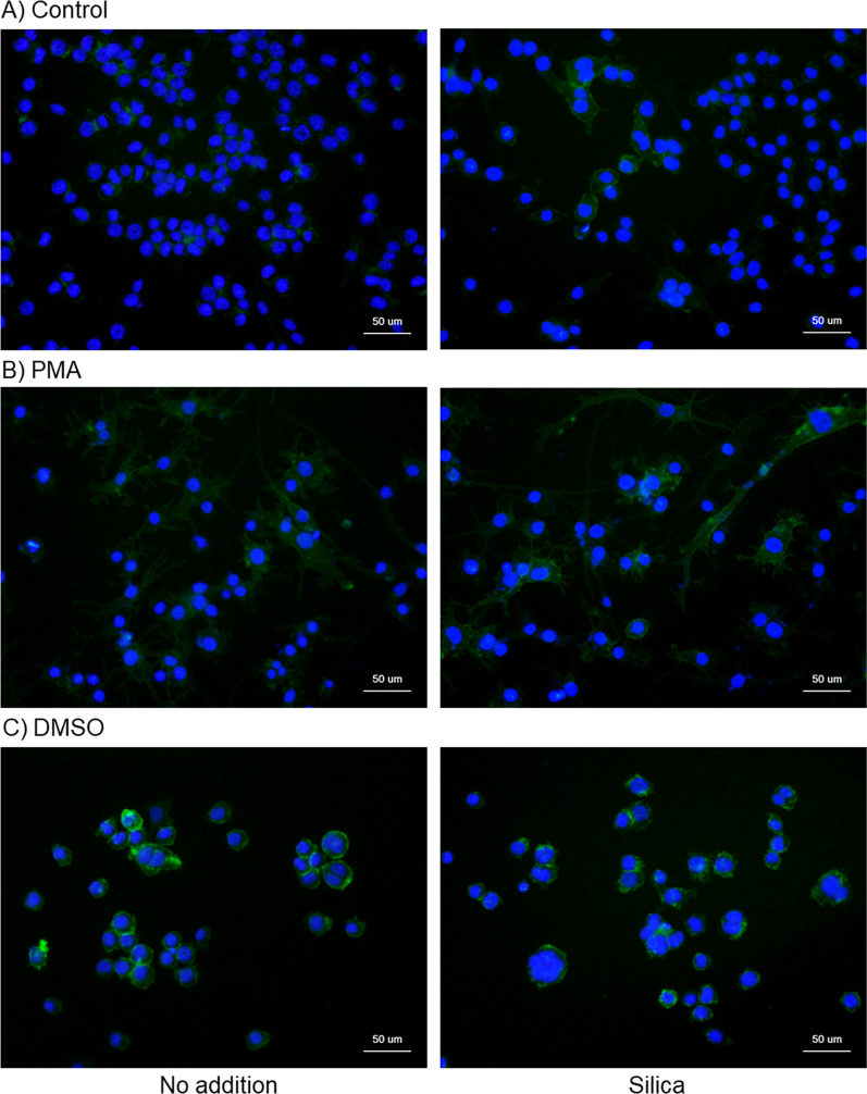
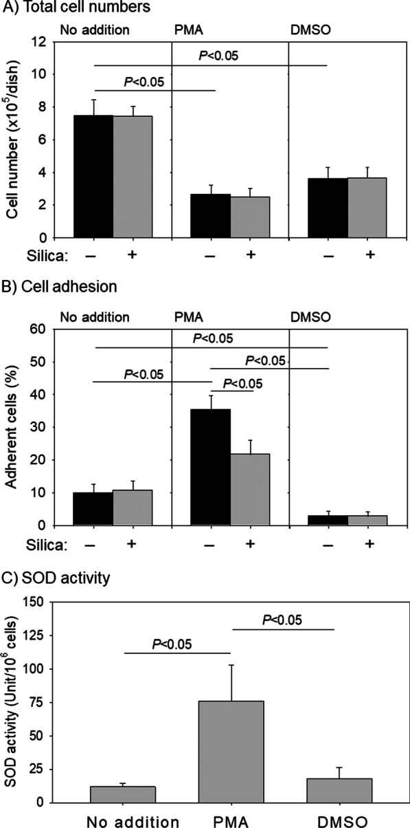
Methods: Human promyelocytic HL60 cells were used either without chemical induction or after differentiation induced using phorbol myristate acetate (PMA) or dimethyl sulfoxide. HL60 cells, osteoblastic MG63, and Balb/c mouse cells were treated with silica MPs, and their surface ultrastructure and numbers were examined using a scanning electron microscope and an automated cell counter, respectively. Differentiation markers, such as acid phosphatase, non-specific esterase, and CD11b, were visualized by cytochemical and immunofluorescent staining, and superoxide dismutase (SOD) activity was quantified.
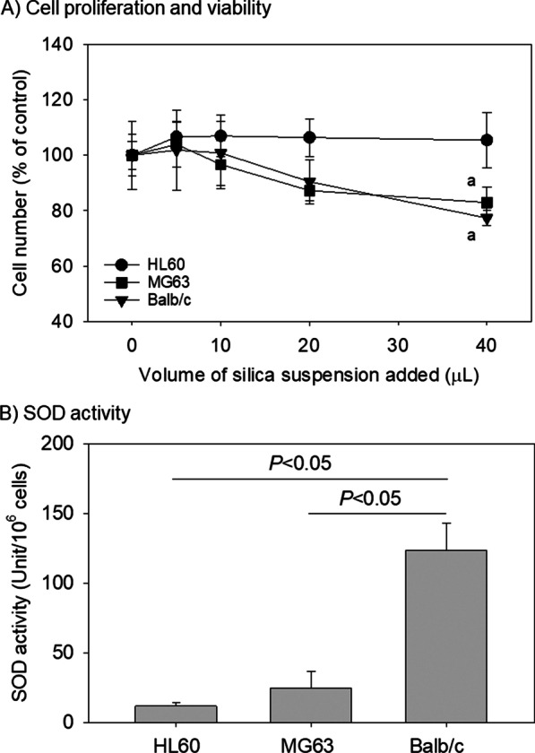
Results: Regardless of SOD activity, silica cytotoxicity was observed in MG63 and Balb/c cells. At sub-toxic doses, silica MPs slightly or moderately upregulated the differentiation markers of the control, PMA-induced monocytic, and dimethyl sulfoxide-induced granulocytic HL60 cells. Although SOD activity was the highest (P < 0.05) in PMA-induced cells, a silica-induced reduction in cell adhesion was observed only in those cells (P < 0.05).
Conclusions: Silica MP contamination of PRF preparations can potentially exacerbate inflammation at implantation sites. Consequently, unless biomedical advantages can be identified, silica-coated plastic blood collection tubes should not be routinely used for PRF preparations.
Keywords: Differentiation; HL60 cells; Silica; Superoxide dismutase; Viability.
© 2022. The Author(s).
Conflict of interest statement
The authors declare no conflict of interest.
Figures


Similar articles
-
Acute cytotoxic effects of silica microparticles used for coating of plastic blood-collection tubes on human periosteal cells.Odontology. 2020 Oct;108(4):545-552. doi: 10.1007/s10266-020-00486-z. Epub 2020 Jan 30. Odontology. 2020. PMID: 31997225 Free PMC article.
-
Evidence for Contamination of Silica Microparticles in Advanced Platelet-Rich Fibrin Matrices Prepared Using Silica-Coated Plastic Tubes.Biomedicines. 2019 Jun 19;7(2):45. doi: 10.3390/biomedicines7020045. Biomedicines. 2019. PMID: 31248187 Free PMC article.
-
Activation of beta 1 integrin fibronectin receptors on HL60 cells after granulocytic differentiation.Blood. 1994 Jan 15;83(2):543-52. Blood. 1994. PMID: 7506954
-
Acute promyelocytic leukemia mutated to radioresistance suppressed monocyte lineage differentiation by phorbol 12-myristate 13-acetate.Leuk Res. 2013 Sep;37(9):1162-9. doi: 10.1016/j.leukres.2013.04.027. Epub 2013 Jun 14. Leuk Res. 2013. PMID: 23773897
-
A technical note on contamination from PRF tubes containing silica and silicone.BMC Oral Health. 2021 Mar 19;21(1):135. doi: 10.1186/s12903-021-01497-0. BMC Oral Health. 2021. PMID: 33740959 Free PMC article. Review.
References
-
- Masuki H, Isobe K, Kawabata H, Tsujino T, Yamaguchi S, Watanabe T, Sato A, Aizawa H, Mourão CF, Kawase T. Acute cytotoxic effects of silica microparticles used for coating of plastic blood-collection tubes on human periosteal cells. Odontology. 2020;108:545–552. doi: 10.1007/s10266-020-00486-z. - DOI - PMC - PubMed
-
- American_Lung_Association. Silicosis. https://www.lungorg/lung-health-diseases/lung-disease-lookup/silicosis. Accessed 12 Mar 2022.
Publication types
MeSH terms
Substances
LinkOut - more resources
Full Text Sources
Research Materials
