. 2022 May 26;185(11):1860-1874.e12.
doi: 10.1016/j.cell.2022.04.024. Epub 2022 May 13.

Host and pathogen response to bacteriophage engineered against Mycobacterium abscessus lung infection

Affiliations

Host and pathogen response to bacteriophage engineered against Mycobacterium abscessus lung infection

Jerry A Nick et al. Cell. .

Abstract

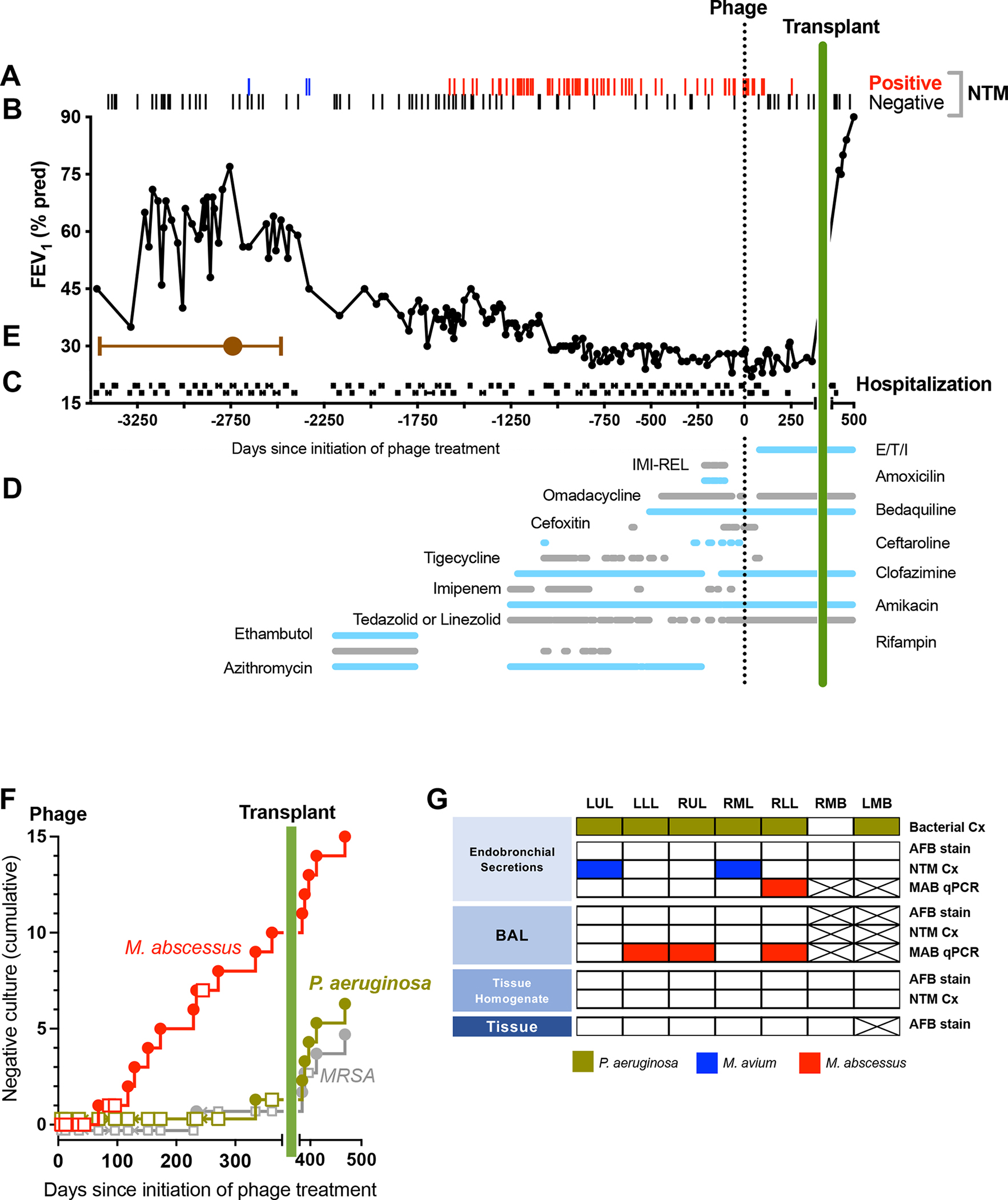
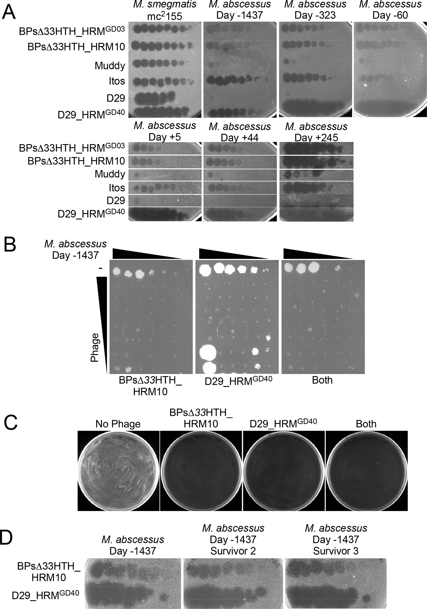
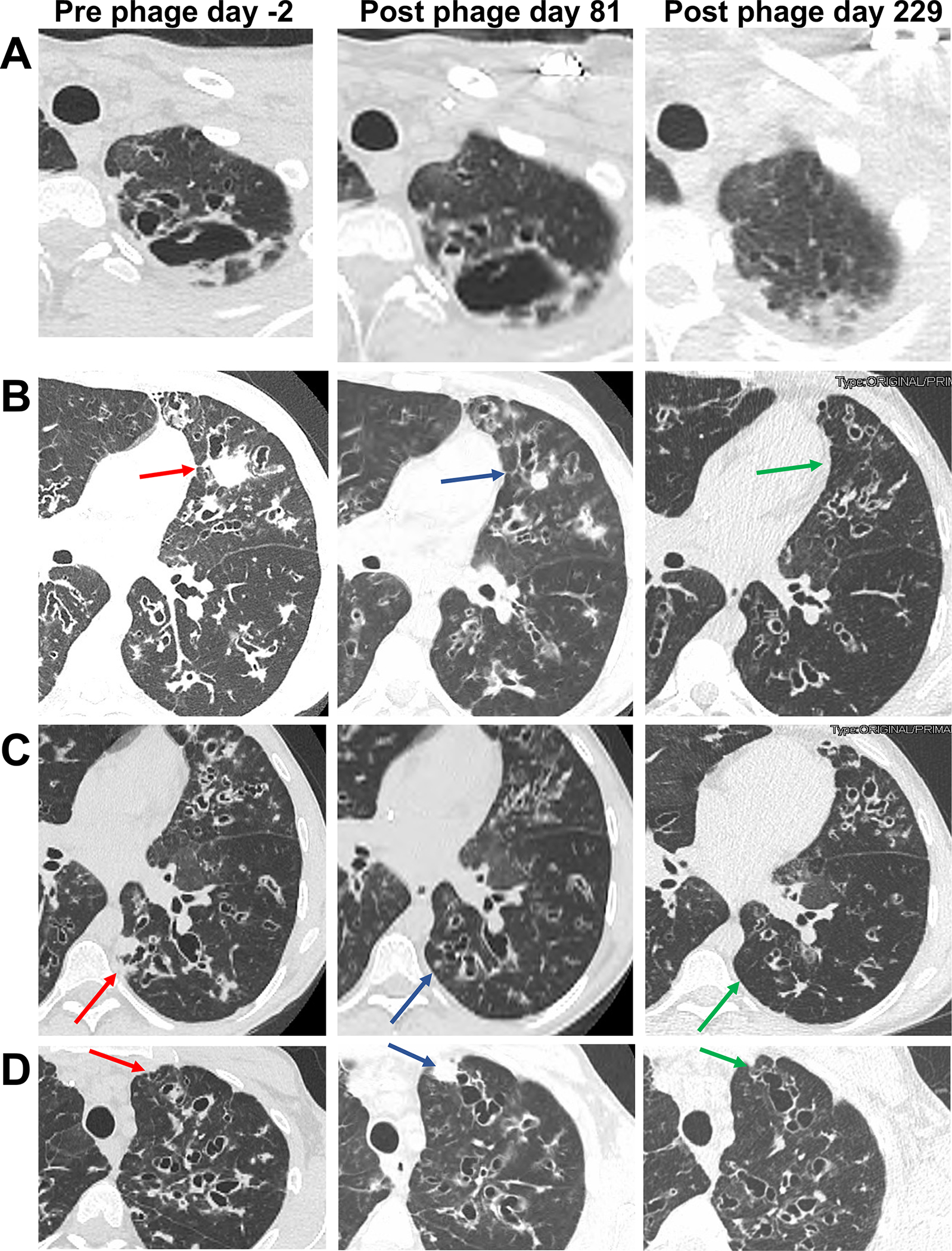
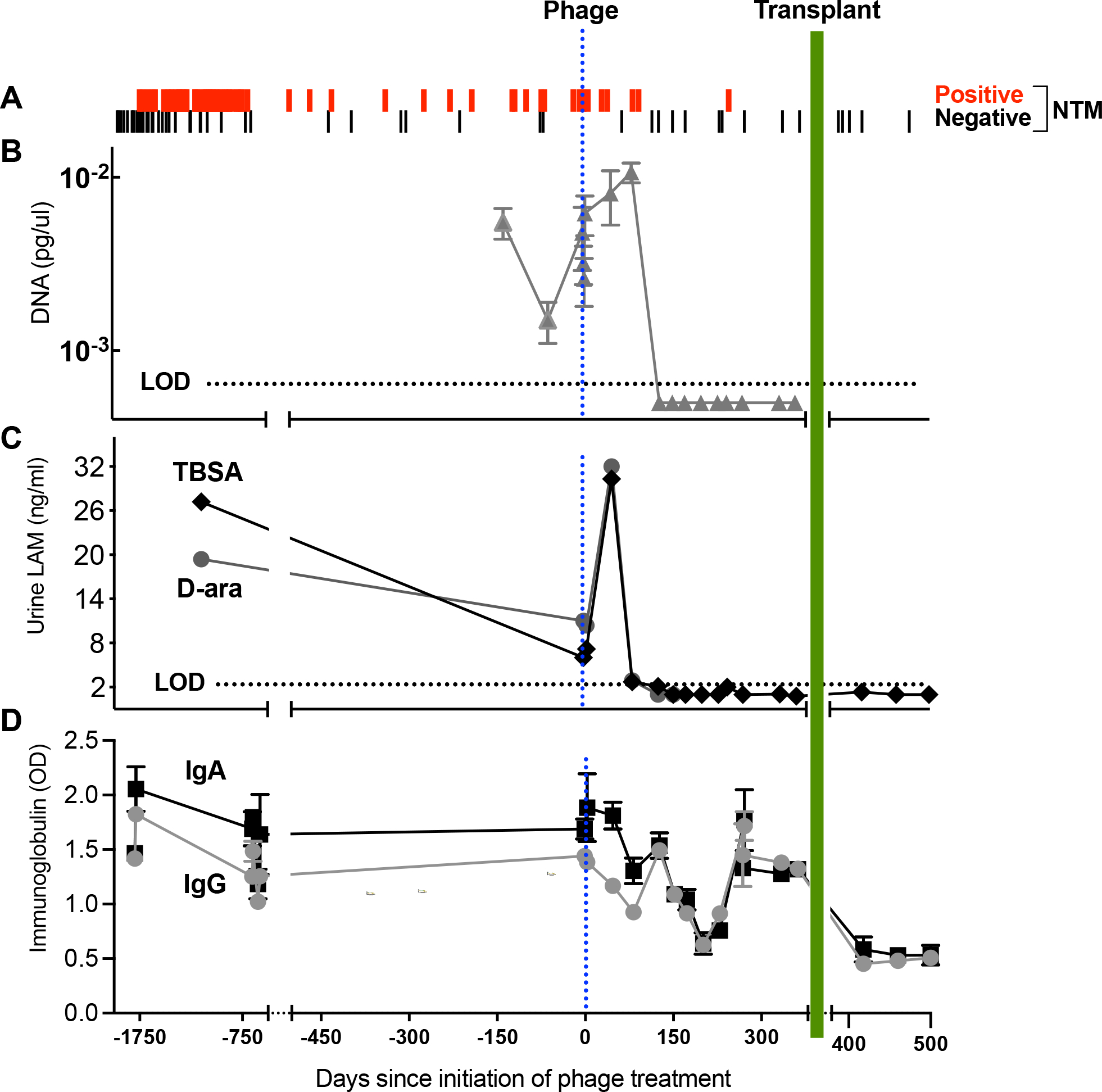
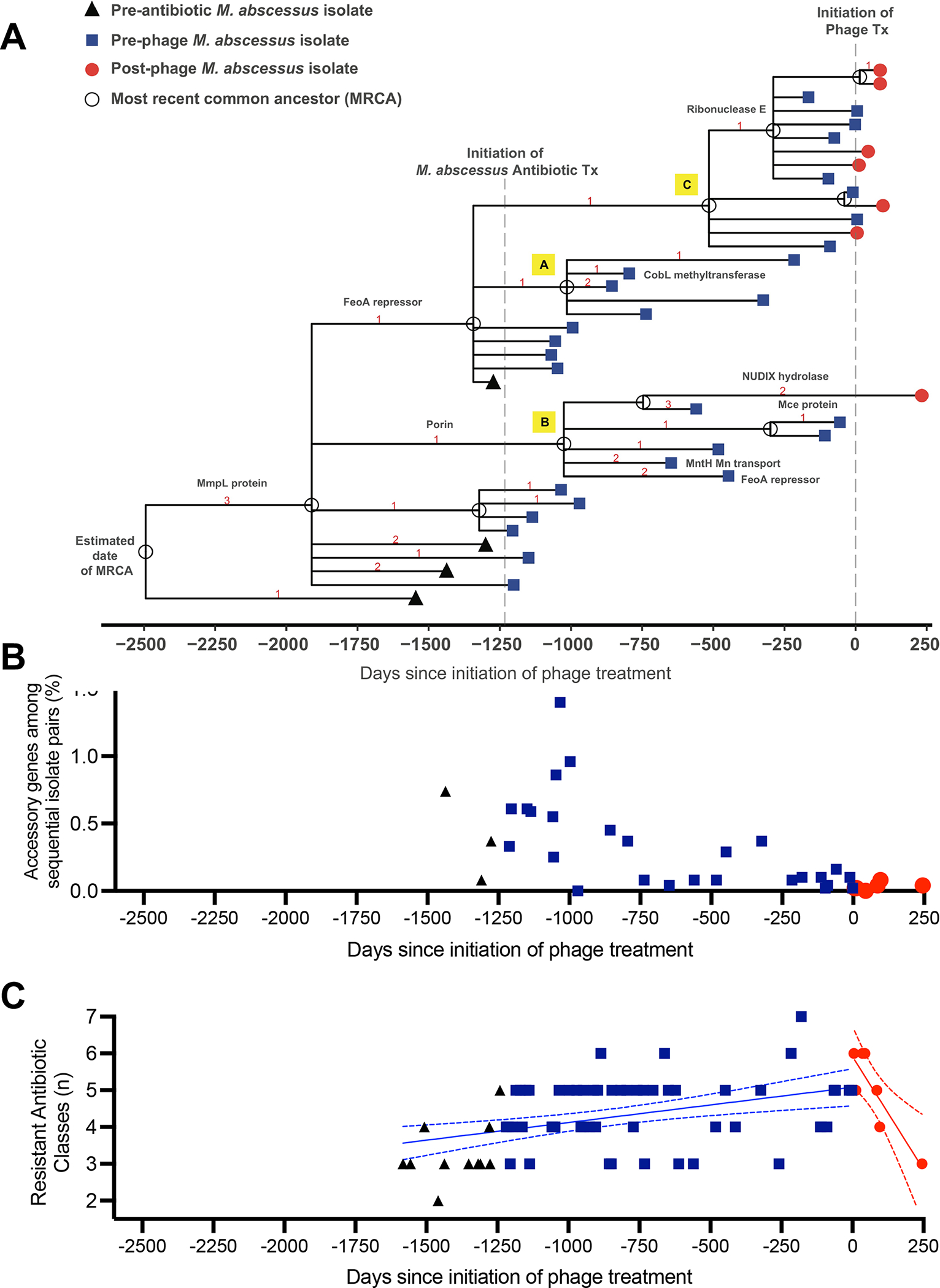
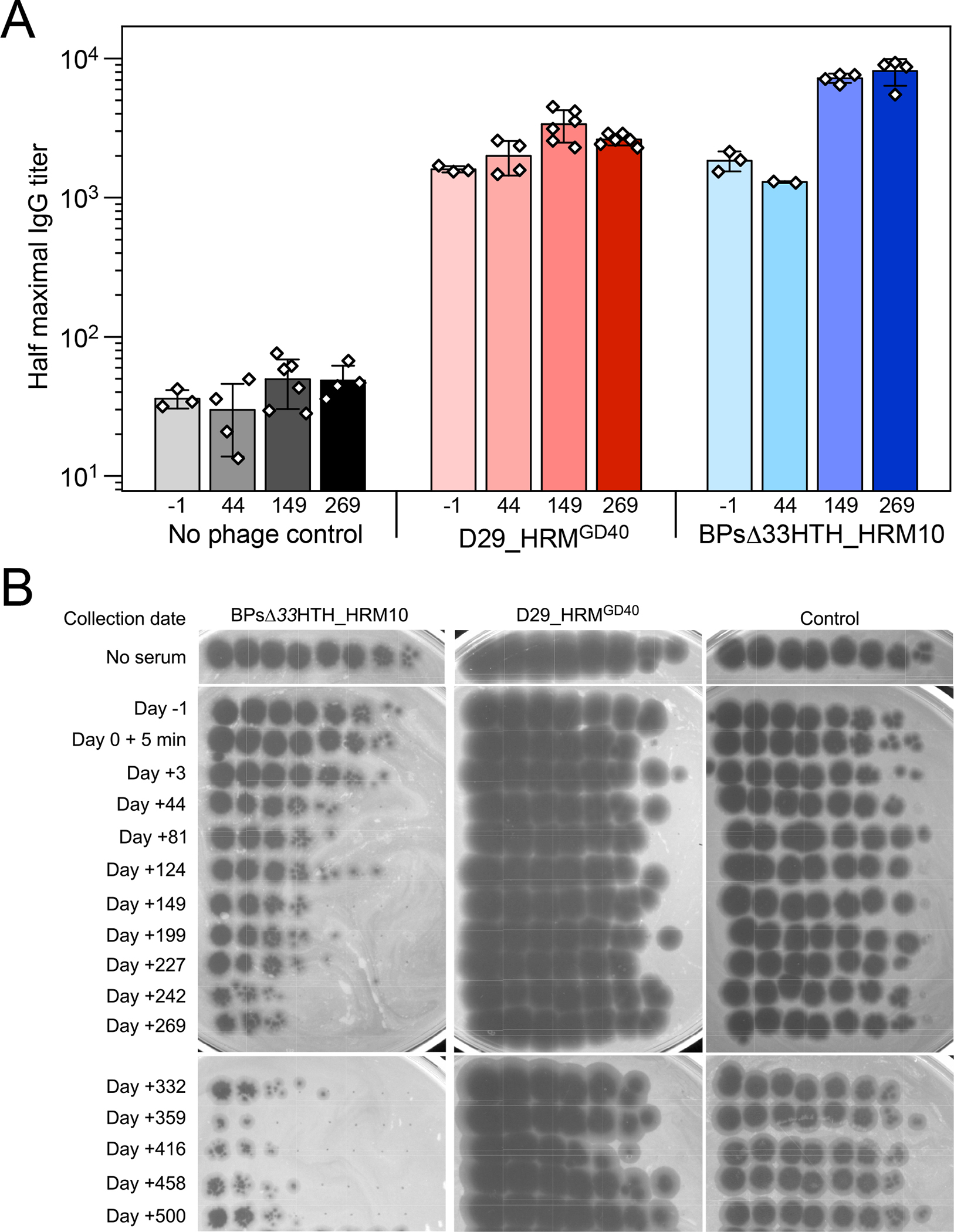
Two mycobacteriophages were administered intravenously to a male with treatment-refractory Mycobacterium abscessus pulmonary infection and severe cystic fibrosis lung disease. The phages were engineered to enhance their capacity to lyse M. abscessus and were selected specifically as the most effective against the subject's bacterial isolate. In the setting of compassionate use, the evidence of phage-induced lysis was observed using molecular and metabolic assays combined with clinical assessments. M. abscessus isolates pre and post-phage treatment demonstrated genetic stability, with a general decline in diversity and no increased resistance to phage or antibiotics. The anti-phage neutralizing antibody titers to one phage increased with time but did not prevent clinical improvement throughout the course of treatment. The subject received lung transplantation on day 379, and systematic culturing of the explanted lung did not detect M. abscessus. This study describes the course and associated markers of a successful phage treatment of M. abscessus in advanced lung disease.

Keywords: CFTR modulator therapy; Mycobacterium abscessus; Mycobacterium avium; NTM; bacteriophages; cystic fibrosis; immunoglobulin; lipoarabinomannan; lung transplant; nontuberculous mycobacterial lung disease.


Conflict of interest statement

Declaration of interests G.F.H. is a compensated consultant for Janssen Inc.

Figures

Figure 1.
Figure 1.. Clinical Summary From Time of Estimated Initial Infection Through Day 500 of Phage Treatment.
Clinical parameters are plotted versus time in days, with initial phage administration labeled as Day 0. (A) Airway culture results (n=155) are depicted with red bars (positive M. abscessus, n=71), blue bars (positive M. avium, n=2) or gray bars (negative culture, n=82). Cultures prior to transplant (day 379) were obtained from expectorated sputum, and following transplant were obtained from BAL. (B) Forced expiratory volume in 1 sec (FEV1, percent predicted) plotted versus days. (C) Hospitalizations (black horizontal lines) plotted versus days. (D) Treatment course. The type and duration of CFTR modulator and various antibiotics (horizontal lines) directed towards M. avium or M. abscessus plotted versus days. Agents used in order of most recent to earliest introduction: 1) Elexacaftor/tezacaftor/ivacaftor (E/T/I), 2) Imipenem-cilastatin-relebactam (IMI-REL), 3) Amoxicillin, 4) Omadacycline, 5) Bedaquiline, 6) Cefoxitin, 7) Ceftaroline, 8) Tigecycline, 9) Clofazimine, 10) Imipenem, 11) Amikacin, 12) Tedizolid or linezolid (oral or IV), 13) Ethambutol, 14) Rifampin, 15) Azithromycin. (E) Timeframe of initial infection. The estimated date of the most common recent ancestor based on divergence dating analysis was pre-phage day -2494 (brown circle) with a 95% Highest Posterior Density (HPD) interval from day -1945 to day -3435 (brown bars). (F) Cumulative negative airway cultures following initiation of phage therapy. The occurrence of negative culture over time was plotted for M. abscessus (negative cultures red circle, positive cultures red square), P. aeruginosa (negative culture green circle, positive culture green square), and MRSA (positive culture grey circle, negative culture grey square). The proportion of negative cultures was greater for M. abscessus compared to P. aeruginosa or MRSA (p=0.007 by Fisher’s Exact Test). (G) Systematic analysis of explanted lung tissue for M. abscessus. Endobronchial secretions, BAL, homogenized tissue and tissue pathology from the left upper lobe (LUL), left lower lobe (LLL), right upper lobe (RUL), right middle lobe (RML), right lower lobe (RLL), right mainstem bronchi (RMB) and left mainstem bronchi (LMB) were analyzed by bacterial culture for usual CF pathogens (Bacterial Cx), AFB stain, NTM cultures (NTM Cx), species-specific qPCR for M. abscessus DNA (MAB qPCR). Positive results are indicated for P. aeruginosa (green box), M. avium (blue box) or M. abscessus (red box). Negative results are indicated by white boxes, and assays not performed by crossed-out box.
Figure 2.
Figure 2.. Phage Therapy Did Not Select for Phage-resistant Isolates
(A) Efficiencies of plating of phages on M. smegmatis mc2155 and six M. abscessus isolates from the subject -1437, -323 and -60 days prior to initiation of phage therapy, as well as 5, 44, and 245 days after therapy started. Phage lysates were 10-fold serially diluted and spotted onto top agar overlays of each strain. Phages BPΔ33HTH_HRMGD03, BPΔ33HTH_HRM10, Muddy, Itos, D29 and D29_HRMGD40 infect each M. abscessus isolate with similar efficiencies to M. smegmatis mc2155. Phage BPΔ33HTH_HRM10, which was used therapeutically previously (Dedrick et al., 2021b; Dedrick et al., 2019), and D29_HRMGD40 were chosen as therapeutic candidates; phage Itos infects efficiently but does not kill the M. abscessus strains tested well. (B) Killing of M. abscessus strain collected -1437 days prior to initiation of phage by BPΔ33HTH_HRM10, D29_HRMGD40 or both phages. Each column has a 10-fold serial dilution of the isolate (left column, 2×10⁵ CFU) and each row has 10-fold serial dilutions of either BPΔ33HTH_HRM10 (top row, 109 PFU), D29_HRMGD40 (top row, 107 PFU) or both phages. (C) Survival assay showing efficient killing of M. abscessus strain collected -1437 days prior to initiation of phage with either BPΔ33HTH_HRM10 or D29_HRMGD40 or both phages together. (D) Phage susceptibility assays of two survivors recovered from a challenge with BPΔ33HTH_HRM10 and D29_HRMGD40 of M. abscessus strain collected -1437 days prior to initiation of phage shows both are fully phage-sensitive. Phages were tested as in panel A.
Figure 3.
Figure 3.. Radiologic Changes During Phage Therapy
(A) Left: Axial CT pre-treatment demonstrates a left apical cavity. Middle: Little change was observed at day 81 of treatment. Right: Near resolution of the cavity was observed at day 229. (B) Left: Axial CT pre-treatment shows widespread bronchiectasis and mucoid impaction, and a consolidative mass in the lingula (red arrow). Middle: At day 81 of treatment the bronchiectasis and mucoid impaction was largely unchanged, but the lingular mass decreased substantially (blue arrow). Right: At day 229 the consolidation was resolved (green arrow), and the mucoid impaction throughout the left mid-lung region was substantially improved. (C) Left: Axial CT pre-treatment shows widespread bronchiectasis and mucoid impaction, and a consolidative mass in the left lower-lobe (red arrow). Middle: At day 81 of treatment the bronchiectasis and mucoid impaction was somewhat improved, and the left lower-lobe mass decreased substantially (blue arrow). Right: At day 229 the consolidation was resolved (green arrow) and bronchiectatic airways were generally reduced in size. (D) Left: Axial CT pre-treatment shows widespread bronchiectasis and mucoid in the left upper-lobe (red arrow). Middle: At day 81 of treatment the bronchiectasis and mucoid impaction was largely unchanged, with the new appearance of a consolidative nodule in the anterior left upper lobe (blue arrow). Right: At day 229 the consolidative nodule present at day 80 was resolved (green arrow).
Figure 4.
Figure 4.. Culture-independent Markers Demonstrate Evidence of M. abscessus Lysis and Host Response.
(A) Airway culture versus date in days pre- or post-phage treatment initiation. Sputum culture results were predominantly positive (red lines) prior to initiation of phage treatment. Post-phage treatment, 6 of 7 cultures were positive through day 96. From day 116 the culture turned predominantly negative (black line) with 9 of 10 negative through day 362. Over this interval a single persister isolate was recovered on day 245. Post-transplant an additional 5 cultures from BAL were negative through day 500 of phage treatment. (B) qPCR analysis of M. abscessus DNA in sputum. The y-axis shows M. abscessus DNA concentrations (pg/ul) extrapolated from a standard curve. (C) Urine LAM. The LAM components D-ara (grey squares) and TBSA (brown diamonds) plotted versus days. Levels of both components peaked 47 days after initiation of phage, and by day 152 were both below LOD, continuing post-transplant. (D) Serum anti-M. abscessus immunoglobulins plotted versus days. Measured immunoglobulins were IgA (black square) and IgG (gray circle). Evidence for a decline in immunoglobulins during each phase of treatment was tested by simple linear regression models fit to IgA and IgG using a three-level categorical variable for pre-phage treatment, phage treatment, and the post-transplant interval as the only covariate. T-tests on the regression coefficients, or linear combinations, were used to compare the mean levels in each period. Compared to pre-phage titers, a significant decline in IgA was detected over the course of phage therapy prior to transplant (p=0.05) while mean IgG values trended lower (p=0.17). Both IgG and IgA declined further post-lung transplant when compared to the year prior on phage treatment (p<0.001 for both).
Figure 5.
Figure 5.. Genetic Diversity and Antibiotic Sensitivity of M. abscessus Isolates Following Phage Therapy
(A) Genetic lineage map of M. abscessus isolates (n=40) over the course of infection and treatment. A time scaled phylogeny was created based on genome wide SNP data using BEAST v1.10.4. The x-axis represents days since the initiation of phage treatment. Isolates are color-coded symbols on branch tips, and the most recent common ancestors (MRCA) are labeled as open circles on nodes. Red numbers indicate the number of nonsynonymous mutations that occurred on each evolutionary branch. Protein annotations for selected mutations are labeled on branches. (B) Gene content comparisons of sequential M. abscessus isolates to assess genomic variation over time. For each pair of sequential isolates (x-axis), pan genome analysis was performed to identify accessory genes unique to only one sample. The y-axis (% accessory genes) is defined as the total # of accessory genes/ sum of all genes from both isolates in the pair. Isolate pairs post-phage were significantly less diverse than isolates pre-phage (p=0.0055 by Wilcoxon 2-sample test). Black triangles represent isolate pairs prior to initiation of M. abscessus antibiotic treatment, blue squares following start of antibiotics, and red circles following initiation of phage therapy. The decrease in accessory genes over time suggests a reduction in genetic diversity and selection for a homogeneous population. (C) Antibiotic resistance of M. abscessus isolates does not increase following phage therapy. Number of resistant antibiotic classes were plotted versus days pre- and post-phage initiation. Black triangles represent isolates prior to initiation of M. abscessus antibiotic treatment, blue squares represent values following start of antibiotics, and red circles following initiation of phage therapy. Simple linear regression demonstrated a significant increase in slope of number of resistant classes over time prior to phage (blue line ± 95%CI, p=0.0007). Following initiation of phage, the slope was significantly negative (red line ± 95%CI, p=0.009).
Figure 6.
Figure 6.. Antibody Binding to Phages Pre- and Post-Phage Treatment Demonstrates Late Development of Anti-phage Neutralizing Antibodies.
(A) Half maximal titers of IgG antibodies in pre-phage serum collected at day -1 and indicated times post-phage initiation for phages BPsΔ33HTH_HRM10 and D29_HRMGD40. Titers for two to six replicates are displayed as white data points on top of a mean bar ± s.d. (B) Serum collected 1 day prior to phage initiation and indicated times post-phage initiation were tested for phage neutralization of BPsΔ33HTH_HRM10, D29_HRMGD40, and a control phage Fionnbharth Δ45–47. Data are representative of duplicate or triplicate assays.

Comment in

References

    1. Aslam S, Lampley E, Wooten D, Karris M, Benson C, Strathdee S, and Schooley RT (2020). Lessons Learned From the First 10 Consecutive Cases of Intravenous Bacteriophage Therapy to Treat Multidrug-Resistant Bacterial Infections at a Single Center in the United States. Open forum infectious diseases 7, ofaa389. - PMC - PubMed
    1. Bryant JM, Brown KP, Burbaud S, Everall I, Belardinelli JM, Rodriguez-Rincon D, Grogono DM, Peterson CM, Verma D, Evans IE, et al. (2021). Stepwise pathogenic evolution of Mycobacterium abscessus. Science 372. - PMC - PubMed
    1. Bryant JM, Grogono DM, Rodriguez-Rincon D, Everall I, Brown KP, Moreno P, Verma D, Hill E, Drijkoningen J, Gilligan P, et al. (2016). Emergence and spread of a human-transmissible multidrug-resistant nontuberculous mycobacterium. Science 354, 751–757. - PMC - PubMed
    1. Chakraborty P, and Kumar A (2019). The extracellular matrix of mycobacterial biofilms: could we shorten the treatment of mycobacterial infections? Microb Cell 6, 105–122. - PMC - PubMed
    1. Champion OL, Karlyshev A, Cooper IAM, Ford DC, Wren BW, Duffield M, Oyston PCF, and Titball RW (2011). Yersinia pseudotuberculosis mntH functions in intracellular manganese accumulation, which is essential for virulence and survival in cells expressing functional Nramp1. Microbiology (Reading) 157, 1115–1122. - PubMed

Publication types

MeSH terms

Substances

LinkOut - more resources