. 2022 Jul:146:103785.
doi: 10.1016/j.ibmb.2022.103785. Epub 2022 May 12.

Functional aspects of evolution in a cluster of salivary protein genes from mosquitoes

Affiliations

Functional aspects of evolution in a cluster of salivary protein genes from mosquitoes

Patricia H Alvarenga et al. Insect Biochem Mol Biol. 2022 Jul.

Abstract

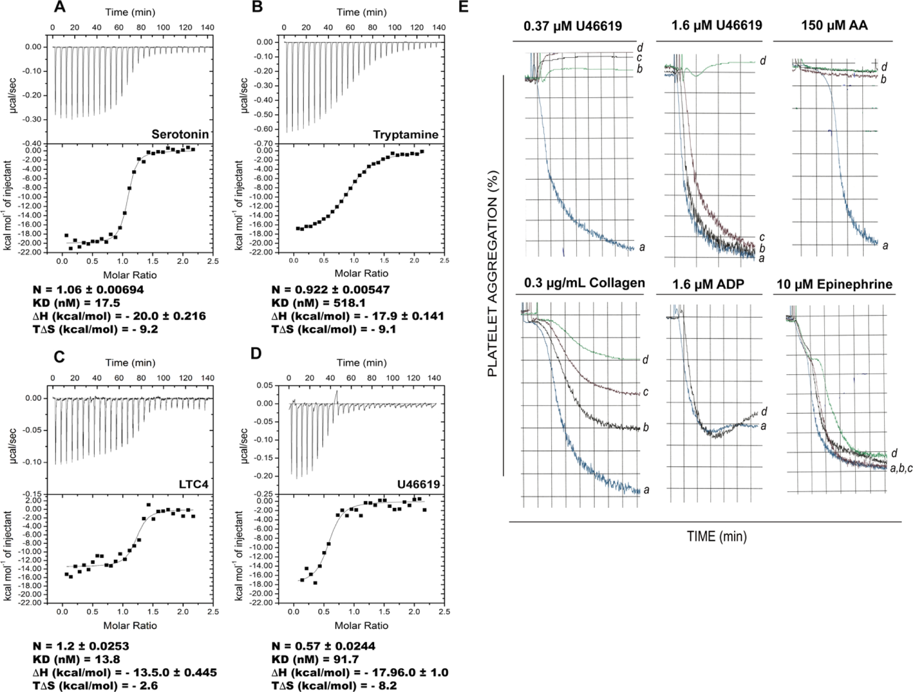
The D7 proteins are highly expressed in the saliva of hematophagous Nematocera and bind biogenic amines and eicosanoid compounds produced by the host during blood feeding. These proteins are encoded by gene clusters expressing forms having one or two odorant-binding protein-like domains. Here we examine functional diversity within the D7 group in the genus Anopheles and make structural comparisons with D7 proteins from culicine mosquitoes in order to understand aspects of D7 functional evolution. Two domain long form (D7L) and one domain short form (D7S) proteins from anopheline and culicine mosquitoes were characterized to determine their ligand selectivity and binding pocket structures. We previously showed that a D7L protein from Anopheles stephensi, of the subgenus Cellia, could bind eicosanoids at a site in its N-terminal domain but could not bind biogenic amines in its C-terminal domain as does a D7L1 ortholog from the culicine species Aedes aegypti, raising the question of whether anopheline D7L proteins had lost their ability to bind biogenic amines. Here we find that D7L from anopheline species belonging to two other subgenera, Nyssorhynchus and Anopheles, can bind biogenic amines and have a structure much like the Ae. aegypti ortholog. The unusual D7L, D7L3, can also bind serotonin in the Cellia species An. gambiae. We also show through structural comparisons with culicine forms that the biogenic amine binding function of single domain D7S proteins in the genus Anopheles may have evolved through gene conversion of structurally similar proteins, which did not have biogenic amine binding capability. Collectively, the data indicate that D7L proteins had a biogenic amine and eicosanoid binding function in the common ancestor of anopheline and culicine mosquitoes, and that the D7S proteins may have acquired a biogenic amine binding function in anophelines through a gene conversion process.

Keywords: Biogenic amine; D7 proteins; Eicosanoids; Evolution; Gene duplication; Hematophagy; Mosquito; Odorant-binding protein; Protein diversity; Saliva.


Conflict of interest statement

Declaration of competing interest

The authors declare that they have no competing interests.

Figures

Figure 1.
Figure 1.. D7 genes are organized in clusters in anopheline and culicine mosquitoes.
Schematic of the representation the pattern of D7 genes distribution and organization in the chromosome maps of six different anopheline species representing various sub genus/series (indicated in parentheses) and the culicine Aedes aegypti. D7L genes are marked with the letter L (1–3) in boxes with purple outline, D7S are represented by the letter “r”(1–5) in boxes with magenta outline and shortened D7S found in Nyssorhynchus species are represented in yellow. In most Anopheles subgenera the D7 cluster contains one or two D7L genes separated by ~7.5 to 7.9 kb from D7L3, which is adjacent to the D7S genes (r1 to r5) that are generally transcribed in the direction (indicated with arrows) opposite to D7L genes. Analysis of available anopheline genomes suggests that all species, regardless of their subgenus, retained D7L2 and D7L3 genes. On the other hand D7L1 genes, believed to arise from D7L2 gene duplication, are just present in series Myzomyia and Pyretophorous from subgenus Cellia and in subgenus Anopheles. The number of D7S genes also varies among the subgenera. All Cellia species retained at least five genes encoding D7S (represented by the letter “r”), and some species (An. stephensi) have a sixth gene, species from the subgenera Anopheles and Nyssorhynchus, consistently show the loss of two and three D7S genes respectively. Details regarding the forms present in each species as well as annotation numbers can be found at Table S1. In Ae. aegypti, representing culicine species, the D7 cluster is arranged similarly but occupies a larger segment of the chromosome. The D7L3 gene present in all Anopheles species is not present in culicines. Culicine D7S were not named like those of Anopheles ones, since the two kinds of D7S are not apparent orthologs. D7 genes whose products have been already characterized in the literature have their respective boxes filled in green (binding) and/or red (no binding) to represent their capacity to bind eicosanoids (in the N-terminal of D7L) and/or serotonin (C-terminal of D7L and D7S). Boxes with no filling color represent genes producing yet uncharacterized proteins, while those filled with purple represent the ones whose proteins structural and functional characterization are being described in the present work, in addition to the Culex quinquefasciatus D7S (D7CQS1) not represented in this scheme. Figure created with BioRender.com.
Figure 2.
Figure 2.. The loss of DS4 leads to the lack of ability of subgenus Cellia D7L1 and L2s to bind biogenic amines -
Comparison of AnSt-D7L1 C-terminal domain and Anopheles gambiae D7r4 published structures (Alvarenga et al., 2010; Mans et al., 2007). A ribbon diagram showing the position of the eight helices (A2-H2) at the C-terminal domain of (A) AnSt-D7L1 and (B) An. gambiae D7r4 containing serotonin. Disulfide bonds are shown as yellow sticks and disulfide bound 4 is labeled as DS4. Detailed view of the amino acids lining (C) AnSt-D7L1 C-terminal domain region and the (D) D7r4 serotonin binding pocket, showing that in AnSt-D7L1 (C) helix H2 is changed in its position and B2 is unwound with R177 partially occupying of the position of serotonin in D7r4 (D). Serotonin is colored magenta, oxygen atoms are shown in red and nitrogen in blue. Hydrogen bonds are represented as dashed red lines.
Figure 3.
Figure 3.. AnDar-D7L2 binds serotonin, cysteinyl leukotrienes and the TXA2 analog U46619 with high affinity and inhibits TXA2-mediated platelet aggregation.
Binding of AnDar-D7L2 to (A) serotonin, (B) tryptamine, (C) LTC4 and (D) U46619 as measured by ITC. The calorimeter cell was filled with recombinant AnDar-D7L2, and experiments were performed at 30°C with successive 10 µL injections of ligand solution. Raw data (measured heats of each injection) are shown in the upper graph of each panel, while binding isotherms along with fitting using single binding site model are shown in lower panels. Binding to serotonin and to U46619 was investigated with 4μM protein and 40μM of ligand, binding to tryptamine was performed with 10 μM protein and 100 μM ligand, and binding to LTC4 was studied with 2 μM protein and 20 μM ligand. Thermodynamic parameters with standard errors were calculated based on each curve using MicroCal software package (Origin 7). Results obtained with other ligands are shown in Fig. S2. (E) The biological significance of AnDar-D7L2 ability to bind U46619 (a TXA2 analog) was further studied by investigating the effect of the recombinant protein on platelet-rich plasma aggregation assays. Platelet-rich plasma was incubated with different concentrations of AnDar-D7L2 for one minute, followed by addition of the respective agonist as shown in the top of each graph. Typical tracings are depicted for each protein concentration as indicated by letters: (a) 0 μM, (b) 0.6 μM, (c) 2 μM and (d) 6 μM. AA: arachidonic acid.
Figure 4.
Figure 4.. AnDar-D7L2 structures and comparison with structures of Ae. aegypti AeD7L1, An. stephensi AnSt-D7L1 and An. gambiae D7r4.
Ribbon diagram of (A) AnDar-D7L2 crystallized in the absence of ligands, (B) AnDar-D7L2 co-crystallized with U46619 (white sticks labeled with U) and serotonin (green, labeled S), and (C) AeD7L1 (PDB: 3DYE) with norepinephrine (yellow sticks labeled as N). On panels A-C the N-terminal domain is colored in dark blue with helices labeled as A-G, while C-terminal domain is colored in cyan with helices labeled as A2-H2. Disulfide bonds are shown as yellow sticks and labeled as DS1–5. (D-E) The N-terminal eicosanoid binding pocket of (D) AnDar-D7L2 and (E) AnSt-D7L1from superimposed views, showing details of their interaction with U46619 (shown in white and indicated as U in both panels) and the amino acid residues lining their respective pockets are labeled and colored blue. (F) C-terminal binding pocket of the AnDar-D7L2-serotonin complex showing interactions with the ligand (represented in green, indicated as S) at the C-terminal domain. Residues lying the pocket and interacting with serotonin are labeled and their carbon atoms are colored cyan. (G) Binding interactions between D7r4 and serotonin, represented in magenta, labeled as S (PDB: 2QEH). Residues lining the pocket and interacting with serotonin are labeled and their carbon atoms are colored light grey. In all panels, ligands are represented as sticks and labeled as U: U46619, S: serotonin, N: norepinephrine. In all stick representations oxygen atoms are shown in red and nitrogen in blue. Hydrogen bonds are represented as dashed red lines.
Figure 5.
Figure 5.. Functional characterization of AnAtr-D7L1 by ITC and analysis of its structural model.
Binding of AnAtr-D7L1 to (A) serotonin, (B) tryptamine, (C) LTC4 and (D) U46619 was analyzed by ITC on a MicroCal VP-ITC instrument. The calorimeter cell was filled with recombinant AnAtr-D7L1 (4 μM), and experiments were performed at 30°C with successive 10 µL injections of ligand solution (40 μM in the syringe). Data were analyzed as in Fig. 3. Results obtained with other ligands are shown in Fig. S4. (E) Serotonin binding pocket structure of an AnAtr-D7L1 model generated using AlphaFold2 (residues labeled in black and represented with carbon atoms colored green) with the structure of An. gambiae D7r4 (residues labeled in blue and represented with carbon atoms in light grey) bound to serotonin shown in magenta (PDB: 2QEH). Oxygen atoms are colored red, nitrogen atoms blue and hydrogen bonds are represented as red dashed lines. (F) Superimposed view of the N-terminal binding site region around helices A and B from the AnAtr-D7L1 model (green) and AnDar-D7L2 (blue) bound to U46619, showing in detail Arg 11 in AnAtr-D7L1 in place of leucine, as well as Phe 50 in AnAtr-D7L1 in place of the Tyr 53 in AnDar-D7L2 shown to be important to stabilize U46619 in the binding pocket by forming a hydrogen bond (red dashed line) with its ω−5 hydroxyl.
Figure 6.
Figure 6.. Functional characterization of An. gambiae D7L3 by ITC and analysis of its structural model.
Binding of An. gambiae D7L3 to (A) serotonin, (B) tryptamine, (C) LTC4 and (D) U46619 was analyzed by ITC on a MicroCal VP-ITC instrument. The calorimeter cell was filled with recombinant D7L3 (4 μM), and experiments were performed at 30°C with successive 10 µL injections of ligand solution (40 μM in the syringe) with 20 seconds duration and spacing of 300 seconds between injections. Data were analyzed as in Fig. 3. Results obtained with other ligands are shown in Fig. S5. (E) Superimposed view of the N-terminal domain of the AlphaFold2 model of D7L3 (teal) with solved structures AnDar-D7L2 (blue) and AnSt-D7L1 (magenta, PDB: 3NHT) bound to U46619 (represented in sticks, with carbons in white and oxygens in red). The insertion loop present in D7L3 as indicated in the figure, occupies part of the N-terminal pocket, explaining why D7L3s are unable to bind eicosanoids. N-terminal helices A and B are labeled. (F) Ribbon diagram comparing the position of the helices (labeled A2-H2) of the C-terminal domain of modeled D7L3 (deep teal) with published structure of An. gambiae D7r4 (light grey) bound to serotonin (magenta sticks). Disulfide bonds are shown in yellow and labeled as DS1–5, corresponding to its order in the structure. (G) Detailed view of the interaction of An. gambiae D7r4 (light grey) with serotonin (PDB:2QEH) superimposed with the C-terminal region of the D7L3 AlphaFold2 model showing the putative C-terminal serotonin binding pocket (teal), demonstrating the conservation of key amino acids lining the binding pocket occupying practically the same positions in both proteins. Key residues present in the pocket are represented as sticks and labeled (position numbers based on D7L3 sequence), serotonin is represented in magenta, oxygen and nitrogen atoms are represented in red and blue, respectively. Hydrogen bonds between An. gambiae D7r4 and serotonin are represented as dashed red lines.
Figure 7.
Figure 7.. Structural and functional comparison of culicine and anopheline D7S proteins.
(A-B) Lack of serotonin binding by culicine D7S as measured by by ITC on a MicroCal VP-ITC instrument. The calorimeter cell was filled with 4 μM of recombinant (A) Ae. aegypti AeD7S1 or (B) Cu. quinquefasciatus D7CQS1, and experiments were performed at 30°C with successive 10 µL injections of serotonin solution (40 μM in the syringe) with 20 seconds duration and spacing of 300 seconds between injections. (C-F) Comparison of culicine and anopheline D7S structures. The crystal structures of recombinant (C) Ae. aegypti AeD7S1 and (D) Culex quinquefasciatus D7CQS1 were determined and compared with that of (E) An. gambiae D7r4 bound to serotonin. Unlike An. gambiae D7r4, and other anopheline D7S (r1-r4), culicine D7S do not have a well-formed biogenic amine binding pocket in accordance with their inability to bind serotonin observed on ITC experiments. (F) AlphaFold2 modeling of one of the An. darlingi shortened D7S (sequence AndarD7SS in Figure S6B), showing that similarly to culicine D7S, it lacks helix H and therefore a properly structured serotonin binding pocket. In all models helices are labeled A2-H2 and disulfide bonds are shown in yellow and labeled as DS followed by a number, corresponding to its order in the structure with respect to the numbers assigned to the corresponding DS in the D7L to facilitate comparison).

Similar articles

Cited by

References

    1. Abdeladhim M, Kamhawi S, Valenzuela JG, 2014. What’s behind a sand fly bite? The profound effect of sand fly saliva on host hemostasis, inflammation and immunity. Infect Genet Evol 28, 691–703. - PMC - PubMed
    1. Adams PD, Afonine PV, Bunkoczi G, Chen VB, Davis IW, Echols N, Headd JJ, Hung LW, Kapral GJ, Grosse-Kunstleve RW, McCoy AJ, Moriarty NW, Oeffner R, Read RJ, Richardson DC, Richardson JS, Terwilliger TC, Zwart PH, 2010. PHENIX: a comprehensive Python-based system for macromolecular structure solution. Acta Crystallogr D Biol Crystallogr 66, 213–221. - PMC - PubMed
    1. Agarwal A, Joshi G, Nagar DP, Sharma AK, Sukumaran D, Pant SC, Parida MM, Dash PK, 2016. Mosquito saliva induced cutaneous events augment Chikungunya virus replication and disease progression. Infect Genet Evol 40, 126–135. - PubMed
    1. Almagro Armenteros JJ, Tsirigos KD, Sonderby CK, Petersen TN, Winther O, Brunak S, von Heijne G, Nielsen H, 2019. SignalP 5.0 improves signal peptide predictions using deep neural networks. Nat Biotechnol 37, 420–423. - PubMed
    1. Alvarenga PH, Francischetti IM, Calvo E, Sa-Nunes A, Ribeiro JM, Andersen JF, 2010. The function and three-dimensional structure of a thromboxane A2/cysteinyl leukotriene-binding protein from the saliva of a mosquito vector of the malaria parasite. PLoS Biol 8, e1000547. - PMC - PubMed

Publication types

MeSH terms

LinkOut - more resources