. 2022 Aug 1:540:215717.
doi: 10.1016/j.canlet.2022.215717. Epub 2022 May 12.

The antidepressant imipramine inhibits breast cancer growth by targeting estrogen receptor signaling and DNA repair events

Affiliations

The antidepressant imipramine inhibits breast cancer growth by targeting estrogen receptor signaling and DNA repair events

Santosh Timilsina et al. Cancer Lett. .

Abstract

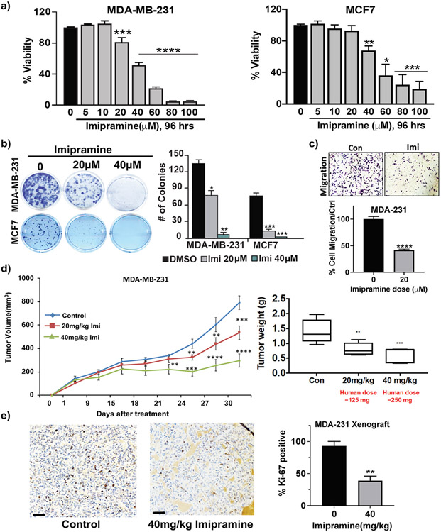
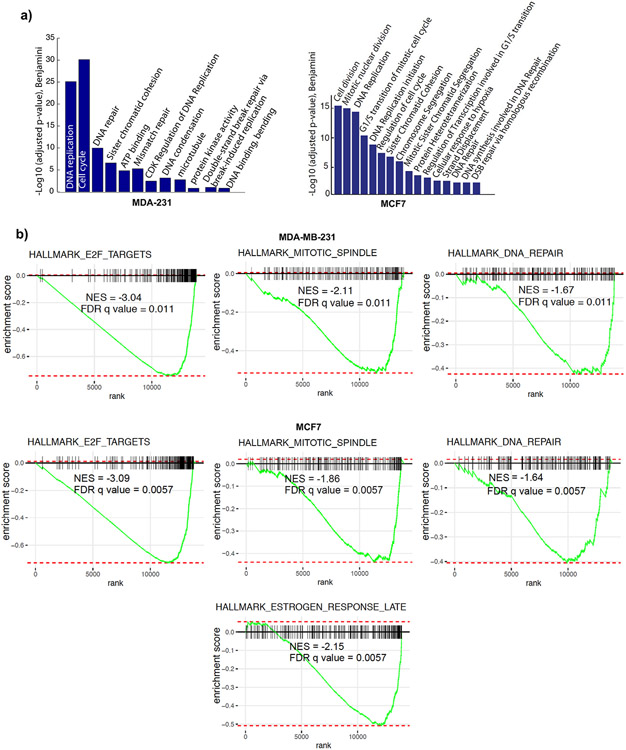
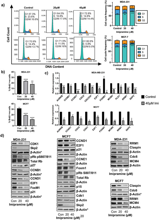
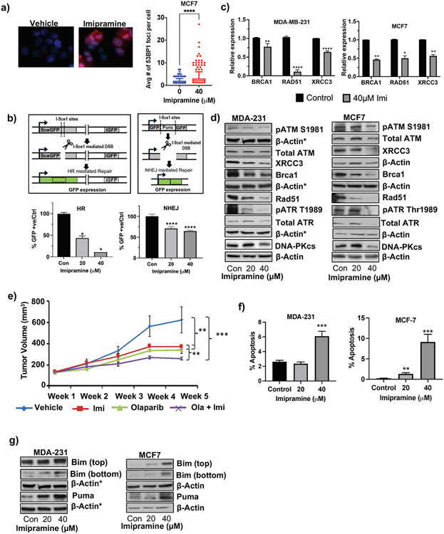
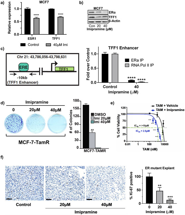
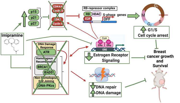
Aberrant activities of various cell cycle and DNA repair proteins promote cancer growth and progression and render them resistant to therapies. Here, we demonstrate that the anti-depressant imipramine blocks growth of triple-negative (TNBC) and estrogen receptor-positive (ER+) breast cancers by inducing cell cycle arrest and by blocking heightened homologous recombination (HR) and non-homologous end joining-mediated (NHEJ) DNA repair activities. Our results reveal that imipramine inhibits the expression of several cell cycle- and DNA repair-associated proteins including E2F1, CDK1, Cyclin D1, and RAD51. In addition, we show that imipramine inhibits the growth of ER + breast cancers by inhibiting the estrogen receptor- α (ER-α) signaling. Our studies in preclinical mouse models and ex vivo explants from breast cancer patients show that imipramine sensitizes TNBC to the PARP inhibitor olaparib and endocrine resistant ER + breast cancer to anti-estrogens. Our studies suggest that repurposing imipramine could enhance routine care for breast cancer patients. Based on these results, we designed an ongoing clinical trial, where we are testing the efficacy of imipramine for treating patients with triple-negative and estrogen receptor-positive breast cancer. Since aberrant DNA repair activity is used by many cancers to survive and become resistant to therapy, imipramine could be used alone and/or with currently used drugs for treating many aggressive cancers.

Keywords: Breast cancer; DNA damage; DNA repair; Drug repurposing; Estrogen receptor; Imipramine.


Figures

Fig. 1.
Fig. 1.. Imipramine inhibits breast cancer growth and progression in vitro and in vivo
(a) MDA-MB-231(left) and MCF7 cells (right) cells treated with vehicle or different doses of imipramine for 96 h and subjected to short-term cell viability assays using Alamar Blue. Bar graph shows percentage of cell viability. (b) Long-term colony formation assay for MDA-MB-231 and MCF7 cells was performed after treatment with vehicle and imipramine for 7 days. The media with vehicle or imipramine was replenished every 3 days. Right: Average number of colonies per group. (c) MDA-MB-231 cells were treated with vehicle or 20 μM imipramine and subjected to transwell migration assays. Bar graphs show the average number of migrated cells per group. (d) Mean tumor volume ± SEM (left) and tumor weight ± SEM (right) for athymic nude mice implanted with MDA-MB-231 cells then treated with vehicle- and imipramine. (e) Immunohistochemical staining was performed on vehicle and imipramine-treated tumors from d (left). Scale bar in the pictures indicate an area of 60 μm. Bar graph shows the percentage of Ki-67 positive cells in each group (right). p-values were calculated using a standard Student t-test. **P < 0.01; ***P < 0.001; ****P < 0.0001.
Fig. 2.
Fig. 2.. Imipramine inhibits the expression of genes involved in DNA repair, cell cycle, and DNA replication
a) Gene Ontology analysis showing biological processes significantly enriched in MDA-MB-231 (left) and MCF7 (right) cells treated with imipramine. (b) Gene Set Enrichment Analysis (GSEA) of the differentially expressed genes showing selected hallmark gene sets negatively enriched in MDA-231 (top) and MCF7 (bottom) cells treated with imipramine. The Normalized Enrichment Score (NES) and FDR q value are displayed.
Fig. 3.
Fig. 3.. Imipramine inhibits cell cycle progression and promotes G1 cell cycle arrest
(a) Representative cell cycle profile of MDA-MB-231(top left) and MCF7 (bottom left) cells. Cells were treated with vehicle or imipramine and subjected to propidium iodide-based cell cycle analysis by flow cytometry. Bar graphs (right) show the percentage of cells in different phases of the cell cycle in MDAMB-231 and MCF7 cells (mean ± SEM, n = 3 independent experiments in triplicate). (b) Bar graphs show % BrdU-positive cells in MDA-MB-231 and MCF7 cells (mean ± SEM, n = 3 independent experiments). (c) qRT-PCR analysis of genes involved in cell cycle and DNA replication pathways in vehicle- and imipramine-treated MDA-MB-231 and MCF7 cells. (d) Western blot analyses of vehicle- and imipramine-treated MDA-MB-231 and MCF7 cells using antibodies against the indicated proteins involved in cell cycle (left) and DNA replication (right), β-actin was used as a loading control. Gel photographs are representative of three or more independent experiments. *The same β-actin was used in multiple blots, p-values were calculated using standard Student t-tests. *P < 0.05 **P < 0.01; ****P < 0.0001.
Fig. 4.
Fig. 4.. Imipramine inhibits DNA repair and promotes apoptosis
a) Representative images showing immunofluorescence in vehicle- and imipramine-treated MDA-MB-231 and MCF7 cells (left) using antibody against 53BP1. Right: Number of 53BP1 foci in vehicle- and imipramine-treated cells (≥100 cells counted/group). 10–90% of values are shown in the box and whisker plots, with remaining values represented as dots. (b) Top left: Schematic illustration of I-SceI endonuclease-based DR-GFP reporter assay to measure doublestrand break (DSB)-induced homologous recombination (HR) repair. Top right: Schematic illustration of I-SceI endonuclease-based EJ5-GFP reporter assay to measure DSB-induced nonhomologous end joining (NHEJ) repair. U2OS-DR-GFP and EJ5-GFP cells were transfected with a pCAGGS vector encoding ISceI endonuclease or empty vector for 24 h, followed by treatment with vehicle or imipramine for 72 h. I-SceI endonuclease causes DSB at DR-GFP or EJ5-GFP loci and if repaired by HR or NHEJ, respectively, results in GFP + cells. (Bottom left) Results of flow cytometry showing percentage of GFP + DR-GFP-U2OS cells after treatment with vehicle or imipramine. (Bottom right) Results of flow cytometry showing percentage of GFP + EJ5-U2OS cells after treatment with vehicle or imipramine. (c) qRT-PCR analysis of genes involved in DNA Repair pathways in vehicle- and imipramine-treated MDA-MB-231 and MCF7 cells. (d) Western blot analyses of proteins involved in DNA repair in MDA-MB-231 and MCF7 cells treated with vehicle or imipramine. β-actin was used as a loading control. Gel photographs are representative of three or more independent experiments. *The same β-actin is shown as the loading control for different proteins probed after stripping the same blot. (e) Mean tumor volume of mice treated with vehicle, imipramine (32 mg/kg body weight), olaparib (50 mg/kg body weight), and imipramine + olaparib (n = 7 mice/group). (f) Bar graphs show percentage of Annexin V positive apoptotic cells in vehicle- and imipramine-treated cells after flow cytometry. (g) Western blot analyses of MDA-MB-231 and MCF7 cells treated with vehicle or imipramine using antibody against pro-apoptotic proteins, β-actin was used as a loading control. Gel photographs are representative of three or more independent experiments. *The same β-actin is shown as the loading control for different proteins probed after stripping the same blot, p- values were calculated using standard Student t-tests. All experiments were repeated at least 3 times and are a representative of 3 biological replicates. *P < 0.05 **P < 0.01; ****P < 0.0001.
Fig. 5.
Fig. 5.. Imipramine inhibits ER-α signaling and sensitizes breast cancer cells to tamoxifen
a) qPCR analysis showing relative expression of ESR1 (gene encoding ER-α) and TFF1 in MCF7 cells treated with vehicle or imipramine for 96 h. (b) Western blot analyses of ER-α and TFF1 in MCF7 cells treated with vehicle or imipramine for 96 h. b-actin was used as a loading control. (c) (left) Schematic illustration of chromatin immunoprecipitation (ChIP) primers flanking the estrogen response element (ERE)/ER-α binding site in the upstream enhancer region of the human TFF1 gene. (Right) Fold change in recruitment/binding of ERα and RNA Pol II to the ERE site on TFF1 enhancer after ChIP-qPCR analysis. (d) Results of long-term colony formation assays of MCF-TamR cells treated with vehicle or imipramine for 7 days. Right: Average number of colonies per group. (e) Short-term viability of MCF7- TamR cells treated with vehicle + tamoxifen or imipramine + tamoxifen, measured by Alamar blue assays. (f) (Left) Representative immunohistochemical images of Ki67 staining of ER mutant explants treated with vehicle or imipramine. Scale bar in the pictures indicate an area of 60 μm (Right) Percent of Ki-67+ cells in vehicle-and imipramine-treated explants. p-values were calculated using standard Student t-tests. All experiments were repeated at least 3 times and are a representative of 3 biological replicates. *P < 0.05 **P < 0.01; ****P < 0.0001. ****p < 0.0001; ***p < 0.001; **p < 0.01; *p < 0.05.
Fig. 6.
Fig. 6.. Imipramine mechanism of action in breast cancers
Model showing imipramine mechanism of action in breast cancers. Red boxes and lines indicate proteins/mechanisms that act to promote cancer growth and progression, while green boxes or lines represent proteins or pathways that inhibit breast cancer growth and progression. Based on our results, we propose that imipramine blocks breast cancer growth and progression by inhibiting cell cycle, DNA Repair, and ER-α signaling. We show that imipramine treatment leads to an increase in CDK inhibitors including p15, p21, and p27, which inhibit the activity of CDK4/6 and CDK2 (marked by X), consequently preventing the hyperphosphorylation and loss of activity of the RB protein. This leads to reduced E2F activity, reduced transcription of S phase genes, and increased G1/S cell cycle arrest. In addition to cell cycle progression, we show that imipramine treatment inhibits DNA Damage Response and DNA Repair pathway (marked by X), by inhibiting key players in the DNA Damage Response/DNA Repair pathway, including ATR, BRCA1, RAD51, and DNA-PKcs. We posit that reduced DNA repair leads to persistent/increased level of DNA damage and consequently apoptosis in imipramine-treated breast cancer cells. We propose that imipramine may block the growth and progression of ER + breast cancers by inhibiting ER-α signaling. Since effectors of DNA repair (such as FOXM1, RAD51 and BRCA1) and ER-a signaling functionally and physically crosstalk, our results suggest that imipramine inhibition of both DNA repair and ER-a signaling amplifies its anti-tumor effects on ER + breast cancers.

Similar articles

Cited by

References

    1. Society AC, Breast Cancer Facts & Figures 2019-2020, American Cancer Society, Inc, Atlanta, 2019.
    1. Waks AG, Winer EP, Breast cancer treatment: a review, JAMA 321 (3) (2019. Jan) 288–300. - PubMed
    1. Luque-Bolivar A, Pérez-Mora E, Villegas VE, Rondón-Lagos M, Resistance and overcoming resistance in breast cancer. Breast cancer, Targets Ther. 12 (2020. Nov) 211–229. - PMC - PubMed
    1. Zhang Z, Zhou L, Xie N, Nice EC, Zhang T, Cui Y, et al., Overcoming cancer therapeutic bottleneck by drug repurposing, Signal Transduct. Targeted Ther 5 (1) (2020. Jul) 1–25, 2020 5:1. - PMC - PubMed
    1. Gallagher EJ, LeRoith D, Obesity and diabetes: the increased risk of cancer and cancer-related mortality, Physiol. Rev 95 (3) (2015. Jul) 727–748. - PMC - PubMed