Case Reports
. 2022 May 13;15(5):e248415.
doi: 10.1136/bcr-2021-248415.

Non-arteritic anterior ischaemic optic neuropathy (NA-AION) and COVID-19 vaccination

Affiliations
Case Reports

Non-arteritic anterior ischaemic optic neuropathy (NA-AION) and COVID-19 vaccination

Srinivasan Sanjay et al. BMJ Case Rep. .

Abstract

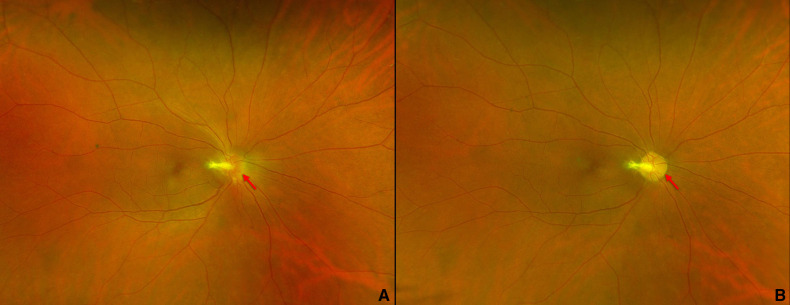
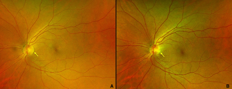
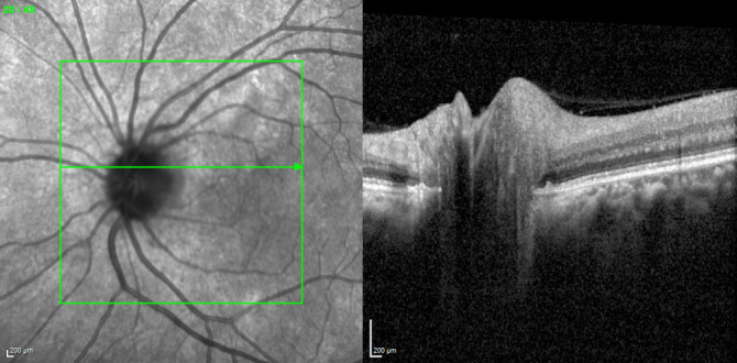
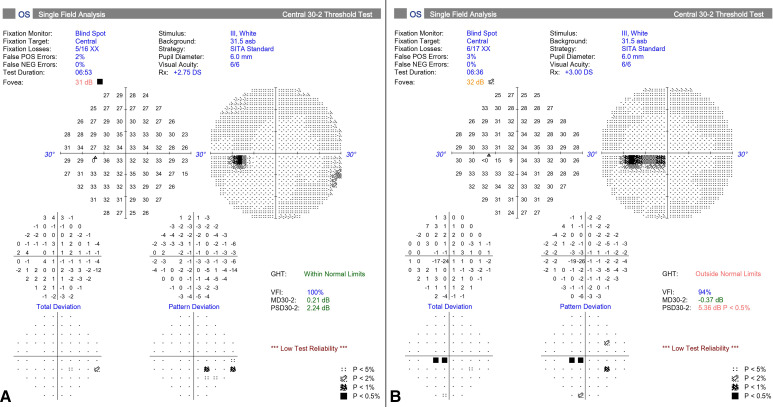
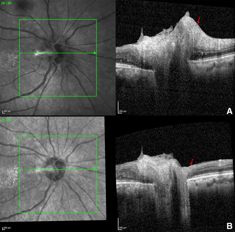
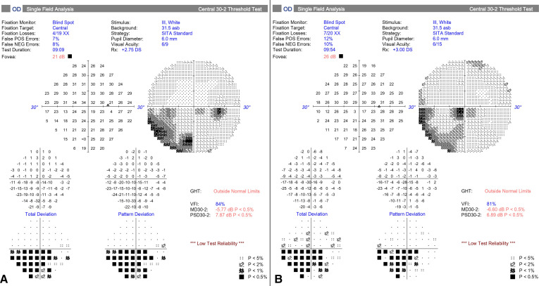
A woman in her 50s presented with diminution of vision in her left eye (OS) 4 days after COVISHIELDTM vaccination. She had been diagnosed with non-arteritic anterior ischaemic optic neuropathy (NA-AION) of right eye (OD) 8 months earlier. The present episode revealed a best-corrected visual acuity (BCVA) of 20/50 in OD and 20/20 in OS with grade 1 relative afferent pupillary defect. Fundus evaluation showed pale disc in OD and temporal disc oedema in OS. Humphrey's visual field analysis showed incomplete inferior altitudinal defect in OD and a centro-caecal scotoma in OS. Systemic investigations were normal. OS was diagnosed with NA-AION. She was started on oral aspirin 75 mg. At 1-month follow-up, disc oedema of OS had resolved with BCVA maintaining at 20/20. The patient was lost to follow-up later. The relationship between the vaccine and the ocular event is temporal with no causal association.

Keywords: COVID-19; Eye; Immunological products and vaccines; Medical management; Neuroopthalmology.


Conflict of interest statement

Competing interests: None declared.

Figures

Figure 1
Figure 1
Infrared photograph of the pupil in bright light at presentation showing relative afferent pupillary defect in left eye.
Figure 2
Figure 2
Fundus photograph of right eye showing (A) crowded hyperaemic disc with peripapillary oedema (red arrow) with fibrous remnant nasally at 8 months earlier (B) fibrous remnant at disc and pale disc with well-defined margins (red arrow) at presentation now.
Figure 3
Figure 3
Fundus photograph of left eye showing (A) well defined disc margin (yellow arrow) at 8 months earlier, (B) blurred temporal disc margin (yellow arrow) with cup disc ratio of 0.2:1 at presentation now.
Figure 4
Figure 4
Spectral domain optical coherence tomography (SD-OCT) of retinal nerve fibre layer image of left eye showing peripapillary swelling temporal to the disc.
Figure 5
Figure 5
Humphrey’s visual field analysis of left eye (OS) showing (A) normal fields at 8 months earlier (B) centrocaecal scotoma at presentation now.
Figure 6
Figure 6
Spectral domain optical coherence tomography image of retinal nerve fibre layer of right eye showing (A) peripapillary swelling (red arrow) with hyperreflective band into vitreous cavity correlating with the fibrous remnant at 8 months earlier, (B) significant reduction in peripapillary swelling (red arrow) at presentation now.
Figure 7
Figure 7
Humphrey’s visual field analysis of right eye (OD) showing (A) incomplete inferior altitudinal scotoma at 8 months earlier, (B) reduction in the extent of inferior altitudinal scotoma at presentation now. GHT, glaucoma hemi field test; NEg, Negative; POS, positive; PSD, pattern standard deviation; VFI, visual field indices.

References

    1. The impact of COVID-19 on global health goals, 2021. Available: https://www.who.int/news-room/spotlight/the-impact-of-covid-19-on-global... [Accessed 21 Jan 2022].
    1. Pichi F, Aljneibi S, Neri P, et al. Association of ocular adverse events with inactivated COVID-19 vaccination in patients in abu dhabi. JAMA Ophthalmol 2021;139:1131–5. 10.1001/jamaophthalmol.2021.3477 - DOI - PMC - PubMed
    1. Goyal M, Murthy SI, Annum S. Bilateral multifocal choroiditis following COVID-19 vaccination. Ocul Immunol Inflamm 2021;29:753–7. 10.1080/09273948.2021.1957123 - DOI - PubMed
    1. Koong LR, Chee WK, Toh ZH, et al. Vogt-Koyanagi-Harada disease associated with COVID-19 mRNA vaccine. Ocul Immunol Inflamm 2021;29:1212–5. 10.1080/09273948.2021.1974492 - DOI - PubMed
    1. Fowler N, Mendez Martinez NR, Pallares BV, et al. Acute-Onset central serous retinopathy after immunization with COVID-19 mRNA vaccine. Am J Ophthalmol Case Rep 2021;23:101136. 10.1016/j.ajoc.2021.101136 - DOI - PMC - PubMed

Publication types