

ATF1/miR-214-5p/ITGA7 axis promotes osteoclastogenesis to alter OVX-induced bone absorption
- PMID: 35568813
- PMCID: PMC9107670
- DOI: 10.1186/s10020-022-00476-7
ATF1/miR-214-5p/ITGA7 axis promotes osteoclastogenesis to alter OVX-induced bone absorption
Abstract
Background: The dynamic balance of osteoblast and osteoclast is critical for bone homeostasis and overactive osteoclastic function may lead to osteoporosis. Activating transcription factor 1 (ATF1) is involved in osteoclastogenesis. However, the detailed mechanisms remain to be explored.
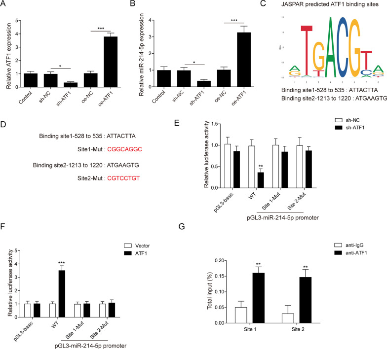
Methods: RAW264.7 cells were used and induced toward osteoclast by RANKL administration. We performed flow cytometry, CCK-8 assay and tartrate-resistant acid phosphatase (TRAP) staining to examine cell apoptosis, proliferation and differentiation of RAW264.7 cells, respectively. Mice were subjected to ovariectomy to induce osteoporosis. Micro CT, HE staining and TRAP staining were performed to evaluate bone loss in the OVX mouse model. Bioinformatics methods, luciferase assays and Chromatin Immunoprecipitation (ChIP) were used to predict and validate the interaction among ATF1, miR-214-5p, and ITGA7.
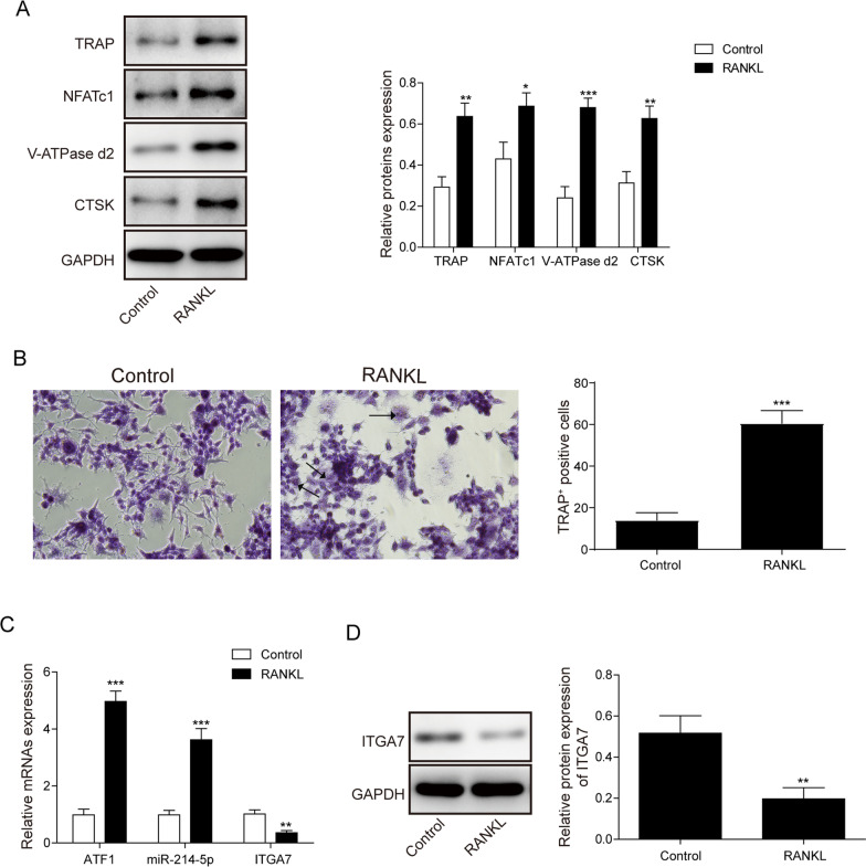
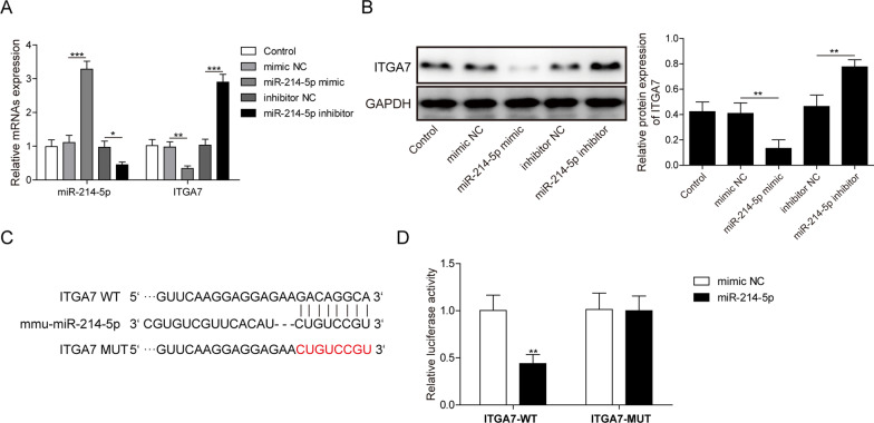
Results: ATF1 and miR-214-5p were up-regulated while ITGA7 was inhibited in RANKL-induced osteoclasts. MiR-214-5p was transcriptionally activated by ATF1. ATF1 knockdown suppressed osteoclast formation by miR-214-5p inhibition. ITGA7 was the direct target of miR-214-5p. Knockdown of miR-214-5p abolished osteoclastogenesis, which was reversed by ITGA7 knockdown. In OVX model, miR-214-5p knockdown suppressed osteoclast differentiation and prevented bone loss.
Conclusion: ATF1/miR-214-5p/ITGA7 axis regulated osteoclast formation both in vivo and in vitro, thereby affecting OVX-induced bone resorption in mice. Knockdown of ATF1 might be a promising strategy to manage osteoporosis.
Keywords: ATF1; ITGA7; Osteoclastogenesis; Osteoporosis; miR-214-5p.
© 2022. The Author(s).
Conflict of interest statement
The authors declare that there is no conflict of interest.
Figures


Similar articles
-
MiR-1224-5p modulates osteogenesis by coordinating osteoblast/osteoclast differentiation via the Rap1 signaling target ADCY2.Exp Mol Med. 2022 Jul;54(7):961-972. doi: 10.1038/s12276-022-00799-9. Epub 2022 Jul 13. Exp Mol Med. 2022. PMID: 35831436 Free PMC article.
-
MiR-221-5p/Smad3 axis in osteoclastogenesis and its function: Potential therapeutic target for osteoporosis.Steroids. 2022 Sep;185:109063. doi: 10.1016/j.steroids.2022.109063. Epub 2022 Jun 11. Steroids. 2022. PMID: 35700796
-
Osteoclast-derived exosomes influence osteoblast differentiation in osteoporosis progression via the lncRNA AW011738/ miR-24-2-5p/ TREM1 axis.Biomed Pharmacother. 2024 Sep;178:117231. doi: 10.1016/j.biopha.2024.117231. Epub 2024 Aug 1. Biomed Pharmacother. 2024. PMID: 39094542
-
miR-128 plays a critical role in murine osteoclastogenesis and estrogen deficiency-induced bone loss.Theranostics. 2020 Mar 4;10(10):4334-4348. doi: 10.7150/thno.42982. eCollection 2020. Theranostics. 2020. PMID: 32292498 Free PMC article.
-
CUL4A-mediated ZEB1/microRNA-340-5p/HMGB1 axis promotes the development of osteoporosis.J Biochem Mol Toxicol. 2023 Aug;37(8):e23373. doi: 10.1002/jbt.23373. Epub 2023 May 30. J Biochem Mol Toxicol. 2023. PMID: 37253097
Cited by
-
Epigenetic regulation in metabolic diseases: mechanisms and advances in clinical study.Signal Transduct Target Ther. 2023 Mar 2;8(1):98. doi: 10.1038/s41392-023-01333-7. Signal Transduct Target Ther. 2023. PMID: 36864020 Free PMC article. Review.
-
Role and Regulation of Transcription Factors in Osteoclastogenesis.Int J Mol Sci. 2023 Nov 10;24(22):16175. doi: 10.3390/ijms242216175. Int J Mol Sci. 2023. PMID: 38003376 Free PMC article. Review.
-
Transcription factor GTF2I regulates osteoclast differentiation through mediating miR-134-5p and MAT2A expressions.J Cell Commun Signal. 2025 Apr 3;19(2):e70010. doi: 10.1002/ccs3.70010. eCollection 2025 Jun. J Cell Commun Signal. 2025. PMID: 40191097 Free PMC article.
-
Spatially distinct otic mesenchyme cells show molecular and functional heterogeneity patterns before hearing onset.iScience. 2023 Aug 29;26(10):107769. doi: 10.1016/j.isci.2023.107769. eCollection 2023 Oct 20. iScience. 2023. PMID: 37720106 Free PMC article.
-
ATF1 and miR-27b-3p drive intervertebral disc degeneration through the PPARG/NF-κB signaling axis.Commun Biol. 2025 May 14;8(1):751. doi: 10.1038/s42003-025-08186-6. Commun Biol. 2025. PMID: 40369110 Free PMC article.
References
Publication types
MeSH terms
Substances
LinkOut - more resources
Full Text Sources
Medical
Molecular Biology Databases
