. 2022 May 14;10(1):75.
doi: 10.1186/s40478-022-01373-0.

Slow motor neurons resist pathological TDP-43 and mediate motor recovery in the rNLS8 model of amyotrophic lateral sclerosis

Affiliations

Slow motor neurons resist pathological TDP-43 and mediate motor recovery in the rNLS8 model of amyotrophic lateral sclerosis

Seong Kwon Hur et al. Acta Neuropathol Commun. .

Abstract

In the intermediate stages of amyotrophic lateral sclerosis (ALS), surviving motor neurons (MNs) that show intrinsic resistance to TDP-43 proteinopathy can partially compensate for the loss of their more disease-susceptible counterparts. Elucidating the mechanisms of this compensation may reveal approaches for attenuating motor impairment in ALS patients. In the rNLS8 mouse model of ALS-like pathology driven by doxycycline-regulated neuronal expression of human TDP-43 lacking a nuclear localization signal (hTDP-43ΔNLS), slow MNs are more resistant to disease than fast-fatigable (FF) MNs and can mediate recovery following transgene suppression. In the present study, we used a viral tracing strategy to show that these disease-resistant slow MNs sprout to reinnervate motor endplates of adjacent muscle fibers vacated by degenerated FF MNs. Moreover, we found that neuromuscular junctions within fast-twitch skeletal muscle (tibialis anterior, TA) reinnervated by SK3-positive slow MNs acquire resistance to axonal dieback when challenged with a second course of hTDP-43ΔNLS pathology. The selective resistance of reinnervated neuromuscular junctions was specifically induced by the unique pattern of reinnervation following TDP-43-induced neurodegeneration, as recovery from unilateral sciatic nerve crush did not produce motor units resistant to subsequent hTDP-43ΔNLS. Using cross-reinnervation and self-reinnervation surgery in which motor axons are disconnected from their target muscle and reconnected to a new muscle, we show that FF MNs remain hTDP-43ΔNLS-susceptible and slow MNs remain resistant, regardless of which muscle fibers they control. Collectively, these findings demonstrate that MN identity dictates the susceptibility of neuromuscular junctions to TDP-43 pathology and slow MNs can drive recovery of motor systems due to their remarkable resilience to TDP-43-driven degeneration. This study highlights a potential pathway for regaining motor function with ALS pathology in the advent of therapies that halt the underlying neurodegenerative process.

Keywords: Amyotrophic lateral sclerosis; Cross-reinnervation surgery; Neurodegeneration; Neuropathology; TDP-43; rNLS8.


Conflict of interest statement

John Q. Trojanowski was a member of the Editorial Board of Acta Neuropathologica. The authors declare that they have no competing interests. Krista J. Spiller is an employee of Janssen R&D, the pharmaceutical company of Johnson & Johnson.

Figures
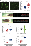
Fig. 1
Fig. 1
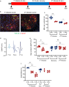
rNLS8 mice recover motor function following hTDP-43ΔNLS suppression after disease onset. a Representative immunofluorescence images of rNLS8 lumbar MNs expressing high levels of cytoplasmic TDP-43 at 6 wks. off Dox compared to low levels of cytoplasmic TDP-43 after 8 wks. recovery following 6 wks. off Dox. Scale bar: 50 μm. The antibody used for immunostaining detects both human and murine TDP-43. b Comparison of immunofluorescence staining intensity for TDP-43 as shown in (a) in the nucleus and cytoplasm of lumbar MNs from rNLS8 mice at 6 wks. off Dox or 6 wks. off Dox followed by 6–8 recovery on Dox. The nuclear area was defined by reference to DAPI staining and the nuclear/cytosolic TDP-43 immunofluorescence intensity ratio was calculated for 40–50 cells per group. The latter metric was compared between groups by unpaired t test; ****p < 0.0001. c TreadScan gait analysis with stance graph for representative rNLS8 mice at 6 wks. off Dox compared to 6 wks. off Dox followed by after 8 wks. recovery on Dox. d Treadmill running speeds of rNLS8 mice after 6 wks. off Dox and after 6 wks. off Dox followed by 6–8 wks. recovery on Dox. N = 9 mice per group, t test; **p = 0.002. e Average traces of compound muscle action potential (CMAP) in the gastrocnemius (GC) muscle of rNLS8 mice after 6 wks. off Dox or 6 wks. off Dox followed by 6–8 wks. recovery on Dox. f Maximum CMAP amplitude in the GC muscle of rNLS8 mice after 6 wks. off Dox or 6 wks. off Dox followed by 10 wks. recovery on Dox (t test, ***p < 0.001). g The proportion of neuromuscular junctions in the fast hindlimb muscle tibialis anterior (TA) innervation in rNLS8 mice after 6 wks. off Dox or 6 wks off Dox followed by 6–8 wks. recovery on Dox (t test; ***p = 0.002)
Fig. 2
Fig. 2
Intrinsically disease-resistant MN subpopulations mediate the reinnervation of vulnerable muscles after disease onset in rNLS8 mice. a The experimental approach used to investigate the identity of MNs that reinnervate the TA muscle during recovery in rNLS8 mice. The retrograde neuronal tracer AAV9-GFP was injected bilaterally in the TA of p4 mice to fluorescently label (in green) the pre-disease motor pool (denoted TA1). Dox was then withdrawn for 6 wks. to activate hTDP-43ΔNLS expression, followed by 8 wks. Dox reintroduction for recovery, after which CTG-594 was injected bilaterally in the TA to fluorescently label (in red) the post-disease motor pool (denoted TA2). Surviving local MNs would thus be labelled with both green and red fluorescence, whereas MNs from adjacent pools expanding their field to reinnervate the TA would be labelled with red fluorescence only. Below are illustrated potential outcomes for the experiment, including the interpretation of TA2 MNs that were either exclusively co-labelled with GFP and CTB-594 or were labelled with CTB-594 alone. b Representative images of lumbar MNs co-labeled with AAV9-GFP and CTB-594 from rNLS8 and non-transgenic (nTg) mice at the endpoint of the experiment outlined in a. Yellow arrows mark MNs labelled with both AAV9-GFP and CTB-594 (i.e., surviving local MNs), while red arrows mark MNs labelled with CTB-594, only (i.e., MNs reinnervating from adjacent motor pools). Scale bar: 30 µm. c Quantification of the proportion of MNs labelled with CTB-594 only (i.e., MNs reinnervating from adjacent motor pools) or both AAV9-GFP and CTB-594 (i.e., surviving local MNs) in lumbar sections from rNLS8 and nTg mice at the endpoint of the experiment outlined in a. Three mice were examined per group, with 20–30 MNs scored per animal. T test, ***p = 0.0003
Fig. 3
Fig. 3
The recovered lower motor circuit in rNLS8 mice is resistant to subsequent hTDP-43ΔNLS challenge. a The experimental approach used to investigate the response of rNLS8 mice to a second instance of hTDP-43ΔNLS expression after recovery from the initial pathological insult. Dox was withdrawn for 6 wks. to initiate a first disease course, after which hTDP-43ΔNLS was suppressed for 10–12 wks. by reintroducing dietary Dox, thus allowing muscle reinnervation by resistant MNs. A second 6-wk. instance of disease was then imposed by withdrawing Dox. Arrows indicate the time points at which mice were analyzed for CMAP, TA innervation and MN counts. b Representative immunofluorescence images of lumbar MNs (VAChT-positive, red) expressing high levels of cytoplasmic hTDP-43 (green) from rNLS8 mice in the first and second disease courses. Scale bar: 100 μm. c Quantitation of the number of MNs per ventral horn in rNLS8 mice at each analysis timepoint in the study outlined in a. The number of animals assayed per timepoint was 4–5, with 10–20 spinal cord sections scored for each animal. T test, ***p < 0.001 for baseline vs. 1st disease course; *p = 0.02 for recovery versus 2nd disease course. d CMAP traces in the GC muscle from rNLS8 mice after the first disease course, after recovery and after the second disease course. e Longitudinal comparison of maximum evoked CMAP amplitude in the GC muscle of rNLS8 mice throughout the experimental time course outlined in (a). The number of animals assayed per timepoint was 5–7. T test, **p = 0.0010 for baseline versus 4 wks. off Dox; ****p < 0.0001 for baseline versus 6 wks. off Dox; other comparisons nonsignificant. f The proportion of intact NMJs in the lateral GC muscle of rNLS8 mice throughout the experimental time course outlined in (a). The number of animals assayed per timepoint was 3–6, with 600–1000 NMJs scored per animal. T test, ***p < 0.001 for baseline versus 4 wks. off Dox; ***p < 0.001 for baseline vs. 6 wks. off Dox; other comparisons nonsignificant
Fig. 4
Fig. 4
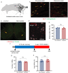
Nonselective degeneration and reinnervation does not alter the susceptibility of the TA muscle to axonal dieback in the presence of pathological hTDP-43ΔNLS. a Schematic representation of a unilateral sciatic nerve crush. DRG dorsal root ganglion, DN distal nerve. b Representative fluorescence images of the TA muscle of nTg mice before and after nerve crush. Immunostaining for SV2 marks nerve endings (red), while labelling with BTX-488 marks motor endplates (green). Scale bar: 100 µm. c Representative image of TA-innervating MNs in the lumbar spinal cord of nTg mice backfilled with CTB-594 on the crushed side (left, red fluorescence) and with CTB-488 on the contralateral side (right, green fluorescence). Yellow arrows mark MNs reinnervating (crushed side) or innervating (non-crushed) motor pools in the TA. Scale bar: 100 µm. d Representative immunofluorescence staining for Mmp9, a marker of fast-fatigable (FF) MNs, in TA-reinnervating MNs (i.e., CTB-594 back-filled; red) on the ipsilateral side of the lumbar spinal cord of nTg mice 10 wks. after unilateral sciatic nerve crush. Scale bar: 100 µm. e Quantification of the proportion of reinnervated FF type MNs (Mmp9-positive) among all CTB-488/CTB-594-positive TA MNs on the ipsilateral and contralateral sides of nTg mice subjected to unilateral sciatic nerve crush. Three animals were assayed, with 30–40 MNs scored per animal. Paired t test, n. s., p = 0.2049. f The experimental approach used to evaluate whether recovery after unilateral sciatic nerve crush in rNLS8 mice ameliorates denervation during subsequent hTDP-43ΔNLS expression. Mice were subjected to unilateral sciatic nerve crush as illustrated in (a), then allowed to recover on Dox for 8–10 wks. Bilateral CMAP and TreadScan analysis were used to confirm full recovery, after which hTDP-43ΔNLS expression was induced by withdrawing Dox for 6 wks. MNs were counted and NMJs analyzed bilaterally at endpoint. g Quantitation of the number of MNs labelled with VaChT per ventral horn in rNLS8 mice analyzed at endpoint in the experiment outlined in (f). Three mice were assayed, with 10 spinal cord sections scored per animal. Paired t test, n. s., p = 0.054 h Quantitation of the proportion of intact NMJs (scored as coincident SV2-positive nerve endings and BTX-488/BTX-594-labeled motor endplates) relative to all BTX-488/BTX-594-labeled motor endplates in the TA muscles on the ipsilateral and contralateral sides of rNLS8 mice at the endpoint of the experiment outlined in (f). Five mice were assayed, with 600–1000 NMJs scored per animal. Paired t test, n. s., p = 0.391
Fig. 5
Fig. 5
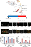
Cross-reinnervation surgery manipulates slow MNs to innervate TA muscle in place of FF MNs. a Schematic representation of unilateral cross reinnervation surgery. b The experimental approach for using cross-reinnervation surgery to investigate the significance of MN identity in axonal regeneration following hTDP-43ΔNLS disease. rNLS8 mice were subjected to unilateral cross-innervation or self-innervation surgery (the latter as a procedural control) and allowed to recover for 6, 10 and 24 wks. Recovery was confirmed using bilateral TA muscle CMAP and TreadScan analysis. At 6, 10 and 24 wks. post-operation, the TA muscle was bilaterally injected with CTB-488 3 days before sacrificed mice to validate the reinnervation surgery. At 10 wks. post-operation, Dox was withdrawn to activate hTDP-43ΔNLS expression for a period of 8 wks. c Representative fluorescence images of sections from the lumbar spinal cord highlighting the ipsilateral (cross-reinnervation surgery; “S”) and contralateral (non-surgical; “N.S.”) sides of rNLS8 mice 10 wks. after surgery. Visible are FF MNs positive for Mmp9 immunostaining (red) and re-innervated TA MNs retrogradely labelled with CTB-488 (green). Mmp9-negative/CTB-488-positive MNs were either slow type or fatigue-resistant TA MNs (white arrows), whereas Mmp9-positive/CTB-488-positive MNs were fast-fatigable TA MNs (yellow arrows). Scale bar: 50 µm. d Representative fluorescence images of sections from the lumbar spinal cord highlighting the ipsilateral (cross-reinnervation surgery; “S”) and contralateral (non-surgical; “N.S.”) sides of rNLS8 mice 10 wks. after surgery. Visible are MNs positive for SK3 immunostaining (red) and re-innervated TA MNs retrogradely labelled with CTB-488 (green). White arrows mark SK3-negative, CTB-488-positive presumptive FF TA MNs. The yellow arrow marks as SK3-positive, CTB-488-positive slow type or fatigue-resistant TA MN. Scale bar: 50 µm. e Quantification of reinnervated FF type TA MNs (Mmp9-positive/CTB-488-positive) as a proportion of total re-innervated TA MNs (all CTB-488-positive) across the study time course outlined in (b). Two-to-four mice were assayed per time point, with 70–80 MNs scored per side. Two-way ANOVA, ***p = 0.0003. f Quantification of reinnervated slow type TA MNs (SK3-positive/CTB-488-positive MNs) as a proportion of total re-innervated TA MNs (all CTB-488-positive) across the study time course outlined in (b). Three-to-five mice were assayed per timepoint, with 70–80 MNs scored per side. Two-way ANOVA, ****p < 0.0001
Fig. 6
Fig. 6
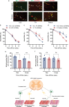
Fast muscle innervated by slow motor units via cross-reinnervation surgery acquire resistance to hTDP-43ΔNLS insult, while slow muscle innervated by fast motor units become susceptible. a Representative fluorescence images of NMJs in the hindlimb lateral GC, TA and soleus muscles on the non-surgical and cross-reinnervation surgery sides of rNLS8 mice after 6 wks. off Dox. Axons were immunostained for SV2 and NFL (red) and motor endplates were labelled with BTX-488 (green). Yellow arrows mark intact NMJs (coincident red and green fluorescence), whereas white arrows mark degenerated NMJs. Scale bar: 50 µm. bd Quantitation of the proportion of NMJs determined to be intact, by fluorescence image analysis as shown in a, in the lateral GC (b; two-way ANOVA, n. s. at each time point), TA (c; two-way ANOVA, *p = 0.019 at 4 wks. off Dox, ****p < 0.0001 at 6 wks. off Dox, **p = 0.0038 at 8 wks. off Dox) and soleus (d; two-way ANOVA, **p = 0.0032 at 6 wks. off Dox; **p = 0.0028 at 8 wks. off Dox) from the cross-reinnervation surgery (S) or non-surgical (N.S.) sides of rNLS8 at the indicated time points after hTDP-43ΔNLS induction. e, f Quantification of Mmp9-positive fast MNs (e; one-way ANOVA, ****p < 0.0001; ***p < 0.001) and SK3-positive slow MNs (f; one-way ANOVA, n. s.) in the lumbar region 3–5 on the non-surgical and surgical sides of rNLS8 mice before and after 8 wks. e or 6 wks. f of hTDP-43ΔNLS expression. g Schematic representation of the principal conclusions from the cross-reinnervation surgery study. SK3-positive slow MNs are resistant to degeneration during hTDP-43ΔNLS insult despite being experimentally forced to reinnervate the fast type, vulnerable TA muscle. Mmp9-positive fast MNs are susceptible to TDP-43 pathology despite being experimentally forced to reinnervate the slow type, resistant soleus muscle

References

    1. Allodi I, Udina E, Navarro X. Specificity of peripheral nerve regeneration: interactions at the axon level. Prog Neurobiol. 2012;98:16–37. doi: 10.1016/j.pneurobio.2012.05.005. - DOI - PubMed
    1. Altman T, Ionescu A, Ibraheem A, Priesmann D. Axonal TDP-43 condensates drive neuromuscular junction disruption through inhibition of local synthesis of nuclear encoded mitochondrial proteins. Nat Commun. 2021;12:6914. doi: 10.1038/s41467-021-27221-8. - DOI - PMC - PubMed
    1. Amado DA, Davidson BL. Gene therapy for ALS: a review. Mol Ther. 2021 doi: 10.1016/j.ymthe.2021.04.008. - DOI - PMC - PubMed
    1. Arnold C. Tailored treatment for ALS poised to move ahead. Nat Med. 2019 doi: 10.1038/d41591-019-00013-w. - DOI - PubMed
    1. Baloh RH, Rakowicz W, Gardner R, Pestronk A. Frequent atrophic groups with mixed-type myofibers is distinctive to motor neuron syndromes. Muscle Nerve. 2007;36:107–110. doi: 10.1002/mus.20755. - DOI - PubMed

Publication types