. 2022 May 14;15(1):112.
doi: 10.1186/s12920-022-01258-0.

Genomic profiling of sporadic multiple meningiomas

Affiliations

Genomic profiling of sporadic multiple meningiomas

E Zeynep Erson-Omay et al. BMC Med Genomics. .

Erratum in

  • Correction: Genomic profiling of sporadic multiple meningiomas.
    Erson-Omay EZ, Vetsa S, Vasandani S, Barak T, Nadar A, Marianayagam NJ, Yalcin K, Miyagishima D, Aguilera SM, Robert S, Mishra-Gorur K, Fulbright RK, McGuone D, Günel M, Moliterno J. Erson-Omay EZ, et al. BMC Med Genomics. 2022 Jun 13;15(1):131. doi: 10.1186/s12920-022-01273-1. BMC Med Genomics. 2022. PMID: 35698142 Free PMC article. No abstract available.

Abstract

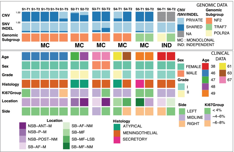
Background: Multiple meningiomas (MMs) rarely occur sporadically. It is unclear whether each individual tumor in a single patient behaves similarly. Moreover, the molecular mechanisms underlying the formation of sporadic MMs and clonal formation etiology of these tumors are poorly understood.

Methods: Patients with spatially separated MMs without prior radiation exposure or a family history who underwent surgical resection of at least two meningiomas were included. Unbiased, comprehensive next generation sequencing was performed, and relevant clinical data was analyzed.

Results: Fifteen meningiomas and one dural specimen from six patients were included. The majority of tumors (12/15) were WHO Grade I; one patient had bilateral MMs, one of which was Grade II, while the other was Grade I. We found 11/15 of our cohort specimens were of NF2-loss subtype. Meningiomas from 5/6 patients had a monoclonal origin, with the tumor from the remaining patient showing evidence for independent clonal formation. We identified a novel case of non-NF2 mutant MM with monoclonal etiology. MMs due to a monoclonal origin did not always display a homogenous genomic profile, but rather exhibited heterogeneity due to branching evolution.

Conclusions: Both NF2-loss and non-NF2 driven MMs can form due to monoclonal expansion and those tumors can acquire inter-tumoral heterogeneity through branched evolution. Grade I and II meningiomas can occur in the same patient. Thus, the molecular make-up and clinical behavior of one tumor in MMs, cannot reliably lend insight into that of the others and suggests the clinical management strategy for MMs should be tailored individually.

Keywords: Clonal formation; Genomics; Sporadic multiple meningiomas.


Conflict of interest statement

The authors declare that they have no conflict of interests.

Figures

Fig. 1
Fig. 1
Schematic summary of the study cohort. First top two panels represent the fraction of private and shared somatic alterations among the lesions of the same patient, CNVs and SNV/INDELs, respectively. Third panel annotation belongs to genomic subgroup with the clonal formation pattern concluded with the study, i.e. MC: Monoclonal formation, IND: Independent formation. Lower group panels summarize the clinical, histological and genomic attributes, such as age, sex, grade, histology, Ki-67 classification and location. SB: Skull Base, NSB: Non-Skull Base, AF: Anterior Fossa, MF: Middle Fossa, M: Midline, NM: Non-Midline, ANT: Anterior, POST: Posterior
Fig. 2
Fig. 2
Representation of case S1. a MRI of 5 lesions, b hematoxylin and eosin (H&E) stain (magnification: 200×) for all 5 lesions, S1-T3: meningioma with psammomatous calcifications. c Phylogeny inferred from the somatic CNV, SNV/INDEL data of the lesions displaying monoclonal formation and branched evolution
Fig. 3
Fig. 3
Representation of case S2. a MRI of the frontal (S2-T2) and the occipital lesion(S2-T1), b hematoxylin and eosin (H&E) stain (magnification: 200×) for both Grade II lesions, S2-T1: with chordoid features marked with black arrows. c Phylogeny inferred from the somatic CNV, SNV/INDEL of the 2 lesions
Fig. 4
Fig. 4
Representation of case S5. a MRI of 2 lesions, b hematoxylin and eosin (H&E) stain (magnification: 200×) for both lesions, S5-T1: secretory meningioma with periodic acid Schiff positive pseudopsammoma bodies, black arrows. c Phylogeny inferred from the somatic CNV, SNV/INDEL of the 2 lesions

References

    1. Tsermoulas G, Turel MK, et al. Management of multiple meningiomas. J Neurosurg. 2018;128(5):1403–1409. doi: 10.3171/2017.2.JNS162608. - DOI - PubMed
    1. Shoshan Y, Chernova O, et al. Radiation-induced meningioma: a distinct molecular genetic pattern? J Neuropathol Exp Neurol. 2000;59(7):614–620. doi: 10.1093/jnen/59.7.614. - DOI - PubMed
    1. Clark VE, Erson-Omay EZ, et al. Genomic analysis of non-NF2 meningiomas reveals mutations in TRAF7, KLF4, AKT1, and SMO. Science. 2013;339(6123):1077–1080. doi: 10.1126/science.1233009. - DOI - PMC - PubMed
    1. Clark VE, Harmanci AS, et al. Recurrent somatic mutations in POLR2A define a distinct subset of meningiomas. Nat Genet. 2016;48(10):1253–1259. doi: 10.1038/ng.3651. - DOI - PMC - PubMed
    1. Harmancı AS, Youngblood MW, et al. Integrated genomic analyses of de novo pathways underlying atypical meningiomas. Nat Commun. 2017;8:14433. doi: 10.1038/ncomms14433. - DOI - PMC - PubMed

Publication types

LinkOut - more resources