Significance of systolic-phase imaging on full-phase ECG-gated CT angiography to detect intimal tears in aortic dissection
- PMID: 35569067
- PMCID: PMC9515039
- DOI: 10.1007/s00380-022-02093-0
Significance of systolic-phase imaging on full-phase ECG-gated CT angiography to detect intimal tears in aortic dissection
Abstract
Purpose: For patients with aortic dissection (AD) and intramural hematoma (IMH), the optimal cardiac phase to detect intimal tears (IT) and ulcer-like projections (ULP) on retrospective electrocardiogram (ECG)-gated computed tomography angiography (CTA) remains unclear. The purpose of this study was to compare the accuracy of retrospective ECG-gated CTA for detecting IT in AD and ULP in IMH between each cardiac phase.
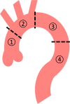
Materials and methods: A total of 75 consecutive patients with AD and IMH of the thoracic aorta were enrolled in this single-center retrospective study. The diagnostic performance to detect IT and ULP in the thoracic aortic regions (including the ascending aorta, aortic arch, and proximal and distal descending aorta) was compared in each cardiac phase on retrospective ECG-gated CTA.
Results: In the systolic phase (20%), the accuracy, sensitivity, and specificity to detect IT in AD was 64% (95% confidence interval [CI] 56-72%), 69% (95%CI 60-78%), and 25% (95%CI 3.3-45%), respectively. In the diastolic phase (70%), the accuracy, sensitivity, and specificity to detect IT in AD was 52% (95%CI 43-60%), 52% (95%CI 42-61%), and 50% (95%CI 25-75%), respectively. The accuracy to detect IT in AD on ECG-gated CTA was significantly higher in the systolic phase than that in the diastolic phase (P = 0.025). However, there were no differences in the accuracy (83%; 95%CI 78-89%), sensitivity (71%; 95%CI 62-80%), or specificity (100%; 95%CI 100%) to detect ULP in IMH between the cardiac cycle phases.
Conclusion: Although it is currently recommended for routine diagnosis of AD and IMH, single-diastolic-phase ECG-gated CTA has risk to miss some IT in AD that are detectable in the systolic phase on full-phase ECG-gated CTA. This information is critical for determining the optimal treatment strategy for AD.
Keywords: Aortic dissection; Intimal tear; Intramural hematoma; Retrospective ECG-gated CTA; Ulcer-like projection.
© 2022. The Author(s).
Conflict of interest statement
Hideki Ota were supported by a research grant from Canon Medical Systems. The other authors report no conflicts of interest.
Figures


Similar articles
-
Detection of the intimal tear in aortic dissection and ulcer-like projection in intramural hematoma: usefulness of full-phase retrospective ECG-gated CT angiography.Jpn J Radiol. 2020 Nov;38(11):1036-1045. doi: 10.1007/s11604-020-01008-1. Epub 2020 Jul 24. Jpn J Radiol. 2020. PMID: 32710132 Free PMC article.
-
A Comparison of Retrospective ECG-Gated CT and Surgical or Angiographical Findings in Acute Aortic Syndrome.Int Heart J. 2023 Sep 30;64(5):839-846. doi: 10.1536/ihj.23-002. Epub 2023 Sep 13. Int Heart J. 2023. PMID: 37704411
-
Clinical and imaging differences between Stanford Type B intramural hematoma-like lesions and classic aortic dissection.BMC Cardiovasc Disord. 2023 Jul 28;23(1):378. doi: 10.1186/s12872-023-03413-6. BMC Cardiovasc Disord. 2023. PMID: 37507680 Free PMC article.
-
Acute aortic syndromes: new insights from electrocardiographically gated computed tomography.Semin Thorac Cardiovasc Surg. 2008 Winter;20(4):340-7. doi: 10.1053/j.semtcvs.2008.11.011. Semin Thorac Cardiovasc Surg. 2008. PMID: 19251175 Review.
-
Tailored treatment modality in acute type A intramural hematoma.J Thorac Cardiovasc Surg. 2023 Nov;166(5):1400-1410. doi: 10.1016/j.jtcvs.2022.01.037. Epub 2022 Feb 3. J Thorac Cardiovasc Surg. 2023. PMID: 35221028 Review.
Cited by
-
Progress of CT aortic angiography combined with coronary artery in the evaluation of acute aortic syndrome.Front Cardiovasc Med. 2022 Nov 21;9:1036982. doi: 10.3389/fcvm.2022.1036982. eCollection 2022. Front Cardiovasc Med. 2022. PMID: 36479572 Free PMC article. Review.
References
-
- Hagan PG, Nienaber CA, Isselbacher EM, Bruckman D, Karavite DJ, Russman PL, Evangelista A, Fattori R, Suzuki T, Oh JK, Moore AG, Malouf JF, Pape LA, Gaca C, Sechtem U, Lenferink S, Deutsch HJ, Diedrichs H, Marcos y Robles J, Llovet A, Gilon D, Das SK, Armstrong WF, Deeb GM, Eagle KA. The international registry of acute aortic dissection (IRAD): new insights into an old disease. JAMA. 2000;283:897–903. doi: 10.1001/jama.283.7.897. - DOI - PubMed
-
- Howard DPJ, Banerjee A, Fairhead JF, Perkins J, Silver LE, Rothwell PM, Oxford Vascular Study Population-based study of incidence and outcome of acute aortic dissection and premorbid risk factor control: 10-year results from the Oxford Vascular Study. Circulation. 2013;127:2031–2037. doi: 10.1161/CIRCULATIONAHA.112.000483. - DOI - PMC - PubMed
-
- Writing group members. Hiratzka LF, Bakris GL, Beckman JA, Bersin RM, Carr VF, Casey DE, Eagle KA, Hermann LK, Isselbacher EM, Kazerooni EA, Kouchoukos NT, Lytle BW, Milewicz DM, Reich DL, Sen S, Shinn JA, Svensson LG, Williams DM. Circulation. 2010;121:e266–e369. - PubMed
-
- Erbel R, Aboyans V, Boileau C, Bossone E, Bartolomeo RD, Eggebrecht H, Evangelista A, Falk V, Frank H, Gaemperli O, Grabenwöger M, Haverich A, Iung B, Manolis AJ, Meijboom F, Nienaber CA, Roffi M, Rousseau H, Sechtem U, Sirnes PA, von Allmen RS, Vrints CJM, ESC Committee for Practice Guidelines ESC Guidelines on the diagnosis and treatment of aortic diseases: Document covering acute and chronic aortic diseases of the thoracic and abdominal aorta of the adult. The Task Force for the Diagnosis and Treatment of Aortic Diseases of the European Society of Cardiology (ESC) Eur Heart J. 2014;35:2873–2926. doi: 10.1093/eurheartj/ehu281. - DOI - PubMed
MeSH terms
LinkOut - more resources
Full Text Sources

