Review
. 2022 Jul:152:110341.
doi: 10.1016/j.ejrad.2022.110341. Epub 2022 May 6.

MR imaging spectrum in COVID associated Rhino-Orbito-Cerebral mucormycosis with special emphasis on intracranial disease and impact on patient prognosis

Affiliations
Review

MR imaging spectrum in COVID associated Rhino-Orbito-Cerebral mucormycosis with special emphasis on intracranial disease and impact on patient prognosis

Apoorva Sehgal et al. Eur J Radiol. 2022 Jul.

Abstract

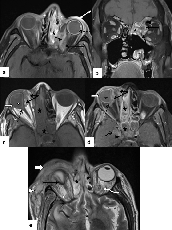
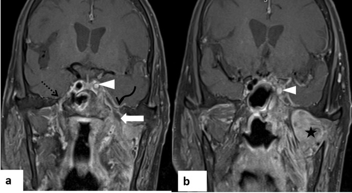
In the wake of the ongoing Coronavirus Disease 2019 (COVID-19) pandemic, a new epidemic of COVID associated mucormycosis (CAM) emerged in India. Early diagnosis and prompt treatment of this deadly disease are of paramount importance in improving patient survival. MRI is the cornerstone of diagnosis of early extrasinus disease, particularly intracranial complications which have traditionally been associated with a high mortality rate. In this review, we depict the sinonasal, perisinus, orbital and intracranial involvement in CAM. Special emphasis is laid on intracranial disease which is categorized into vascular, parenchymal, meningeal, bony involvement and perineural spread. Vascular complications are the most common form of intracranial involvement. Some unusual yet interesting imaging findings such as nerve abscesses involving the optic, trigeminal and mandibular nerves and long segment vasculitis of the internal carotid artery extending till its cervical segment are also illustrated. In our experience, patient outcome in CAM (survival rate of 88.5%) was better compared to the pre-pandemic era. Presence of intracranial disease also did not affect prognosis as poorly as traditionally expected (survival rate of 82.8%). Involvement of brain parenchyma was the only subset of intracranial involvement that was associated with higher mortality (p value 0.016). The aim of this review is to familiarise the reader with the MR imaging spectrum of CAM with special focus on intracranial complications and a brief account of their impact on patient prognosis in our experience.

Keywords: COVID associated mucormycosis; Fungal abscess; Intracranial complication; Invasive fungal sinusitis; Magnetic Resonance Imaging; Perineural spread; Rhinoorbitocerebral mucormycosis.


Conflict of interest statement

The authors declare that they have no known competing financial interests or personal relationships that could have appeared to influence the work reported in this paper.

Figures

Fig. 1
Fig. 1
Rhino-orbital involvement in two patients with CAM. Axial T2W MR image (a) reveals heterogeneously hyperintense mucosal thickening in left ethmoid sinus (arrow) with extrasinus extension into left orbit (dashed arrow). Non-specific mucosal thickening is noted in right ethmoid sinus (arrowhead). In another patient with CAM, post debridement, coronal T1 (b) and T2FS MR images (c) show mucosal thickening in right maxillary sinus with paramagnetic fungal elements within appearing isointense on T1 and markedly hypointense on T2W image (arrow). Left ethmoid sinusitis is also seen (arrowhead) with extension into inferomedial extraconal left orbit (dashed arrow).
Fig. 2
Fig. 2
‘Black turbinate sign’ and oroantral fistula in CAM. Post contrast coronal (a) and axial T1FS MR images (b) reveal lack of contrast enhancement in right middle turbinate (arrowhead) and maxillary sinus mucosa (dashed arrow)-‘black turbinate sign’. Axial DWI (c) shows hyperintensity with signal drop on corresponding ADC map (d) suggestive of diffusion restriction in the regions of the non-enhancing sinonasal mucosa. Extrasinus extension is noted in the form of hard palate (curved arrows in a) and periantral fat involvement (arrow in b,c,d). Coronal (e) and axial post contrast MR images (f) in the same patient show extensive involvement of the hard palate (curved arrow) with oroantral fistula on right (straight arrow) and ulceration in the adjacent palatal mucosa. Soft tissue abscesses are noted along the right cheek (arrowhead). High power (x400) hematoxylin and eosin stained section of the sinonasal mucosa (g) shows irregular branching fungal hyphae (arrow) in a background of necrosis.
Fig. 3
Fig. 3
Early anterior and posterior periantral involvement in CAM. Axial T2W (a) and post contrast T1FS MR images (b) show left maxillary sinusitis (*) with anterior and posterior periantral fat involvement (dashed arrow). Heterogeneity and enhancement are also seen in the left pterygopalatine fossa (arrow).
Fig. 4
Fig. 4
Carotid space involvement in CAM. Axial post contrast T1FS MR image (a), DWI (b) and corresponding ADC map (c) show mucosal disease in left maxillary sinus (curved arrow) and nasal cavity (*)with a peripherally enhancing abscess in left posterior periantral space (double arrow) and nasopharyngeal mucosal involvement (arrowhead). Inferiorly, axial post contrast image (d), DWI (e) and ADC map (f) reveal the involvement of the left oropharyngeal mucosal (dashed arrow) and carotid space (arrow). Note the characteristic restriction of diffusion in the walls of the left internal carotid artery (arrow in e, f) suggestive of inflammation of the vessel wall-vasculitis.
Fig. 5
Fig. 5
Early orbital involvement in CAM. Axial (a) and coronal (b) T2FS MR images in a patient with CAM, post debridement, show right ethmoid (*) and maxillary sinusitis (dashed arrow) with inflammatory soft tissue and heterogeneity in inferomedial aspect of extraconal space of right orbit (arrowhead). Note the involvement of the extraocular muscles- superior oblique, medial and inferior recti (arrow) which appear bulky with surrounding fat stranding. Mucosal thickening is also noted in left ethmoid and maxillary sinuses.
Fig. 6
Fig. 6
Extensive orbital involvement in three patients with CAM. In a patient with CAM, post debridement, axial (a) and coronal (b) T1FS MR images depict left ethmoid and maxillary sinusitis (*) with a peripherally enhancing abscess in medial extraconal space of left orbit (arrow) and inflammatory changes in the medial rectus (curved arrow) and superior oblique (arrowhead) muscles. Associated left choroidal detachment (dashed arrow) with suprachoroidal collection and inflammatory changes in the preseptal space (double arrow) are seen. Axial pre- (c) and post contrast T1W MR images (d) in another patient show heterogeneously enhancing inflammatory soft tissue in medial extraconal and intraconal spaces of right orbit (arrow) with resultant deformation of the right ocular globe (dashed arrow). Associated uveoscleral thickening (white block arrow) and T1 hyperintensity of the vitreous (white *) is seen in right globe as compared to left suggestive of panophthalmitis. Note made of right ethmoid and sphenoid sinusitis (black *) with right ICA thrombosis (arrowhead) and enhancing soft tissue along the paracavernous dura (curved arrow). Axial T2W MR image (e) in a third patient shows extensive right pre-septal (white block arrow) and post-septal cellulitis (white dashed arrow) with inflammation extending into the peri-orbital region (arrowhead) and posterior tenting of the ocular globe giving the ‘guitar pick sign’ (black arrow) suggestive of orbital compartment syndrome. Note made of bilateral ethmoid sinusitis (*) with left post-septal cellulitis (curved arrow).
Fig. 7
Fig. 7
Orbital apex disease with optic nerve ischemia in CAM. Axial T2FS (a) and post contrast T1FS MR images (b) in a patient with CAM, post debridement, reveal left posterior ethmoid sinusitis (*) with inflammatory changes in the intraconal space of left orbit (dashed arrow) encasing the optic nerve and extending posteriorly till the orbital apex (arrow). Axial T2FS image at a lower level (c) shows involvement of the left cavernous sinus (arrow) which appears bulky with wall thickening in the ipsilateral ICA (double arrow) suggestive of vasculitis. Axial TOF angiography image (d) depicts non-visualization of the left ophthalmic artery suggestive of thrombosis in the presence of a patent but mildly attenuated ipsilateral ICA (arrow). Note the normal right ophthalmic artery (dashed arrow). Axial DWI (e) and corresponding ADC map (f) show diffusion restriction in the left intraorbital (dashed arrow) and pre-chiasmatic intracranial optic nerve (curved arrow) indicating resultant optic nerve ischemia.
Fig. 8
Fig. 8
Parenchymal and optic nerve abscess with ICA thrombosis in CAM. Axial post-contrast T1FS image (a), DWI (b) and corresponding ADC map (c) show a rim-enhancing abscess with restricted diffusion involving the left temporal lobe (black arrow in a, b, c) and prechiasmatic left optic nerve (white arrow in a, b, c). The centre of the abscess cavity shows higher mean ADC value of 0.72 × 10-3mm2/s (ROI 1) as compared to its wall which has a mean ADC value of 0.45 × 10-3mm2/s (ROI 2). Parenchymal abscess is seen to extend into the left orbit (curved arrow in d) and cavernous sinus with ICA thrombosis (dashed arrow in d, e) on axial post-contrast (d) and TOF angiography images (e). Note the long segment thrombosis of left ICA extending to its cervical segment with associated vessel wall thickening on axial T2W image (arrowhead in f). Left ethmoid and sphenoid sinusitis (* in d) with involvement of masticator space (double arrow in f) is noted.
Fig. 9
Fig. 9
Anterior circulation infarct in CAM. Axial T2 (a) and coronal FLAIR MR images (b) reveal hyperintensity involving the left cerebral hemisphere and gangliothalamic complex with loss of grey-white matter differentiation (block arrow), gyriform cortical T1 hyperintensity (white arrow) on sagittal T1W image (c) and diffusion restriction (arrowhead) on DWI (d) and corresponding ADC map (e). There is complete non-visualization of left ICA, ACA and MCA (dashed arrow) on TOF angiography image (f) suggestive of complete left ICA thrombosis with infarction and cortical laminar necrosis.
Fig. 10
Fig. 10
Posterior circulation infarct in CAM. MR images demonstrate acute pontine infarct (dashed arrow) appearing hypointense on sagittal T1 W (a), hyperintense on axial T2W image (b) with diffusion restriction on DWI (c) and no post contrast enhancement (e). Soft tissue surrounding the basilar artery is seen in the prepontine cistern (black arrow in a,b,c,d), appearing hyperintense on T1, hypointense on T2 with diffusion restriction on DWI and blooming on SWI (d). However, basilar artery (white arrow in b, e, f) shows normal flow void, calibre and enhancement on T2 W (b), TOF angiography (f) and post-contrast image (e) respectively. Note made of T2 hypointense contents in sphenoid sinus and bilateral posterior ethmoid air cells (* in b) with focal pachymeningitis along left temporal lobe (arrowhead in e) and involvement of left temporal fossa (dashed arrow in e).
Fig. 11
Fig. 11
Mucor emboli in CAM. Coronal FLAIR image (a) shows multiple, tiny, round hyperintense mucor emboli (arrow) at grey-white matter junction of bilateral cerebral hemispheres with diffusion restriction on DWI (b).
Fig. 12
Fig. 12
Dural abscess in CAM. Coronal T1W (a) and post contrast coronal (b) and axial (c) T1FS MR images reveal sinonasal disease in bilateral ethmoid (*), left frontal sinus (thin white arrow) and nasal cavity (double arrow) with extension into bilateral orbits (dashed arrow). There is non-visualization of the cribriform plate of ethmoid (curved arrow) with contiguous intracranial disease spread in the form of few peripherally enhancing dural based (white block arrow) and parenchymal (arrowhead) granulomas/ abscesses in bilateral basifrontal regions.
Fig. 13
Fig. 13
Skull base osteomyelitis in two patients with CAM. Axial T2W (a) and post-contrast T1FS image (b) in a patient with CAM show marrow signal alteration in right greater wing of sphenoid (arrow in a) with associated enhancing phlegmonous collection extending into the extraconal orbit, intracranial extradural space and temporal fossa (arrowhead, double arrow and block arrow in b respectively). Note made of bilateral ethmoid sinusitis (*).Sagittal T1 (c) and post contrast axial T1FS MR images (d) in another patient with CAM post orbital exenteration, depict marrow signal alteration involving the clivus appearing hypointense on T1 with post contrast enhancement (arrow in c,d). Right ICA thrombosis (arrowhead in d) and left ethmoid sinusitis (* in d) is noted. High power (x400) hematoxylin and eosin stained section of the sinonasal mucosa (e) in second patient shows angioinvasion by fungal hyphae (arrow). High power (x400) hematoxylin and eosin stained section of nerve (f) in the second patient shows perineural invasion by fungal hyphae (arrow).
Fig. 14
Fig. 14
Frontal bone osteomyelitis in CAM. Axial STIR (a), post contrast coronal (b,c) and sagittal (d) MR images depict marrow signal alteration with heterogeneous post contrast enhancement in the frontal bone (arrow) and bilateral orbital roofs (dashed arrow) with associated enhancing subperiosteal (double arrow) and dural soft tissue (curved arrow) suggestive of osteomyelitis with pachymeningitis. Note made of left maxillary sinusitis (block arrow) with periantral inflammation (arrowhead).
Fig. 15
Fig. 15
Perineural spread along left maxillary and vidian nerves in CAM. Axial post-contrast MIP images (a,b) show thickening and enhancement of the left maxillary nerve in the region of the foramen rotundum and inferior orbital fissure (white solid and dashed arrow in a respectively) and vidian nerve (double white arrow in b). Coronal post contrast MPR image depicts the thickened nerves in the left foramen rotundum and vidian canal (arrow and double arrow respectively). Note made of normal right maxillary and vidian nerves (black and white arrowheads in a,b,c respectively). Ethmoid (L > R) and sphenoid sinusitis is noted (*).
Fig. 16
Fig. 16
Perineural spread along left ophthalmic nerve in CAM. Coronal T1W (a) and post contrast coronal (b) and sagittal (c) MR images reveal irregular thickening and enhancement of the left ophthalmic nerve (arrow) along the orbital roof above the superior rectus-levator palpebrae superioris complex indicating perineural spread of disease. Note the normal appearance of fat along the orbital roof on right (arrowhead in a,b). Left ethmoid sinusitis (* in a,b) along with orbital disease (curved arrow in a,b) and intraparenchymal brain abscess (dashed arrow in c) are noted.
Fig. 17
Fig. 17
Perineural spread with nerve abscess along left mandibular nerve in CAM. Coronal post contrast T1FS images (a,b) show rim enhancing abscess along the left mandibular nerve (arrow in a) with widening of the foramen ovale (curved arrow in a) and resultant acute denervation changes in muscles of mastication (* in b). Left cavernous sinus involvement with ICA thrombosis (arrowhead) is also seen. Note the normal right mandibular nerve (dashed arrow in a).
Fig. 18
Fig. 18
Extensive perineural spread along the right trigeminal and facial nerves in CAM. Coronal post contrast T1FS MR images (a,b,c) from anterior to posterior show perineural spread along the right maxillary division at the level of the infraorbital foramen and cavernous sinus (arrow in a,b). Disease spread is also seen along the right ophthalmic division (dashed arrow in b), mandibular division (arrowhead in c) with denervation changes in the muscles of mastication (* in b,c). Axial post contrast image (d) demonstrates a rim enhancing abscess involving the Meckel’s cave (curved arrow), cisternal segment (arrowhead) and pontine nucleus (double arrow) of the trigeminal nerve. Post contrast MIP images (e,f) show enhancement and thickening in the expected location of the greater superficial petrosal nerve (dashed arrow), extending into the genu (thin arrow), along the tympanic (thick arrow), mastoid (arrowhead in f) and cisternal (curved arrow) segments of the right facial nerve suggestive of perineural spread. Double arrow in f marks the nerve abscess involving the trigeminal nucleus.

Similar articles

Cited by

References

    1. Elinav H., Zimhony O., Cohen M.J., Marcovich A.L., Benenson S. Rhinocerebralmucormycosis in patients without predisposing medical conditions: a review of the literature. Clin. Microbiol. Infect. 2009;15(7):693–697. - PubMed
    1. Patel A., Kaur H., Xess I., Michael J.S., Savio J., Rudramurthy S., Singh R., Shastri P., Umabala P., Sardana R., Kindo A., Capoor M.R., Mohan S., Muthu V., Agarwal R., Chakrabarti A. A multicentre observational study on the epidemiology, risk factors, management and outcomes of mucormycosis in India. Clin. Microbiol. Infect. 2020;26(7) - PubMed
    1. Sen M., Honavar S.G., Bansal R., Sengupta S., Rao R., Kim U., et al. Epidemiology, clinical profile, management, and outcome of COVID–19–associated rhino–orbital–cerebral mucormycosis in 2826 patients in India – Collaborative OPAI–IJO Study on Mucormycosis in COVID–19 (COSMIC), Report 1. Indian J. Ophthalmol. 2021;69:1670–1692. - PMC - PubMed
    1. Meher R., Wadhwa V., Kumar V., Shisha Phanbuh D., Sharma R., Singh I., Rathore P.K., Goel R., Arora R., Garg S., Kumar S., Kumar J., Agarwal M., Singh M., Khurana N., Sagar T., Manchanda V., Saxena S. COVID associated mucormycosis: A preliminary study from a dedicated COVID Hospital in Delhi. Am. J. Otolaryngol. 2022;43(1):103220. - PMC - PubMed
    1. Pai V., Sansi R., Kharche R., Bandili S.C., Pai B. Rhino-orbito-cerebral Mucormycosis: Pictorial Review. Insights into imaging. 2021;12(1):1–7. - PMC - PubMed