

Cross-linked actin networks (CLANs) affect stiffness and/or actin dynamics in transgenic transformed and primary human trabecular meshwork cells
- PMID: 35569518
- PMCID: PMC11029344
- DOI: 10.1016/j.exer.2022.109097
Cross-linked actin networks (CLANs) affect stiffness and/or actin dynamics in transgenic transformed and primary human trabecular meshwork cells
Abstract
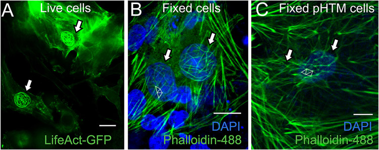
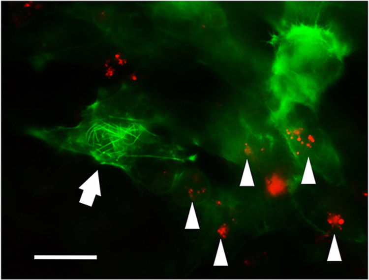
Cross-linked actin networks (CLANs) in trabecular meshwork (TM) cells may contribute to increased IOP by altering TM cell function and stiffness. However, there is a lack of direct evidence. Here, we developed transformed TM cells that form spontaneous fluorescently labelled CLANs. The stable cells were constructed by transducing transformed glaucomatous TM (GTM3) cells with the pLenti-LifeAct-EGFP-BlastR lentiviral vector and selection with blasticidin. The stiffness of the GTM3-LifeAct-GFP cells were studied using atomic force microscopy. Elastic moduli of CLANs in primary human TM cells treated with/without dexamethasone/TGFβ2 were also measured to validate findings in GTM3-LifeAct-GFP cells. Live-cell imaging was performed on GTM3-LifeAct-GFP cells treated with 1 μM latrunculin B or pHrodo bioparticles to determine actin stability and phagocytosis, respectively. The GTM3-LifeAct-GFP cells formed spontaneous CLANs without the induction of TGFβ2 or dexamethasone. The CLAN containing cells showed elevated cell stiffness, resistance to latrunculin B-induced actin depolymerization, as well as compromised phagocytosis, compared to the cells without CLANs. Primary human TM cells with dexamethasone or TGFβ2-induced CLANs were also stiffer and less phagocytic. The GTM3-LifeAct-GFP cells are a novel tool for studying the mechanobiology and pathology of CLANs in the TM. Initial characterization of these cells showed that CLANs contribute to at least some glaucomatous phenotypes of TM cells.
Keywords: Actin; Cross-linked actin networks; Glaucoma; Latrunculin; Phagocytosis; Stiffness; Trabecular meshwork.
Copyright © 2022 Elsevier Ltd. All rights reserved.
Figures


Similar articles
-
Characterization, Enrichment, and Computational Modeling of Cross-Linked Actin Networks in Transformed Trabecular Meshwork Cells.Invest Ophthalmol Vis Sci. 2025 Feb 3;66(2):65. doi: 10.1167/iovs.66.2.65. Invest Ophthalmol Vis Sci. 2025. PMID: 40009371 Free PMC article.
-
Evaluation of Cross-Linked Actin Networks (CLANs) in Human Trabecular Meshwork Cells and Tissues.Methods Mol Biol. 2025;2858:1-15. doi: 10.1007/978-1-0716-4140-8_1. Methods Mol Biol. 2025. PMID: 39433662
-
Cross-linked actin networks (CLANs) in glaucoma.Exp Eye Res. 2017 Jun;159:16-22. doi: 10.1016/j.exer.2017.02.010. Epub 2017 Feb 24. Exp Eye Res. 2017. PMID: 28238754 Free PMC article. Review.
-
Characterization, enrichment, and computational modeling of cross-linked actin networks in trabecular meshwork cells.bioRxiv [Preprint]. 2024 Aug 21:2024.08.21.608970. doi: 10.1101/2024.08.21.608970. bioRxiv. 2024. Update in: Invest Ophthalmol Vis Sci. 2025 Feb 03;66(2):65. doi: 10.1167/iovs.66.2.65. PMID: 39229235 Free PMC article. Updated. Preprint.
-
Trabecular meshwork stiffness in glaucoma.Exp Eye Res. 2017 May;158:3-12. doi: 10.1016/j.exer.2016.07.011. Epub 2016 Jul 19. Exp Eye Res. 2017. PMID: 27448987 Free PMC article. Review.
Cited by
-
In vitro comparison of human and murine trabecular meshwork cells: implications for glaucoma research.Sci Rep. 2024 Sep 23;14(1):22002. doi: 10.1038/s41598-024-73057-9. Sci Rep. 2024. PMID: 39313534 Free PMC article.
-
A Novel Mouse Model of TGFβ2-Induced Ocular Hypertension Using Lentiviral Gene Delivery.Int J Mol Sci. 2022 Jun 21;23(13):6883. doi: 10.3390/ijms23136883. Int J Mol Sci. 2022. PMID: 35805889 Free PMC article.
-
GSK3β Inhibitors Inhibit TGFβ Signaling in the Human Trabecular Meshwork.Invest Ophthalmol Vis Sci. 2024 Aug 1;65(10):3. doi: 10.1167/iovs.65.10.3. Invest Ophthalmol Vis Sci. 2024. PMID: 39087933 Free PMC article.
-
Inhibition of TGF-β2-Induced Trabecular Meshwork Fibrosis by Pirfenidone.Transl Vis Sci Technol. 2023 Nov 1;12(11):21. doi: 10.1167/tvst.12.11.21. Transl Vis Sci Technol. 2023. PMID: 37975842 Free PMC article.
-
Identification of matrix stiffness-related molecular subtypes in HCC via integrating multi-omics analysis and machine learning algorithms.J Transl Med. 2025 Jul 1;23(1):716. doi: 10.1186/s12967-025-06733-7. J Transl Med. 2025. PMID: 40598539 Free PMC article.
References
-
- 2000. The Advanced Glaucoma Intervention Study (AGIS): 7. The relationship between control of intraocular pressure and visual field deterioration.The AGIS Investigators. Am J Ophthalmol 130, 429–440. - PubMed
-
- Baqué S, Guinovart JJ, Ferrer JC, 1997. Glycogenin, the primer of glycogen synthesis, binds to actin. FEBS Letters 417, 355–359. - PubMed
-
- Bermudez JYWH, Patel GC, Yan LJ, Clark AF, Mao W, 2016. Two-dimensional differential in-gel electrophoresis (2D-DIGE) reveals proteins associated with cross-linked actin networks in human trabecular meshwork cells. J Clin Exp Ophthalmol 7.
-
- Briscoe BJ, Sebastian KS, Adams MJ, 1994. The effect of indenter geometry on the elastic response to indentation. Journal of Physics D: Applied Physics 27, 1156–1162.
Publication types
MeSH terms
Substances
Grants and funding
LinkOut - more resources
Full Text Sources
Medical
Research Materials
