Review
. 2022 May 29:1209:339338.
doi: 10.1016/j.aca.2021.339338. Epub 2021 Dec 1.

Toward a next-generation diagnostic tool: A review on emerging isothermal nucleic acid amplification techniques for the detection of SARS-CoV-2 and other infectious viruses

Affiliations
Review

Toward a next-generation diagnostic tool: A review on emerging isothermal nucleic acid amplification techniques for the detection of SARS-CoV-2 and other infectious viruses

Md Mamunul Islam et al. Anal Chim Acta. .

Abstract

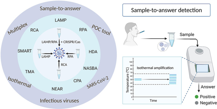
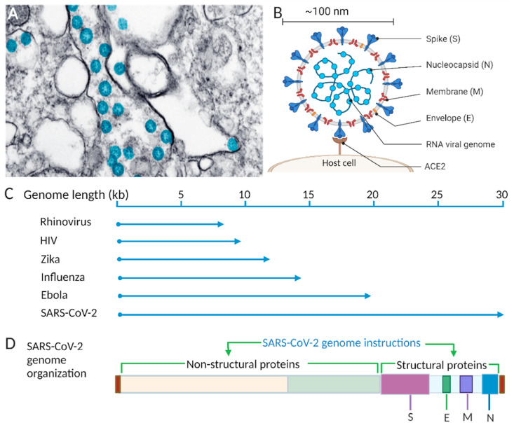
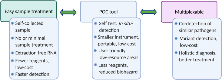
As the COVID-19 pandemic continues to affect human health across the globe rapid, simple, point-of-care (POC) diagnosis of infectious viruses such as SARS-CoV-2 remains challenging. Polymerase chain reaction (PCR)-based diagnosis has risen to meet these demands and despite its high-throughput and accuracy, it has failed to gain traction in the rapid, low-cost, point-of-test settings. In contrast, different emerging isothermal amplification-based detection methods show promise in the rapid point-of-test market. In this comprehensive study of the literature, several promising isothermal amplification methods for the detection of SARS-CoV-2 are critically reviewed that can also be applied to other infectious viruses detection. Starting with a brief discussion on the SARS-CoV-2 structure, its genomic features, and the epidemiology of the current pandemic, this review focuses on different emerging isothermal methods and their advancement. The potential of isothermal amplification combined with the revolutionary CRISPR/Cas system for a more powerful detection tool is also critically reviewed. Additionally, the commercial success of several isothermal methods in the pandemic are highlighted. Different variants of SARS-CoV-2 and their implication on isothermal amplifications are also discussed. Furthermore, three most crucial aspects in achieving a simple, fast, and multiplexable platform are addressed.

Keywords: CRISPR/Cas; Infectious viruses detection; Isothermal amplification; Pandemic; SARS-CoV-2 detection; Variants.


Conflict of interest statement

There is no competing interests.

Figures

Image 1
Graphical abstract
Fig. 1
Fig. 1
Timeline of the major events of the COVID-19 pandemic. WHO, World Health Organization; FDA, Food and Drug Administration; CDC, Centers for Disease Control and Prevention. Created with BioRender.com.
Fig. 2
Fig. 2
SARS-CoV-2 morphology and genomic illustrations. A) Transmission electron microscope image of SARS-CoV-2 viral particles (blue) [15], B) Schematic of SARS-CoV-2 with structural proteins, and its genome, binding interaction between host cell and virus spike protein, C) Comparison of the SARS-CoV-2 viral genome with genomes of other common RNA viruses. D) SARS-CoV-2 genome organization showing the relative positions of nonstructural and structural proteins. Created with BioRender.com. (For interpretation of the references to color in this figure legend, the reader is referred to the Web version of this article.)
Fig. 3
Fig. 3
Viral epidemiology of SARS-CoV-2. A) Transmission, B) Clinical symptoms, C) Diagnosis, and D) Disease dynamics and test viability. Figure D is reprinted by permission from [Springer Nature] [Nature Reviews Genetics], Testing at scale during the COVID-19 pandemic, Tim R. Mercer et al., [31]. Copyright 2021. Created with BioRender.com.
Fig. 4
Fig. 4
Simple representation of the LAMP mechanism. In the first step, both inner and outer primer pairs and polymerase enzyme generate dumbbell-like structures from the target. Then, in cycling amplification step, the two inner primers and polymerase further amplify these structures. The elongation steps generate various sizes of the products (three arrows are used to represent various reaction pathways. The two Loop primers are also used in the elongation step. (Reproduced with permission from Ref. [64]). Created with BioRender.com.
Fig. 5
Fig. 5
RT-LAMP assay to detect SARS-CoV-2. The LAMP primer mix contain three pairs of target-specific primers: two forward primers, two backward primers, and two loop primers. The LAMP reaction mix contained DNA polymerase, reverse transcriptase, isothermal buffer, and signal reporter. A simple heating block can be used to amplify the target region of the viral RNA using colorimetric detection. The image on the right shows the time-dependent color change, adapted from Dao Thi et al., [125]. Reprinted with permission from AAAS. Created with BioRender.com. (For interpretation of the references to color in this figure legend, the reader is referred to the Web version of this article.)
Fig. 6
Fig. 6
Schematic of the nicking endonuclease amplification reaction (NEAR). Two main steps are involved in the NEAR: duplex formation (left) and amplification (right). Key components of NEAR are a pair of target-specific primers (P1, P2) and two enzymes: strand-displacing polymerase and nicking endonuclease enzymes. Figure adapted with permission from Ref. [140]. Created with BioRender.com.
Fig. 7
Fig. 7
A) Simple mechanism of NASBA. B) Schematic of the single-tube experimental setup to detect SARS-CoV-2 RNA from saliva samples via NASBA-based INSIGHT. Figure adapted with permission from Ref. Wu, Q. et al., [146]. Created with BioRender.com.
Fig. 8
Fig. 8
Schematic of recombinase polymerase amplification (RPA) [161]. A) RPA mechanism (reproduced from Ref. [161] with permission from the Royal Society of Chemistry). B) A simple workflow of a typical RT-RPA. The amplification mix contains a pair of primers, recombinase, loading factors, reverse transcriptase, DNA polymerase, accessory proteins, and single-strand binding proteins that are available in commercial kits. Created with BioRender.com.
Fig. 9
Fig. 9
Schematic of RCA-based detection based on synthetic complementary DNA of SARS-CoV-2. After two rounds of RCA, the amplified product was detected with an optomagnetic sensing platform. MNP-magnetic nano particles. Figure adapted with permission from Ref. [175].
Fig. 10
Fig. 10
Simple illustration of signal-mediated amplification of RNA technology (SMART). Target-initiated 3WJ complex formation and subsequent extension and in vitro transcribed RNA product generation are accomplished by synergy between two enzymes-DNA polymerase and T7 RNA polymerase. The 3′ end of the template probe is modified (x) by phosphorylation to prevent extension by DNA polymerase. Created with BioRender.com.
Fig. 11
Fig. 11
Schematic of helicase-dependent amplification (HDA). HDA relies on helicase enzyme to unwind the target. A pair of primers, DNA polymerase, and single strand binding proteins ensure rapid accumulation of HDA products. Created with BioRender.com.
Fig. 12
Fig. 12
Schematic of cross priming amplification (CPA). CPA relies on Cross primer and several Displacement primers. CPA consist initial products (step I), stem-loop products (step II), and subsequent amplification of the final products (step III). Adapted with permission from Ref. Xu, G. et al., [194].
Fig. 13
Fig. 13
Class 2 Cas enzymes used in the CRISPR/Cas system. A) The CRISPR/Cas9 system uses a two-part gRNA (tracrRNA, crRNA (top)) or an sgRNA (bottom) with a linker between tracrRNA and crRNA. B) sgRNA-directed endonuclease activity of Cas9 on target dsDNA. C) Collateral cleavage and target cleavage activity of Cas12. D) Collateral cleavage and target (ssDNA) cleavage activity of Cas14. D) Collateral cleavage and target (ssRNA) cleavage activity of Cas13. All Cas enzymes are drawn by showing their relative size (created with BioRender.com). Figures B–E adapted with permission from Ref. [239].
Fig. 14
Fig. 14
Simpler representation of a typical isothermal amplification based CRISPR/Cas detection system for viral nucleic acids. Typically, LAMP or RPA based amplification is involved to amplify target for subsequent class 2 Cas enzyme activity by Cas12 and Cas13. The system involves “Collateral cleavage” based qualitative and quantitative detection. Created with BioRender.com.
Fig. 15
Fig. 15
Flowchart of different commercial isothermal-based detection systems of SARS-CoV-2. A) ID NOW COVID-19 detection by Abbott. Sample processing units (left) contain a sample receiver, transfer cartridge, and test base. The sample receiver containing lysis buffer is used treat patient sample. Following quick mixing, the transfer cartridge is used to transfer the treated sample manually to the test base containing a lyophilized NEAR mix. Amplification and fluorescence-based detection are performed by the ID NOW instrument (right). Adapted with permission from Ref. [140]. B) “All-In-One Test Kit”—a commercial POC testing system developed by Lucira Health. The system involves a user-friendly, simple workflow of “self-test” virus detection on a small and portable device. (adapted with permission) [304].
Fig. 16
Fig. 16
Characteristics of a next-generation diagnostic tool with the three most important criteria: easy sample treatment, POC tool, and multiplexable system.
Fig. 17
Fig. 17
Smartphone-based handheld POC instrument. A) Simple workflow of SARS-CoV-2 sample preparation by brief heat treatment and transfer of the thermally lysed sample to a syringe. Another syringe was loaded with LAMP reaction mix. B) Photograph of the microfluidic chip integrated POC tool. C) Disposable microfluidic cartridge. Figure adapted with permission from Ganguli et al., [83].
Fig. 18
Fig. 18
Schematic showing amino acid changes to the spike (S) protein in the four variants of concern (VOCs) of SARS-CoV-2-Alpha, Beta, Gamma, and Delta [349]. S1 and S2 are the two functional domains of the S protein. RBD-receptor-binding domain, FCS-furin cleavage site.

Similar articles

Cited by

References

    1. Andersen K.G., Rambaut A., Lipkin W.I., Holmes E.C., Garry R.F. The proximal origin of SARS-CoV-2. Nat. Med. 2020;26:450–452. - PMC - PubMed
    1. Virus Origin/Origins of the SARS-CoV-2 Virus. https://www.who.int/health-topics/coronavirus/origins-of-the-virus
    1. Chen N., et al. Epidemiological and clinical characteristics of 99 cases of 2019 novel coronavirus pneumonia in Wuhan, China: a descriptive study. Lancet Lond. Engl. 2020;395:507–513. - PMC - PubMed
    1. Li Q., et al. Early transmission dynamics in Wuhan, China, of novel coronavirus–infected pneumonia. N. Engl. J. Med. 2020 doi: 10.1056/NEJMoa2001316. - DOI - PMC - PubMed
    1. Coronavirus Disease (COVID-19) – World Health Organization. https://www.who.int/emergencies/diseases/novel-coronavirus-2019