. 2022 May 15;17(1):274.
doi: 10.1186/s13018-022-03170-w.

Electrophysiological and pathological changes in the vastus medialis and vastus lateralis muscles after early patellar reduction and nerve growth factor injection in rabbits with patellar dislocation

Affiliations

Electrophysiological and pathological changes in the vastus medialis and vastus lateralis muscles after early patellar reduction and nerve growth factor injection in rabbits with patellar dislocation

Yu Wu et al. J Orthop Surg Res. .

Abstract

Background: Patellar dislocation can cause a series of changes in the trochlear groove and patella. However, the influence of patellar dislocation on the medialis (VM) and vastus lateralis (VL) muscles and whether nerve growth factor (NGF) is beneficial to proprioceptive rehabilitation for patellar dislocation are unknown. The purpose of this study was to investigate the effects on VM and VL after the injection of NGF and early reduction in rabbits for patellar dislocation with electrophysiological and pathological analysis.

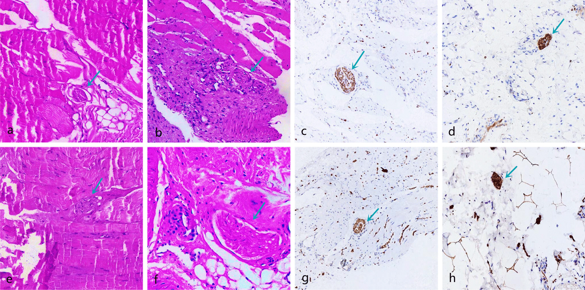
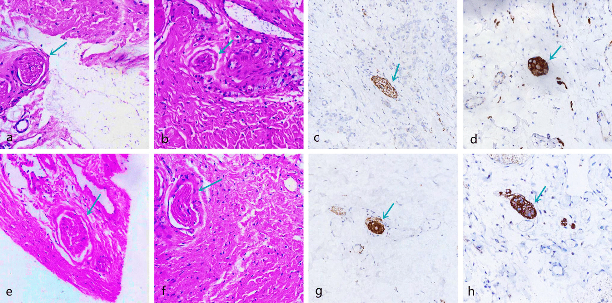
Methods: Sixty 2-month-old rabbits were randomly divided into four groups (15 rabbits in each group). Rabbits in Group 1, Group 2, and Group 3 underwent patellar dislocation surgery, and rabbits in Group 4 underwent sham surgery. One month later, patellar reduction was performed in Groups 1 and 2. NGF was injected into the rabbits of Group 1. The electrophysiological and pathological changes in VM and VL were analyzed at 1 month and 3 months after patellar reduction.

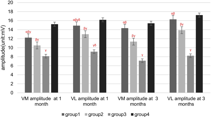
Results: The electrophysiological and pathological indices in Groups 1 and 2 were significantly different from those in Group 3 at 1 and 3 months after patellar reduction. There were significant differences between NGF injection Group 1 and Group 2 without NGF injection. There was no significant difference between Group 1 and Group 4 at 3 months after patellar reduction.

Conclusions: Patellar dislocation can cause abnormal electrophysiological and pathological effects on VM and VL. Patellar reduction should be performed as early as possible, and NGF injection may be beneficial to the rehabilitation of proprioception.

Keywords: Electrophysiological; Nerve growth factor (NGF); Patellar dislocation; Proprioceptor.


Conflict of interest statement

The authors declare that they have no competing interests.

Figures

Fig. 1
Fig. 1
Schematic of the experimental protocol
Fig. 2
Fig. 2
a At the insertion point of the femur, the medial retinacular band and joint capsule were cut, and the lateral joint capsule and lateral retinacular band were overlapped with two stitches. b The lateral joint capsule and lateral retinacular band were loosened, and the medial retinacular band and joint capsule were sutured to reduce the patella and kept in the trochlear tract
Fig. 3
Fig. 3
VM EMG images at 3 months after patella reduction. a Group 1. b Group 2. c Group 3. d Group 4
Fig. 4
Fig. 4
VM SEPs images at 3 months after patella reduction. a Group 1. b Group 2. c Group 3. d Group 4
Fig. 5
Fig. 5
Four groups regarding the latency period of EMG at different detection times. α P < 0.05 versus Group 2 in the same month; β P < 0.05 versus Group 3 in the same month; γ P < 0.05 versus Group 4 in the same month; δ P < 0.05 versus 3 months in the same group
Fig. 6
Fig. 6
Four groups regarding the amplitude of EMG at different detection times. α P < 0.05 versus Group 2 in the same month; β P < 0.05 versus Group 3 in the same month; γ P < 0.05 versus Group 4 in the same month; δ P < 0.05 versus 3 months in the same group
Fig. 7
Fig. 7
Four groups regarding the latency period of SEPs at different detection times. α P < 0.05 versus Group 2 in the same month; β P < 0.05 versus Group 3 in the same month; γ P < 0.05 versus Group 4 in the same month; δ P < 0.05 versus 3 months in the same group
Fig. 8
Fig. 8
Four groups regarding the amplitude of SEPs at different detection times. α P < 0.05 versus Group 2 in the same month; β P < 0.05 versus Group 3 in the same month; γ P < 0.05 versus Group 4 in the same month; δ P < 0.05 versus 3 months in the same group
Fig. 9
Fig. 9
Four groups regarding the number of proprioceptors at different detection times. α P < 0.05 versus Group 2 in the same month; β P < 0.05 versus Group 3 in the same month; γ P < 0.05 versus Group 4 in the same month; δ P < 0.05 versus 3 months in the same group
Fig. 10
Fig. 10
Pathology of Group 1; the green arrow refers to the proprioceptors. The morphology was basically normal, and the membrane was dense without obvious atrophied or deformed. a, b VM and VL (HE × 100) at 1 month after patella reduction; c, d VM and VL (immunofluorescence × 100) at 1 month after patella reduction. e, f VM and VL (HE × 100) at 3 months after patella reduction; g, h VM and VL (immunofluorescence × 100) at 3 months after patella reduction
Fig. 11
Fig. 11
Pathology of Group 2; the green arrow refers to the proprioceptors. The proprioceptors were widened and arranged loosely, and the outer thin-walled connective tissue sac changed irregularly. a, b VM and VL (HE × 100) at 1 month after patella reduction; c, d VM and VL (immunofluorescence × 100) at 1 month after patella reduction. e, f VM and VL (HE × 100) at 3 months after patella reduction; g, h VM and VL (immunofluorescence × 100) at 3 months after patella reduction
Fig. 12
Fig. 12
Pathology of Group 3; the green arrow refers to the proprioceptors. The proprioceptors were arranged in disorder and loose, the long axis direction from parallel to irregular, atrophied, deformed, and cleavage. a, b VM and VL (HE × 100) at 1 month after patella reduction; c, d VM and VL (immunofluorescence × 100) at 1 month after patella reduction. e, f VM and VL (HE × 100) at 3 months after patella reduction; g, h VM and VL (immunofluorescence × 100) at 3 months after patella reduction
Fig. 13
Fig. 13
Pathology of Group 4; the green arrow refers to the proprioceptors. The proprioceptors were round or oval with regular shapes and smooth edges. a, b VM and VL (HE × 100) at 1 month after patella reduction; c, d VM and VL (immunofluorescence × 100) at 1 month after patella reduction. e, f VM and VL (HE × 100) at 3 months after patella reduction; g, h VM and VL (immunofluorescence × 100) at 3 months after patella reduction

Similar articles

References

    1. Frosch S, Balcarek P, Walde TA, Schuttrumpf JP, Wachowski MM, Ferleman KG, et al. The treatment of patellar dislocation: a systematic review. Z Orthop Unfall. 2011;149(6):630–645. doi: 10.1055/s-0030-1250691. - DOI - PubMed
    1. Hsiao M, Owens BD, Burks R, Sturdivant RX, Cameron KL. Incidence of acute traumatic patellar dislocation among active-duty United States military service members. Am J Sports Med. 2010;38(10):1997–2004. doi: 10.1177/0363546510371423. - DOI - PubMed
    1. Li W, Wang Q, Wang F, Zhang Y, Ma L, Dong J. Femoral trochlear dysplasia after patellar dislocation in rabbits. Knee. 2013;20:485–489. doi: 10.1016/j.knee.2013.05.016. - DOI - PubMed
    1. Wang S, Ji G, Yang X, Wang X, Wang R, Li M, et al. Femoral trochlear groove development after patellar subluxation and early reduction in growing rabbits. Knee Surg Sports Traumatol Arthrosc. 2016;24:247–253. doi: 10.1007/s00167-014-3372-z. - DOI - PubMed
    1. Niu J, Qi Q, Niu Y, et al. Patella morphological alteration after patella instability in growing rabbits. J Orthop Surg Res. 2017;12:106–112. doi: 10.1186/s13018-017-0615-y. - DOI - PMC - PubMed

Substances

LinkOut - more resources