Case Reports
. 2022 May 6;17(7):2353-2361.
doi: 10.1016/j.radcr.2022.04.003. eCollection 2022 Jul.

Pulmonary arteriovenous malformation and inherent complications with solitary lung nodule biopsy-literature overview and case report

Affiliations
Case Reports

Pulmonary arteriovenous malformation and inherent complications with solitary lung nodule biopsy-literature overview and case report

Cung-Van Cong et al. Radiol Case Rep. .

Abstract

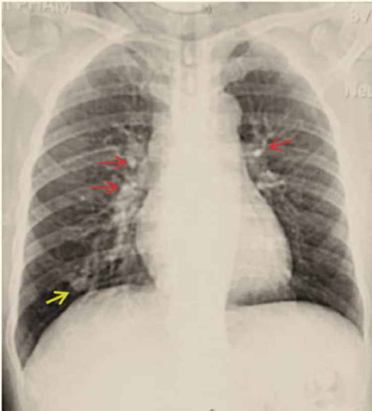
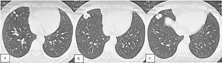
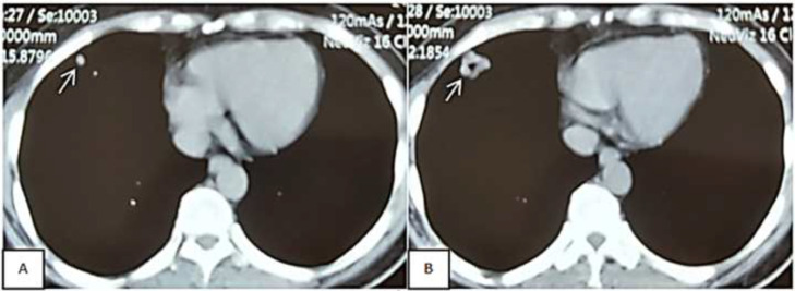
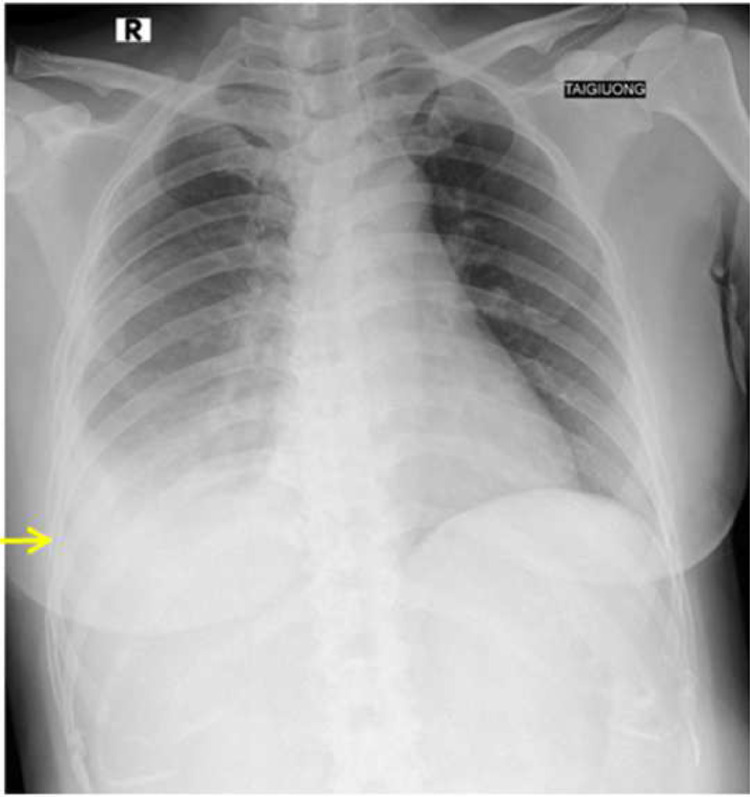
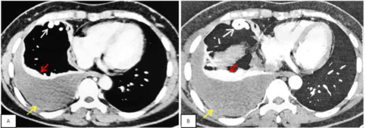
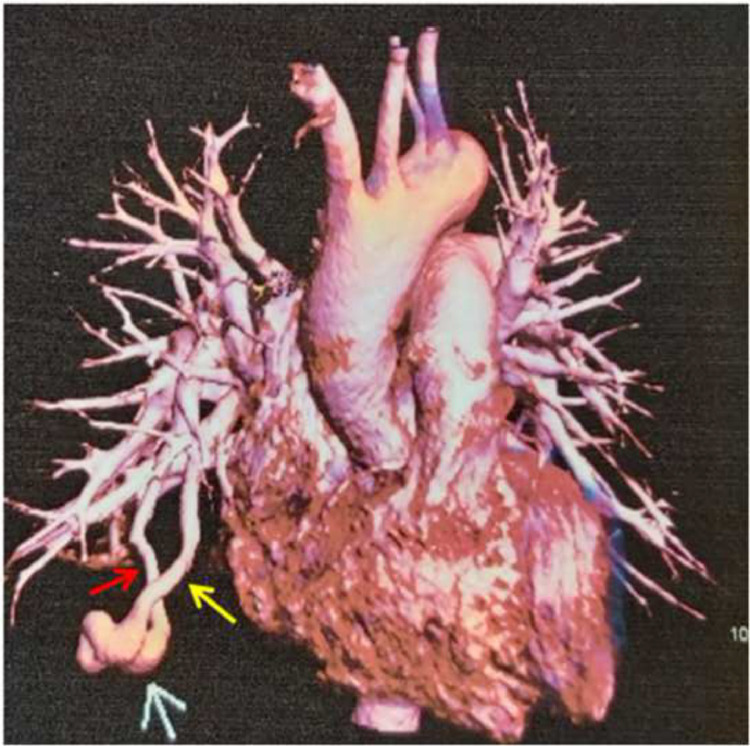
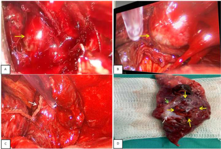
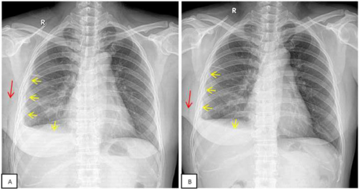
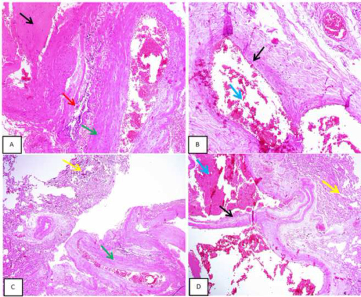
Pulmonary arteriovenous malformation, also known as an arteriovenous fistula, is typically a congenital disease caused by structural deficiencies, particularly the lack of capillary wall development, leading to the abnormal dilation of the pulmonary capillaries. The majority of pulmonary arteriovenous malformation cases are associated with Rendu-Osler-Weber syndrome, also known as hereditary hemorrhagic telangiectasia. Pulmonary arteriovenous malformation rarely occurs due to chest trauma. Pulmonary arteriovenous malformations are long-lasting and often first diagnosed in adults. More than two-thirds of pulmonary arteriovenous malformation lesions are found in the lower lung lobe and the subpleural area, and the vast majority of cases present with the monofocal form. The initial diagnosis is often based on the identification of a solitary pulmonary nodule. However, a solitary nodule detected on chest computed tomography that is not correctly diagnosed as pulmonary arteriovenous malformation, even after intravenous contrast injection, can lead to the performance of a transthoracic biopsy. Biopsy of pulmonary arteriovenous malformations can lead to stroke occurrence, during which the patient often presents with severe pleural bleeding, which can have lifelong consequences if not immediately treated. We report a case of pulmonary arteriovenous malformation that was discovered incidentally in an adult patient who underwent non-contrast computed tomography. Misdiagnosis occurred, and transthoracic lung biopsy was performed. Complications were discovered late, and the patient underwent surgical pulmonary arteriovenous malformation removal and was treated for hemothorax.

Keywords: Hemothorax; Hereditary hemorrhagic Telangiectasia; Pulmonary arteriovenous malformation; Rare diseases; Vascular malformation.


Figures

Fig 1 –
Fig. 1
Posteroanterior chest radiograph taken during the initial medical examination at the local hospital. A faint nodule was visible in the lower third of the right lung (yellow arrow). A few calcifications in the hilar regions were visible on both sides (red arrow).
Fig 2 –
Fig. 2
Chest computed tomography of the lung window. Slices are displayed from top to bottom. A–C: Nodular, multi-segment appearance near the periphery of the right middle right lung lobe (white arrows), sized 22 × 18 mm. The nodule boundary with the surrounding lung parenchyma is quite clear.
Fig 3 –
Fig. 3
Chest computed tomography of the mediastinal window. Slices are displayed from top to bottom. (A and B) The nodules have the same shape as shown in Fig. 2 (white arrows).
Fig 4 –
Fig. 4
Chest X-ray film, taken with a mobile X-ray machine at the patient's bed in an anterior–posterior position (the patient was laying on their back, with no gas pocket in the stomach). A uniform opacity was observed in the lower third of the right lung, and the diaphragm was not visible, resulting in suspected right pleural effusion (yellow arrow).
Fig 5 –
Fig. 5
Chest computed tomography with intravenous contrast, showing two consecutive slices from top to bottom. (A, B) A single nodule appears in the middle lobe with strong contrast enhancement (white arrows). The contrast density is similar to that of the cardiac chambers. Right pleural effusion, 60-mm fluid layer (yellow arrow) with passive pleural atelectasis (red arrow).
Fig 6 –
Fig. 6
Volume rendering technique image of the patient from the front, which clearly indicates the PAVM sac (white arrow), pulmonary artery (red arrow), and pulmonary vein drainage (yellow arrow).
Fig 7 –
Fig. 7
Image of right middle lobectomy containing the PAVM sac and the postoperative specimen mass. (A, B) Exposing the middle lobe and the PAVM sac from the outside (yellow arrow). (C) Stapler apex image after middle lobe resection (white arrow). (D) Postoperative specimen mass clearly shows the pulmonary artery (green arrow), draining pulmonary vein (white arrow), and PAVM sac (yellow arrows).
Fig 8 –
Fig. 8
Chest X-ray films from 5 and 7 days after surgery. (A) Postoperative radiograph on the fifth day showed right pleural thickening (yellow arrow) and air in the subcutaneous tissue of the right chest wall (red arrow). (B) Postoperative film on the seventh day showed right pleural thickening (yellow arrow) and only trace amounts of subcutaneous air in the right chest wall (red arrow). On both films, right pleural effusion was suspected.
Fig 9 –
Fig. 9
Postoperative microscopic histopathology results. Template code: G22-0033. (A–D) Microscopic sections showed lung tissue (yellow arrows) exhibiting areas of thin and partially thick vascular channels of various sizes (black arrow), lined by endothelial cells and filled with red blood cells, surrounded by extensive intraalveolar hemorrhages (blue arrow) and focal areas of atelectasis. This area is in close proximity to the bronchus with a narrowed lumen (red arrow) and accompanying medium-sized arteries (green arrow) and predominantly closer to the thick pleural lining. Moderate plasmalymphocytic and hemosiderin macrophages are noted within the peribronchial and some alveolar spaces, indicative of the old bleeding area. No fibrocalcific nodules or evidence of chronic granulomatous disease was identified. No malignant cells were found. The conclusion was vascular abnormalities consistent with the vascular malformation of PAVM.

Similar articles

Cited by

References

    1. Richard Webb W, Higgins CB. Thoracic imaging: pulmonary and cardiovascular radiology, Wolters KluwerPhiladelphia, The United States of America, 2017; 3 E; p. 275–305.
    1. Liu S, Xu X, Qin Y, Zhao X. Rare ruptured pulmonary arteriovenous malformation mimicking lung tumor in a girl. Anatol J Cardiol. 2021;25(7):E30. doi: 10.5152/AnatolJCardiol.2021.74790. PMID: 34236335. - DOI - PMC - PubMed
    1. Lin YP, Liang CH, Wu FZ. Pulmonary arteriovenous malformation mimicking a lung mass. Br J Hosp Med (Lond) 2019;80(8) doi: 10.12968/hmed.2019.80.8.ii. iiPMID: 31437038. - DOI - PubMed
    1. Takase Y, Nakano S, Yamaki E, Kawashima Pulmonary arteriovenous malformation with metal allergy. BMJ Case Rep. 2021;14(3) doi: 10.1136/bcr-2020-240275. PMID: 33692060. - DOI - PMC - PubMed
    1. Gungor S, Yakar HI. Usefulness of dynamic fluorodeoxyglucose positron emission tomography/computed tomography in diagnosing pulmonary arteriovenous malformation mimicking a lung tumour. Interact Cardiovasc Thorac Surg. 2021;33(4):665–667. doi: 10.1093/icvts/ivab130. PMID: 33954790. - DOI - PMC - PubMed

Publication types

LinkOut - more resources