

Low Turnover Renal Osteodystrophy With Abnormal Bone Quality and Vascular Calcification in Patients With Mild-to-Moderate CKD
- PMID: 35570986
- PMCID: PMC9091581
- DOI: 10.1016/j.ekir.2022.02.022
Low Turnover Renal Osteodystrophy With Abnormal Bone Quality and Vascular Calcification in Patients With Mild-to-Moderate CKD
Abstract
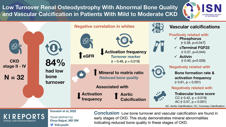
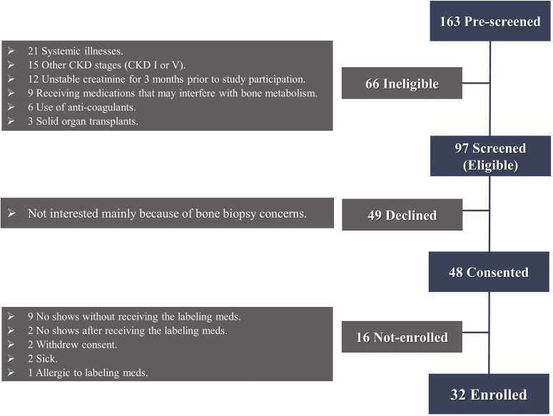
Introduction: Limited information is available on renal osteodystrophy (ROD) and vascular calcification (VC) during early chronic kidney disease (CKD). This study was designed to evaluate ROD and VC in 32 patients with CKD stages II to IV.
Methods: Patients underwent dual-energy X-ray absorptiometry (DXA) for assessment of bone mineral density (BMD) and trabecular bone score (TBS), thoracic computed tomography for VC scoring using the Agatston method, and anterior iliac crest bone biopsy for mineralized bone histology, histomorphometry, and Fourier transform infrared spectroscopy (FTIR). Classical and novel bone markers were determined in the blood.
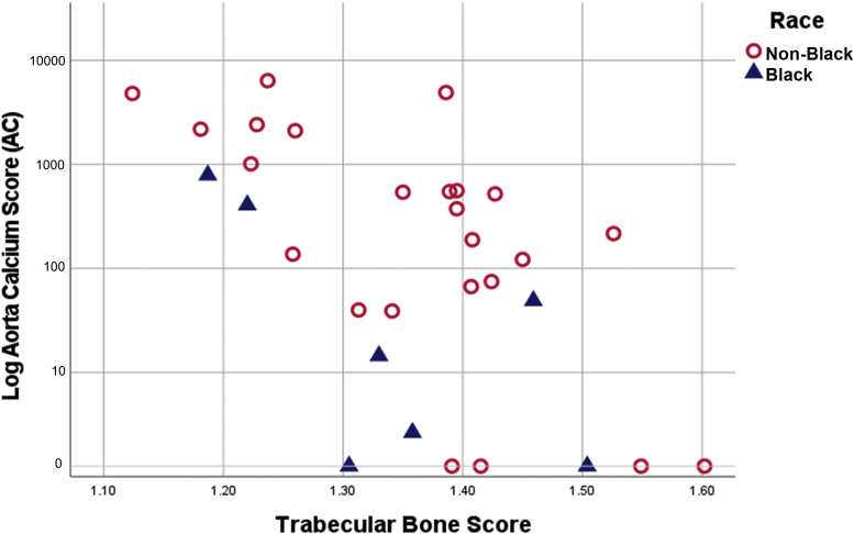
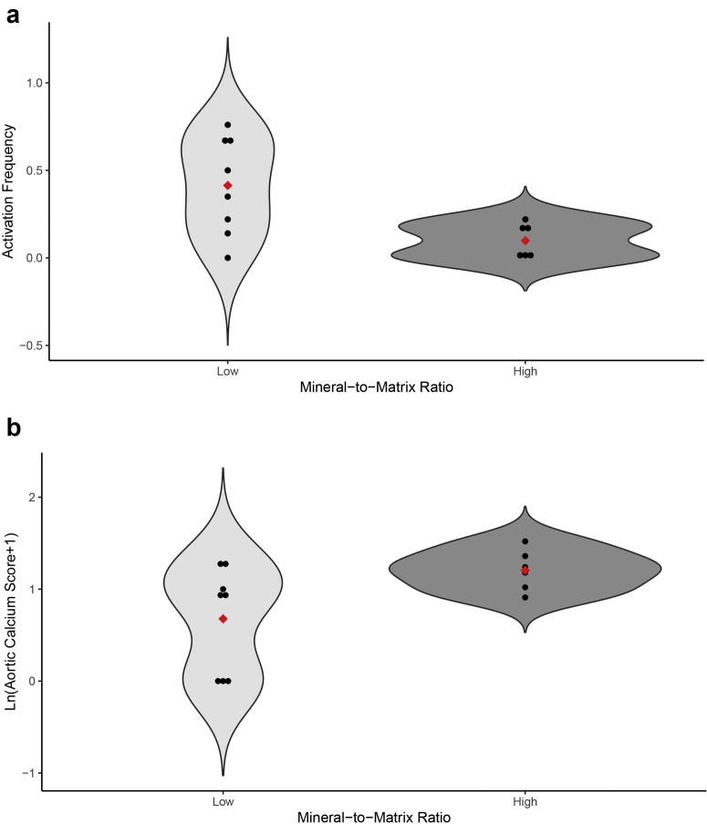
Results: Mean estimated glomerular filtration rate (eGFR) was 44 ± 16 ml/min per 1.73 m2. Of the patients, 84% had low bone turnover. In Whites, eGFR correlated negatively with the turnover parameter activation frequency (Ac.f) (r -0.48, P = 0.019) and with parameters of bone formation. Most patients had VC (>80%) which correlated positively with levels of phosphorus, c-terminal fibroblast growth factor-23, and activin. Aortic calcifications (ACs) correlated negatively with bone formation rate (BFR) and Ac.f (rho -0.62, -0.61, P < 0.001). TBS correlated negatively with coronary calcification (rho -0.42, P = 0.019) and AC (rho -0.57, P = 0.001). These relationships remained after adjustment of age. The mineral-to-matrix ratio, an FTIR metric reflecting bone quality, was negatively related to Ac.f and positively related to AC.
Conclusion: Low bone turnover and VC are predominant in early stages of CKD. This is the first study demonstrating mineral abnormalities indicating reduced bone quality in these stages of CKD.
Keywords: DXA; bone biopsy; bone quality; cardiovascular calcification; renal osteodystrophy; trabecular bone score.
© 2022 International Society of Nephrology. Published by Elsevier Inc.
Figures
Similar articles
-
Effects of unfractionated heparin on renal osteodystrophy and vascular calcification in chronic kidney disease rats.Bone. 2014 Jan;58:168-76. doi: 10.1016/j.bone.2013.10.010. Epub 2013 Oct 18. Bone. 2014. PMID: 24145307
-
Aortic vascular calcification is inversely associated with the trabecular bone score in patients receiving dialysis.Bone. 2018 Aug;113:118-123. doi: 10.1016/j.bone.2018.05.014. Epub 2018 May 29. Bone. 2018. PMID: 29775762
-
FGF-23 and sclerostin in serum and bone of CKD patients.Clin Nephrol. 2023 May;99(5):209-218. doi: 10.5414/CN111111. Clin Nephrol. 2023. PMID: 36970967 Free PMC article.
-
[Diagnosis and treatment of osteoporosis in patients with chronic kidney disease : Joint guidelines of the Austrian Society for Bone and Mineral Research (ÖGKM), the Austrian Society of Physical and Rehabilitation Medicine (ÖGPMR) and the Austrian Society of Nephrology (ÖGN)].Wien Med Wochenschr. 2023 Oct;173(13-14):299-318. doi: 10.1007/s10354-022-00989-0. Epub 2022 Dec 21. Wien Med Wochenschr. 2023. PMID: 36542221 Free PMC article. Review. German.
-
Chronic Kidney Disease with Mineral Bone Disorder and Vascular Calcification: An Overview.Life (Basel). 2024 Mar 21;14(3):418. doi: 10.3390/life14030418. Life (Basel). 2024. PMID: 38541742 Free PMC article. Review.
Cited by
-
Application of artificial intelligence to chronic kidney disease mineral bone disorder.Clin Kidney J. 2024 Jun 6;17(6):sfae143. doi: 10.1093/ckj/sfae143. eCollection 2024 Jun. Clin Kidney J. 2024. PMID: 38899159 Free PMC article. Review.
-
Updates in the chronic kidney disease-mineral bone disorder show the role of osteocytic proteins, a potential mechanism of the bone-Vascular paradox, a therapeutic target, and a biomarker.Front Physiol. 2023 Jan 26;14:1120308. doi: 10.3389/fphys.2023.1120308. eCollection 2023. Front Physiol. 2023. PMID: 36776982 Free PMC article. Review.
-
Low Turnover Bone Disease in Early CKD Stages.Kidney Int Rep. 2022 Apr 18;7(6):1445. doi: 10.1016/j.ekir.2022.03.037. eCollection 2022 Jun. Kidney Int Rep. 2022. PMID: 35685312 Free PMC article. No abstract available.
-
Effect of Antidiabetic Drugs on Bone Health in Patients with Normal Renal Function and in Chronic Kidney Disease (CKD): Insight into Clinical Challenges in the Treatment of Type 2 Diabetes.J Clin Med. 2023 Nov 23;12(23):7260. doi: 10.3390/jcm12237260. J Clin Med. 2023. PMID: 38068310 Free PMC article. Review.
-
Relative comparison of chronic kidney disease-mineral and bone disorder rat models.Front Physiol. 2023 Feb 3;14:1083725. doi: 10.3389/fphys.2023.1083725. eCollection 2023. Front Physiol. 2023. PMID: 36818435 Free PMC article.
References
-
- US Department of Health and Human Services, Center for Disease Control and Prevention Chronic Kidney Disease in the United States, 2019. US Department of Health and Human Services, Centers for Disease Control and Prevention. https://www.cdc.gov/kidneydisease/pdf/2019_National-Chronic-Kidney-Disea... Published 2019.
Grants and funding
LinkOut - more resources
Full Text Sources
Research Materials
Miscellaneous
