. 2022 Apr 27:13:889226.
doi: 10.3389/fphar.2022.889226. eCollection 2022.

Curcumin Restrains Oxidative Stress of After Intracerebral Hemorrhage in Rat by Activating the Nrf2/HO-1 Pathway

Affiliations

Curcumin Restrains Oxidative Stress of After Intracerebral Hemorrhage in Rat by Activating the Nrf2/HO-1 Pathway

Chenyang Duan et al. Front Pharmacol. .

Abstract

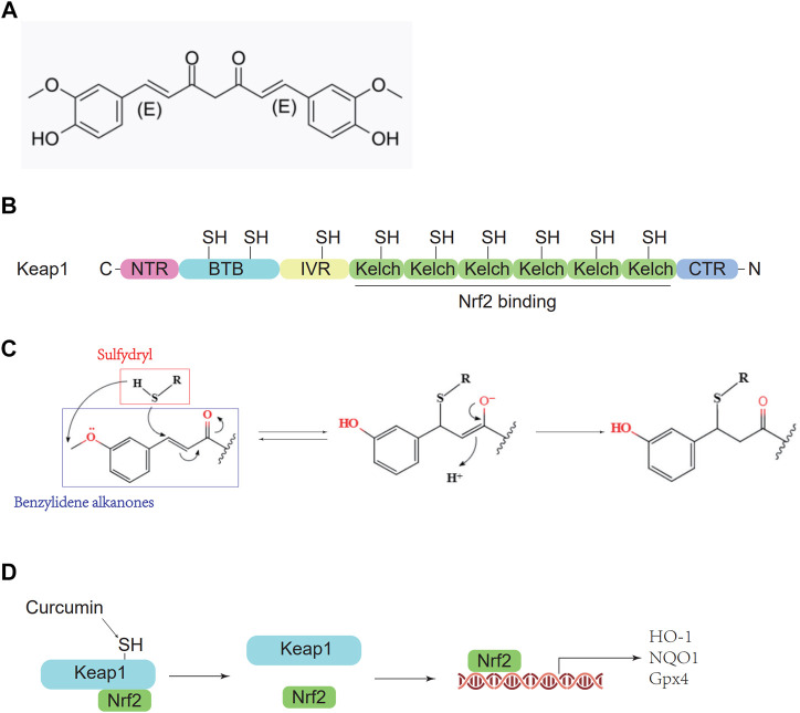
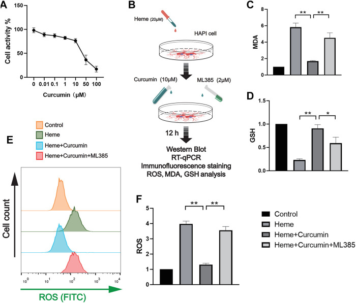
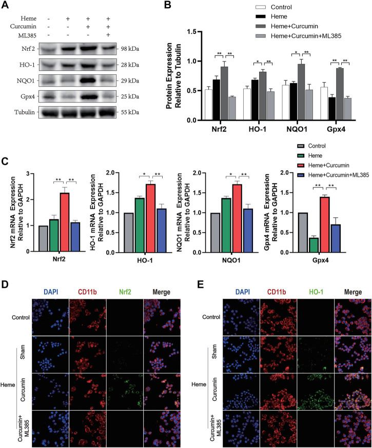
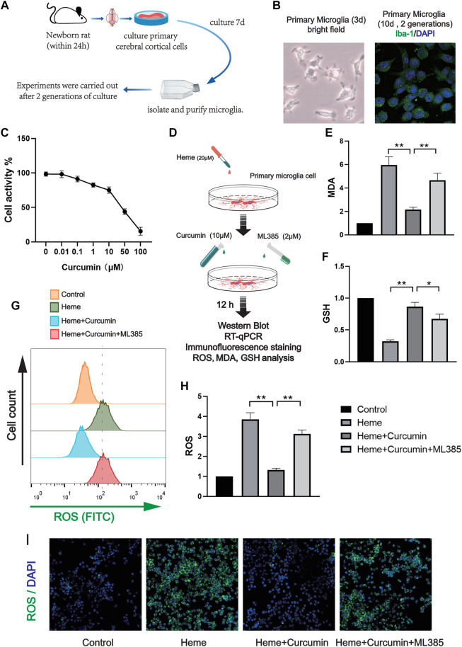
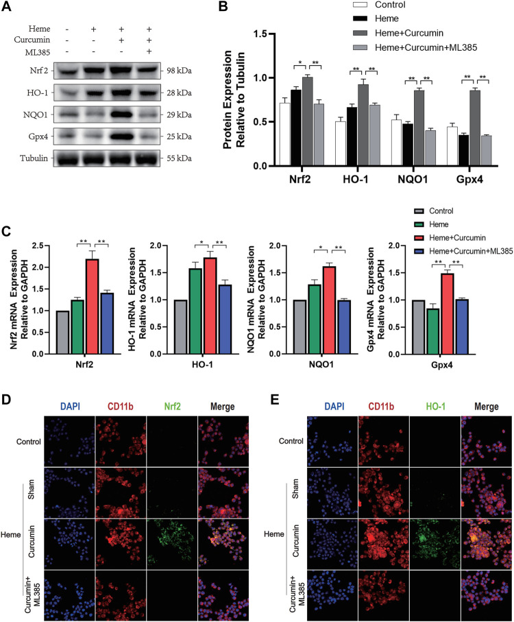
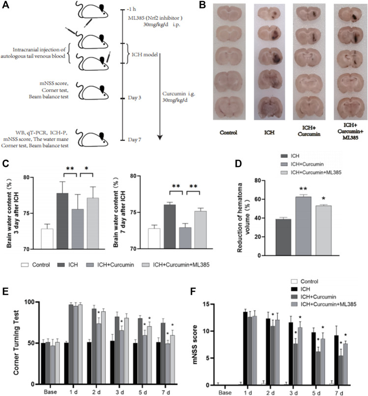
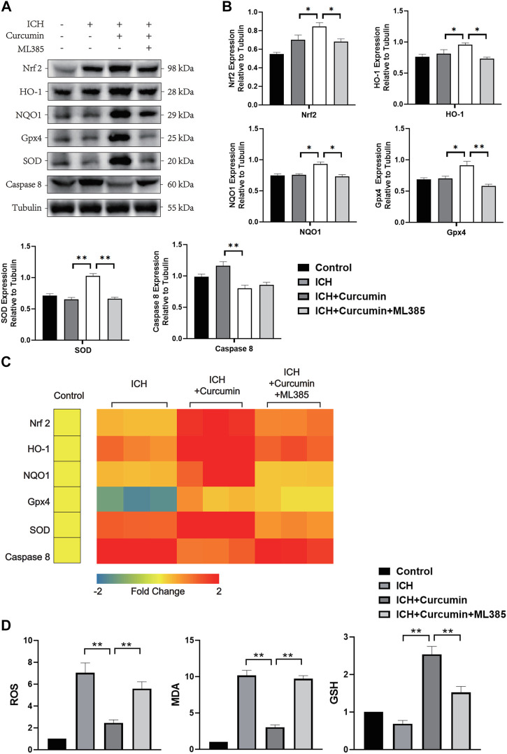
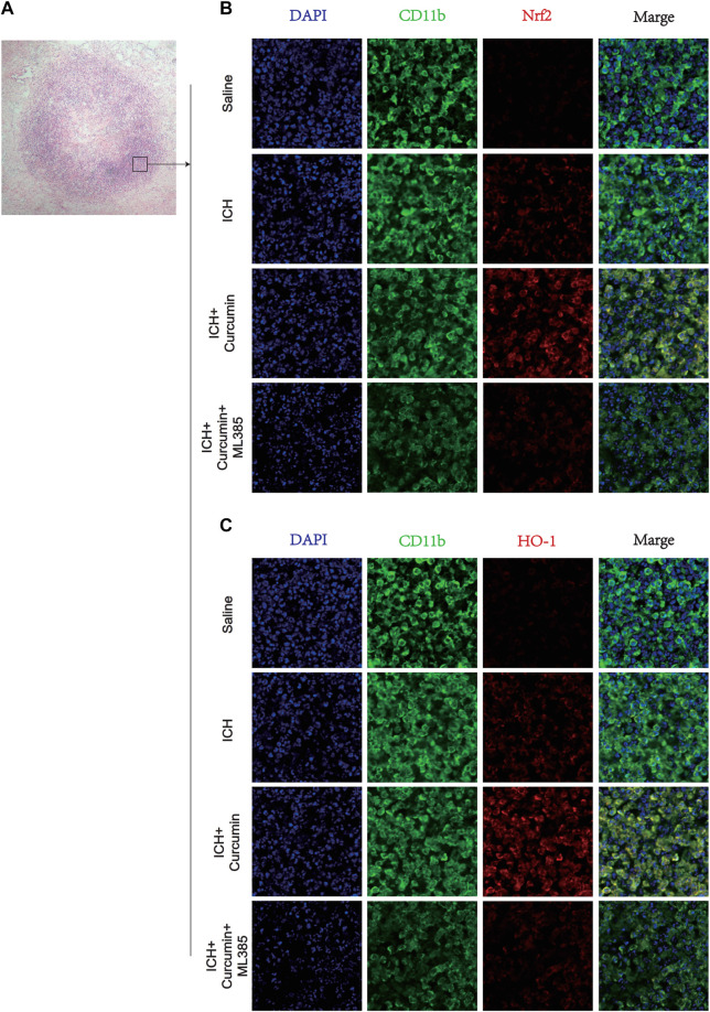
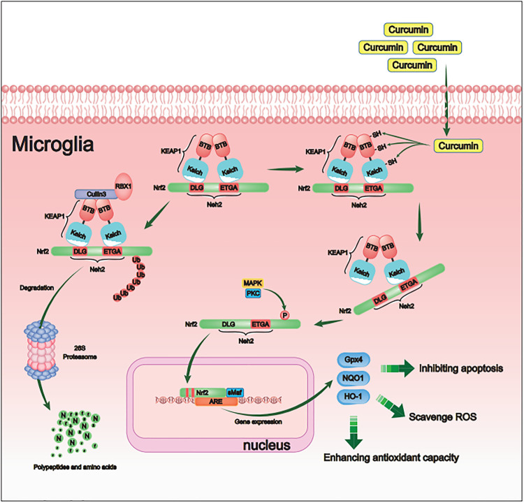
Intracerebral hemorrhage (ICH), a severe hemorrhagic stroke, induces cerebral oxidative stress and severe secondary neurological injury. Curcumin was demonstrated to inhibit oxidative stress in the brain after ICH. However, the pharmacological mechanism needs further research. We used an intrastriatal injection of autologous blood to make the rat ICH model, and then the rat was treated with curcumin (100 mg/kg/day). Modified Neurological Severity Score (mNSS) and corner test results showed that curcumin could significantly promote the neurological recovery of ICH rats. Meanwhile, curcumin could substantially reduce ROS and MDA in the tissues around intracranial hematoma and prevent GSH depletion. To explore the pharmacological molecular mechanism of curcumin, we used HAPI cells and primary rat cortical microglia for in vitro experiments. In vitro, heme-treated cells were used as the cell model of ICH to explore the molecular mechanism of inhibiting oxidative stress by curcumin treatment. The results showed that curcumin significantly inhibited heme-induced oxidative stress, decreased intracellular ROS and MDA, and promoted Nrf2 and its downstream antioxidant gene (HO-1, NQO1, and Gpx4) expression. These results suggest that curcumin inhibits oxidative stress by activating the Nrf2/HO-1 pathway. Here, our results indicate that curcumin can promote the inhibition of oxidative stress in microglia by activating the Nrf2/HO-1 pathway and promoting neurological recovery after ICH, providing a new therapeutic target for clinical treatment of ICH.

Keywords: HO-1; Nrf2; ROS; curcumin; hematoma; intracerebral hemorrhage; microglia; oxidative stress.


Conflict of interest statement

The authors declare that the research was conducted in the absence of any commercial or financial relationships that could be construed as a potential conflict of interest.

Figures

FIGURE 1
FIGURE 1
Pharmacological molecular mechanisms of curcumin. (A) The chemical structure of Curcumin. (B) Domain architecture of Keap1. (C) Chemical reaction sketch described curcumin’s enhanced reactivity of the SH group. (D) Curcumin inhibits Keap1 capture and binds Nrf2.
FIGURE 2
FIGURE 2
Curcumin was confirmed to promote the antioxidant function of HAPI cells. (A) The activity of the HAPI cells treated with curcumin at different concentrations for 12 h was determined, and the cellular activity of the cells was evaluated by the CCK-8 analysis. (B) In vitro experimental procedures. (C,D) Based on the CCK-8 analysis, the heme-induced HAPI cells were treated with curcumin (10 μM) for 12 h, then the levels of MDA and GSH were determined. (E) After ROS in each group were labeled by FITC, flow cytometry results were performed. (F) ROS levels in HAPI cells were measured with a microplate reader. Data are shown as the means ± SD of n = 3 samples/group and at least three independent experiments were performed. Statistical analyses were conducted using two-way ANOVA. *, p < 0.01 and **, p < 0.01.
FIGURE 3
FIGURE 3
Curcumin could activate the Nrf2/HO-1 pathway in HAPI cells. (A) Western blot analysis of each group of HAPI cells. (B) ImageJ software was used to further quantify the gray values of Western blot bands. (C) The expression of the Nrf2, HO-1, NQO1, and Gpx4 was detected using RT–qPCR and compared with that of an internal reference gene (GAP). (D) The HAPI cell membrane protein CD11b was labeled with Texas Red, and Nrf2 was marked by FITC. (E) The HAPI cell membrane protein CD11b was labeled with Texas Red, and HO-1 was marked by FITC. The data are shown as the means ± SD of n = 3 samples/group and at least three independent experiments were performed. Statistical analyses were conducted using two-way ANOVA. *, p < 0.01 and **, p < 0.01.
FIGURE 4
FIGURE 4
Curcumin was confirmed to promote the antioxidant function of rat primary microglia. (A) The activity of the primary microglia treated with curcumin at different concentrations for 12 h was determined, and the cellular activity of the cells was evaluated by the CCK-8 analysis. (B) In vitro experimental procedures. (C,D) Based on the CCK-8 analysis, heme-induced primary microglia were treated with curcumin (10 μM) for 12 h, then the levels of MDA and GSH were determined. (E) After ROS in each group were labeled by FITC, flow cytometry results were performed. (F) ROS levels in primary microglia were measured with a microplate reader. (I) Intracellular ROS levels were analyzed using the ROS Assay Kit and the results were presented using confocal fluorescence images, in which FITC labeled intracellular ROS. The data are shown as the means ± SD of n = 3 samples/group and at least three independent experiments were performed. The statistical analyses were conducted using the two-way ANOVA. *, p < 0.01 and **, p < 0.01.
FIGURE 5
FIGURE 5
Curcumin could activate the Nrf2/HO-1 pathway in rat primary microglia. (A) Western blot analysis of each group of primary microglia. (B) ImageJ software was used to further quantify the gray values of the western blot bands. (C) The expression of the Nrf2, HO-1, NQO1, and Gpx4 was detected using RT–qPCR and compared with that of an internal reference gene (GAP). (D) The primary microglial membrane protein CD11b was labeled with Texas Red, and Nrf2 was marked by FITC. (E) The primary microglial membrane protein CD11b was labeled with Texas Red, and HO-1 was marked by FITC. The data are shown as the means ± SD of n = 3 samples/group and at least three independent experiments were performed. The statistical analyses were conducted using the two-way ANOVA. *, p < 0.01 and **, p < 0.01.
FIGURE 6
FIGURE 6
In vivo experimental procedures and neurological function evaluation. (A) Graphical abstract of the in vivo study. (B) The coronal sections of the brain tissue were prepared following sucrose dehydration (5 mm), to evaluate intracranial hematoma. (C) The cerebral water content was measured 3 and 7 d after ICH. (D) The reduction in the hematoma volume is reported as a percentage. (E) Statistical analysis of the corner turn test. (F) The mNSS score of rats in each group. The data are shown as the means ± SD of n = 3 samples/group and at least three independent experiments were performed. Statistical analyses were conducted using two-way ANOVA. Compared with ICH group, *, p < 0.01 and **, p < 0.01. Compared with ICH + Curcumin group, ★, p < 0.01.
FIGURE 7
FIGURE 7
In vivo experiments were performed to evaluate the function of curcumin activation of the Nrf2/HO-1 pathway. (A) The western blot analysis of Nrf2, HO-1, NQO1, Gpx4, SOD, and Caspase8 in the surrounding tissues of hematoma at 7 d after ICH. (B) ImageJ software was used to further quantify the gray values of Western blot bands. (C) Heat map of the mRNA abundance of genes in the Nrf2 signaling pathway, including Nrf2, HO-1, NQO1, Gpx4, SOD, and Caspase8. (D) The kit was used to detect the MDA, ROS, and GSH content in each group of the surrounding tissues of hematoma (the specific detection method has been described in detail above). Data are shown as the means ± SD of n = 3 samples/group and at least three independent experiments were performed. The statistical analyses were conducted using the two-way ANOVA. *, p < 0.01 and **, p < 0.01.
FIGURE 8
FIGURE 8
Immunofluorescence staining of microglia in the surrounding hematoma at 7 days post ICH. (A) H&E staining in the tissue surrounding the hematoma. (B) The microglial membrane protein CD11b was labeled with FITC, and the membrane protein Nrf2 was labeled with Texas Red. (C) The microglial membrane protein CD11b was labeled with FITC, and the membrane protein HO-1 was labeled with Texas Red. n = 3/group. The data are from at least three such independent experiments.
FIGURE 9
FIGURE 9
Schematic diagram of the molecular mechanism by which curcumin promotes antioxidant activity of intracranial microglia after intracerebral hemorrhage. By activating the Nrf2/ARE pathway, curcumin promoted the expression of many downstream antioxidant-related genes (including HO-1, NQO1, and Gpx4). Therefore, curcumin inhibited the cerebral oxidative injury after ICH.

References

    1. Aguilar M. I., Freeman W. D. (2010). Spontaneous Intracerebral Hemorrhage. Semin. Neurol. 30 (5), 555–564. 10.1055/s-0030-1268865 - DOI - PubMed
    1. Banerjee S., Chakravarty A. R. (2015). Metal Complexes of Curcumin for Cellular Imaging, Targeting, and Photoinduced Anticancer Activity. Acc. Chem. Res. 48 (7), 2075–2083. 10.1021/acs.accounts.5b00127 - DOI - PubMed
    1. Chen J., Sanberg P. R., Li Y., Wang L., Lu M., Willing A. E., et al. (2001). Intravenous Administration of Human Umbilical Cord Blood Reduces Behavioral Deficits after Stroke in Rats. Stroke 32 (11), 2682–2688. 10.1161/hs1101.098367 - DOI - PubMed
    1. Chen Q. M. (2021). Nrf2 for Cardiac protection: Pharmacological Options against Oxidative Stress. Trends Pharmacological Sciences 42, 729–744. 10.1016/j.tips.2021.06.005 - DOI - PMC - PubMed
    1. Deng S., Liu S., Jin P., Feng S., Tian M., Wei P., et al. (2021). Albumin Reduces Oxidative Stress and Neuronal Apoptosis via the ERK/Nrf2/HO-1 Pathway after Intracerebral Hemorrhage in Rats. Oxid Med. Cel Longev 2021, 8891373. 10.1155/2021/8891373 - DOI - PMC - PubMed