. 2022 Apr 18;3(3):100111.
doi: 10.1016/j.xhgg.2022.100111. eCollection 2022 Jul 14.

De novo variants of CSNK2B cause a new intellectual disability-craniodigital syndrome by disrupting the canonical Wnt signaling pathway

Affiliations

De novo variants of CSNK2B cause a new intellectual disability-craniodigital syndrome by disrupting the canonical Wnt signaling pathway

Maria Asif et al. HGG Adv. .

Abstract

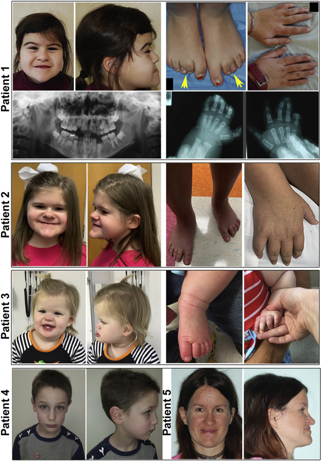
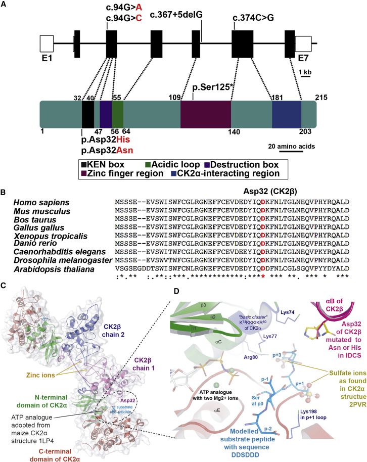
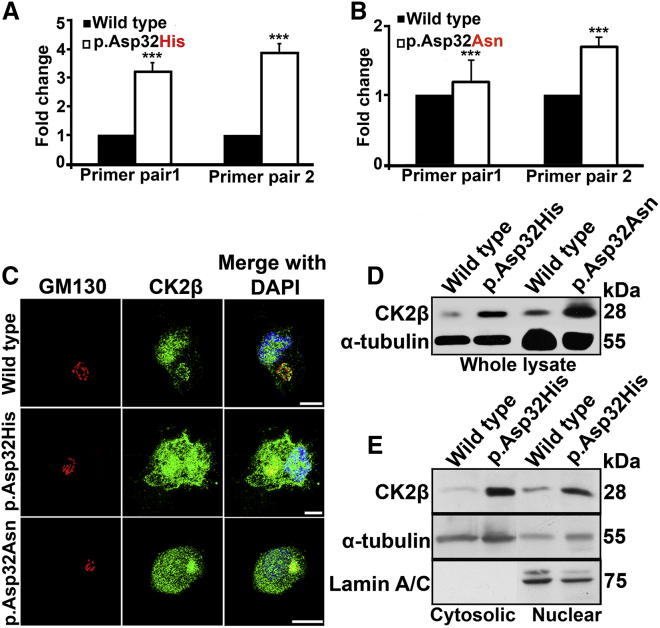
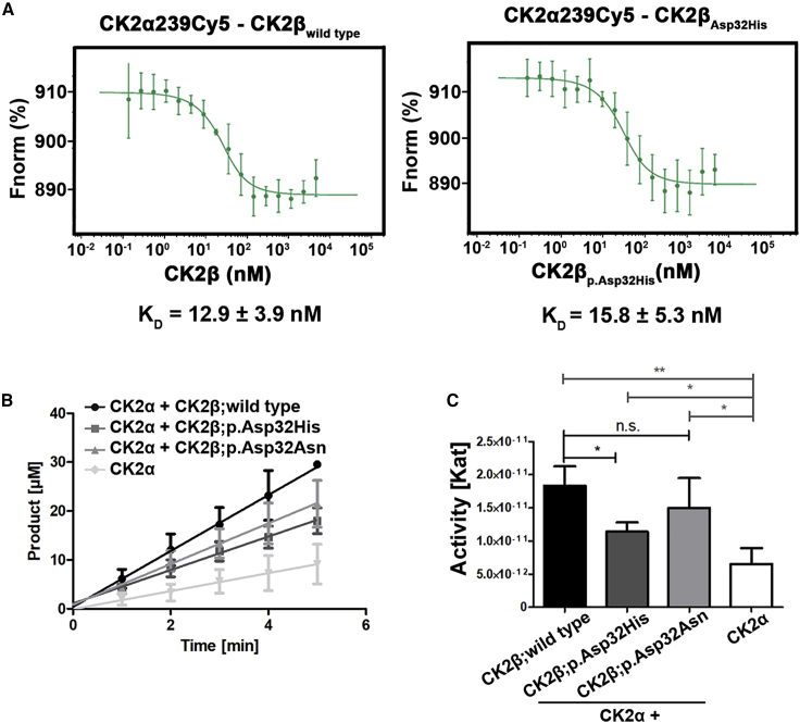
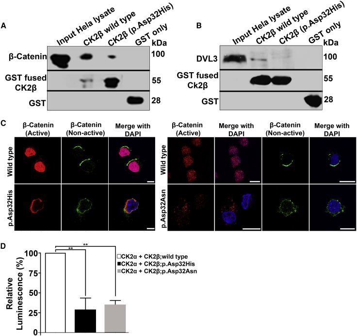
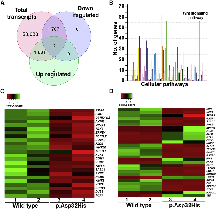
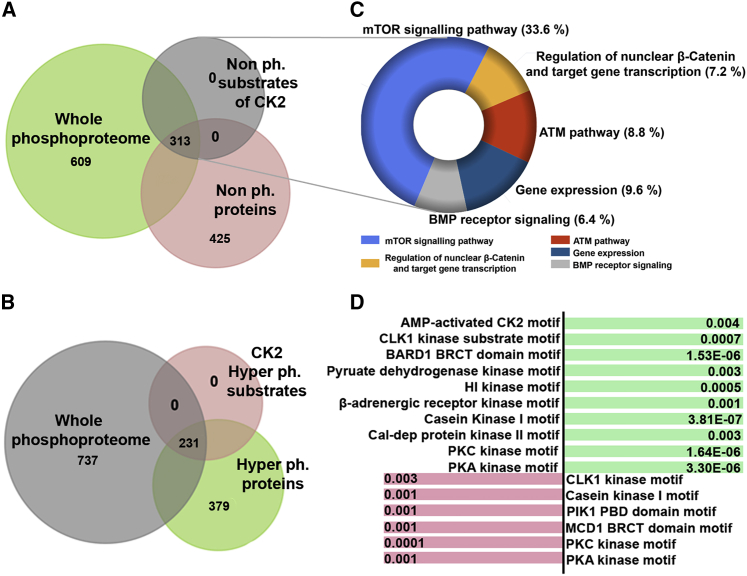
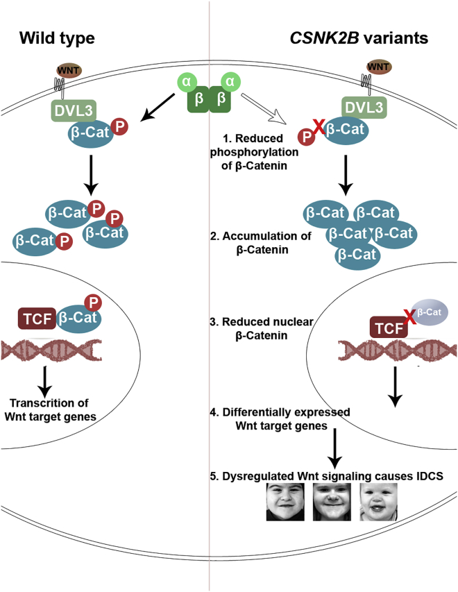
CSNK2B encodes for casein kinase II subunit beta (CK2β), the regulatory subunit of casein kinase II (CK2), which is known to mediate diverse cellular pathways. Variants in this gene have been recently identified as a cause of Poirier-Bienvenu neurodevelopmental syndrome (POBINDS), but functional evidence is sparse. Here, we report five unrelated individuals: two of them manifesting POBINDS, while three are identified to segregate a new intellectual disability-craniodigital syndrome (IDCS), distinct from POBINDS. The three IDCS individuals carried two different de novo missense variants affecting the same codon of CSNK2B. Both variants, NP_001311.3; p.Asp32His and NP_001311.3; p.Asp32Asn, lead to an upregulation of CSNK2B expression at transcript and protein level, along with global dysregulation of canonical Wnt signaling. We found impaired interaction of the two key players DVL3 and β-catenin with mutated CK2β. The variants compromise the kinase activity of CK2 as evident by a marked reduction of phosphorylated β-catenin and consequent absence of active β-catenin inside nuclei of the patient-derived lymphoblastoid cell lines (LCLs). In line with these findings, whole-transcriptome profiling of patient-derived LCLs harboring the NP_001311.3; p.Asp32His variant confirmed a marked difference in expression of genes involved in the Wnt signaling pathway. In addition, whole-phosphoproteome analysis of the LCLs of the same subject showed absence of phosphorylation for 313 putative CK2 substrates, enriched in the regulation of nuclear β-catenin and transcription of the target genes. Our findings suggest that discrete variants in CSNK2B cause dominant-negative perturbation of the canonical Wnt signaling pathway, leading to a new craniodigital syndrome distinguishable from POBINDS.

Keywords: CK2; CK2α; CK2β; CSNK2B; DVL3; GestaltMatcher; POBINDS; Wnt signaling; intellectual disability-craniodigital syndrome; whole transcriptome profiling; whole-phosphoproteome profiling; β-catenin.


Conflict of interest statement

The authors declare no competing interests.

Figures

Figure 1
Figure 1
Clinical presentation of patients with CSNK2B variants Front and side views of subjects 1–3 diagnosed with IDCS show strikingly similar facial gestalt, whereas subjects 4 and 5 diagnosed with POBINDS show vivid difference of facial gestalt, and no obvious digital or limb anomaly was recorded as in IDCS. Digital anomalies of IDCS patients are shown in two panels on right side. For subject 1, arrows show brachydactyly 2–5 and syndactyly 2–3 of the digits of the feet. Notably, preoperative radiographs of the hands and teeth are shown for subject 1.
Figure 2
Figure 2
CSNK2B variants identified in IDCS patients (A) Upper panel: genomic structure of human CSNK2B. The seven exons of CSNK2B are displayed by boxes, which are drawn to scale (1 kb = 1 cm). The filled boxes depict the open reading frame, and the open boxes show the UTRs. The introns are drawn as connecting lines of arbitrary length. Vertical lines on filled boxes indicate the positions of the variants. Lower panel: 215-amino-acid-long CK2β protein composed of five domains along with identified variants are shown. The protein structure is constructed according to the indicated scale bar. Figure is recreated from Bibby and Litchfield (2005). (B) Cross-species alignment showing conservation of Asp32 of CK2β. Note that Asp32 is conserved in all species. Asterisk (∗) is used for the conserved residues, colon (:) for conservative changes, and dot (.) for semiconservative changes. (C) Structural overview of the CK2α2β2 holoenzyme along with a modeled substrate peptide and the ATP analogue AMPPNP. Dotted circle shows the wild-type amino acid Asp32 and substrate peptide in its close proximity. (D) Zoomed picture focuses on the critical neighborhood around Asp32 of CK2β illustrating the IDCS-associated variants Asp32Asn and Asp32His. Two sulfate ions visible in the human CK2α structure PDB: 2PVR were drawn in ball-and-sticks representation; as outlined in the Material and methods section, these sulfate ions served as an orientation to model the p+1 and p+3 side chains of the substrate peptide DDSDDD (blue carbon atoms) into the active site.
Figure 3
Figure 3
Quantification of CSNK2B transcript and encoded protein product along with cellular localization in LCLs (A) Quantitative real-time PCR data showing 3-fold increased expression of CSNK2B mRNA in patient (GenBank: NM_001320.7:c.94G>C (p.Asp32His)) as compared with wild type. CSNK2B mRNA amounts were quantified relative to control sample. ∗∗∗p = 0.0001 calculated by Student’s t test. n = 3; error bars represent standard deviation (SD). (B) Graph shows 2-fold increased expression of CSNK2B mRNA of subject (GenBank: NM_001320.7:c.94G>A (p.Asp32Asn)) resulting from amplifying a second set of primers as compared with wild type. ∗∗∗p = 0.0001 calculated by Student’s t test. n = 3; error bars represent SD. (C) Confocal microscopy images showing increased amount of CK2β (green) in nuclei and in cytoplasm of subject-derived (NP_001311.3; p.Asp32His and NP_001311.3; p.Asp32Asn) LCLs as compared with wild type. GM130 (red) serves as marker of Golgi apparatus, and DAPI (blue) indicates staining of nucleus. Scale bar: 10 μm. (D) Immunoblotting shows an increased amount of CK2β in whole-cell lysates obtained from mutant (NP_001311.3; p.Asp32His and NP_001311.3; p.Asp32Asn) LCLs versus wild type. α-Tubulin is used as loading controls. (E) Immunoblots show an increased amount of CK2β in cytosolic and nuclear fraction in NP_001311.3; p.Asp32His mutant versus wild type. Loading controls are α-tubulin and lamin A/C, for cytosol and nucleus, respectively.
Figure 4
Figure 4
Interaction of CK2 subunits and kinase activity of mutated beta subunits (A) Graph shows interaction of CK2 subunits analyzed by microscale thermophoresis (MST). Graph in the left panel shows KD value (12.9 ± 3.9 nM) in wild-type CK2β bound to CK2α239Cy5. n = 3. Graph in the right panel shows KD values (15.8 ± 5.3 nM) in mutant CK2β (NP_001311.3; p.Asp32His) bound to CK2α239Cy5. n = 5. (B) Graph shows the amount of phosphorylated substrate peptide in the given time from capillary electrophoresis assay. Note that CK2 containing CK2β: NP_001311.3; p.Asp32His shows decreased amount of phosphorylated peptide as compared with wild type, whereas CK2 containing NP_001311.3; p.Asp32Asn shows no significant differences. The color and shape key denoting all the samples given in the figure. n = 3; error bars represent SD. (C) Kinase activity of CK2 wild type and mutants based on capillary electrophoresis assay. Bar graph shows kinase activity of CK2 containing wild-type and mutant beta subunits (NP_001311.3; p.Asp32His and NP_001311.3; p.Asp32Asn). Note that the activity of CK2 containing NP_001311.3; p.Asp32His is reduced as compared with wild type, but CK2 containing NP_001311.3; p.Asp32Asn variant does not show any significant difference. ∗p ≤ 0.05, ∗∗p ≤ 0.01, nsp ˃ 0.05 (Student’s t test). n = 3; error bars represent SD. ns, non-significant.
Figure 5
Figure 5
Impacts of variants on the dynamics of β-catenin and DVL3 (A) Pull-down assay from HeLa total protein extracts indicates reduced interaction of endogenous β-catenin with mutant (NP_001311.3; p.Asp32His) GST-tagged CK2β as compared with wild type. GST serves as negative control. Bands of approximately 85 kDa of β-catenin were observed on western blot after probing with rabbit monoclonal β-catenin antibody. GST-fused proteins were visualized by probing the membrane with in-house-generated mouse monoclonal GST antibody. (B) Pull-down assay shows reduced interaction of DVL3 with GST-fused CK2β mutant (NP_001311.3; p.Asp32His) as compared with wild-type. GST was used as negative control. Bands of approximately 78 kDa of DVL3 were observed by rabbit monoclonal DVL3 antibody. (C) Immunofluorescence shows decreased amount of active β-catenin (red) in nuclei of CK2β: NP_001311.3; p.Asp32His (left panel) and CK2β: NP_001311.3; p.Asp32Asn (right panel) LCLs as compared with the wild type. Localization pattern of non-active β-catenin (green) remains the same in wild-type and both mutant LCLs. DAPI (blue) indicates staining of nucleus. Scale bar, 5 μm (left panel); 10 μm (right panel). (D) Graph showing reduced kinase activity of CK2 carrying mutants (NP_001311.3; p.Asp32His and NP_001311.3; p.Asp32Asn) of CK2β as compared with wild type measured by ADP-Glo assay. Note that β-catenin was used as substrate. Error bars represent SD; n = 3. ∗∗p ≤ 0.01 (Student’s t test).
Figure 6
Figure 6
Whole-transcriptome profiling of patient LCLs (NP_001311.3; p.Asp32His) (A) Venn diagram showing total number of transcripts and those differentially regulated. Note that the fold change (FC) > 1, p < 1 for 58,038 transcripts and FC > 2, p < 0.05 for upregulated and downregulated transcripts. (B) Graph showing pathway enrichment of differentially expressed genes obtained from RNA sequencing (RNA-seq) data performed in NP_001311.3; p.Asp32His mutant LCLs and age- and sex-matched control. Note that two of the highest peaks are related to the genes involved in the immune response pathway and Wnt signaling. x axis shows the number of genes, and y axis shows cellular pathways. FC > 5 and p < 0.05. (C) Heatmaps of differentially expressed Wnt target genes observed in NP_001311.3; p.Asp32His mutant LCLs compared with control. Color key shows the level of upregulation or downregulation: green shows higher expression as compared with bright red. (D) Heatmaps of differentially expressed transcription regulators observed in NP_001311.3; p.Asp32His mutant LCLs compared with control.
Figure 7
Figure 7
Whole-phosphoproteome profiling of patient LCLs (NP_001311.3; p.Asp32His) (A) Venn diagram showing whole phosphoproteome of mutant (NP_001311.3; p.Asp32His) and wild-type LCLs. Note that among 1,347 (green) proteins, 425 (pink) are non-phosphorylated. These 425 contain 313 putative CK2 substrates motifs (shown in gray). The FC is >2; q value is 0.05. (B) Venn diagram showing whole phosphoproteome of NP_001311.3; p.Asp32His mutant and wild-type LCLs. Note that among 1,347 proteins shown in gray, 379 are hyper-phosphorylated (shown in green). These 379 contain 231 substrates of CK2 (shown in pink). The FC is >2; q value is 0.05. (C) Donut graph generated by Funrich shows the percent of genes involved in a biological pathway. Note that each pathway is color coded and the key is given. (D) The diagram presents substrate motifs of different kinases that are hyper-phosphorylated (shown in green) and hypo-phosphorylated (shown in pink). The respective p values of enriched motifs are written corresponding to each kinase.
Figure 8
Figure 8
CK2 is the crucial member of the canonical Wnt signaling pathway Mutant CK2 has reduced the phosphorylation rate of β-catenin, which thus fails to translocate to the nucleus and consequently affects the regulation of Wnt target genes. This disruption of Wnt signaling leads to the phenotype seen in the studied patient.

References

    1. Toriello H.V., Higgins J.V. Craniodigital syndromes: report of a child with Filippi syndrome and discussion of differential diagnosis. Am. J. Med. Genet. 1995;55:200–204. doi: 10.1002/ajmg.1320550210. - DOI - PubMed
    1. Hussain M.S., Battaglia A., Szczepanski S., Kaygusuz E., Toliat M.R., Yigit G., Beleggia F., Tinschert S., Clayton-Smith J., Vasudevan P., et al. Mutations in CKAP2L, the human homolog of the mouse Radmis gene, cause Filippi syndrome. Am. J. Hum. Genet. 2014;95:622–632. doi: 10.1016/j.ajhg.2014.10.008. - DOI - PMC - PubMed
    1. Balasubramanian M., Lord H., Levesque S., Guturu H., Thuriot F., Sillon G., Bowen J., Calhoun A.R., Viskochil D.H., Bejerano G., et al. DDD Study Chitayat syndrome: hyperphalangism, characteristic facies, hallux valgus and bronchomalacia results from a recurrent c. 266A> G p.(Tyr89Cys) variant in the ERF gene. J. Med. Genet. 2017;54:157–165. doi: 10.1136/jmedgenet-2016-104143. - DOI - PubMed
    1. Kaygusuz E., Khayyat A.I.A., Abdullah U., Budde B.S., Asif M., Ahmed I., Makhdoom E.U.H., Sur Erdem I., Baig J.M., Khan M.M.A., et al. A 24 generation old founder mutation impairs splicing of RBBP8 in Pakistani families affected with Jawad syndrome. Clin. Genet. 2021;100:486–488. doi: 10.1111/cge.14028. - DOI - PubMed
    1. Woods C.G., Crouchman M., Huson S.M. Three sibs with phalangeal anomalies, microcephaly, severe mental retardation, and neurological abnormalities. J. Med. Genet. 1992;29:500–502. - PMC - PubMed