

Vagus Nerve Stimulation as a Potential Therapy in Early Alzheimer's Disease: A Review
- PMID: 35572001
- PMCID: PMC9098960
- DOI: 10.3389/fnhum.2022.866434
Vagus Nerve Stimulation as a Potential Therapy in Early Alzheimer's Disease: A Review
Abstract
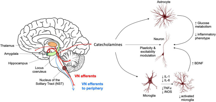
Cognitive dysfunction in Alzheimer's disease (AD) is caused by disturbances in neuronal circuits of the brain underpinned by synapse loss, neuronal dysfunction and neuronal death. Amyloid beta and tau protein cause these pathological changes and enhance neuroinflammation, which in turn modifies disease progression and severity. Vagal nerve stimulation (VNS), via activation of the locus coeruleus (LC), results in the release of catecholamines in the hippocampus and neocortex, which can enhance synaptic plasticity and reduce inflammatory signalling. Vagal nerve stimulation has shown promise to enhance cognitive ability in animal models. Research in rodents has shown that VNS can have positive effects on basal synaptic function and synaptic plasticity, tune inflammatory signalling, and limit the accumulation of amyloid plaques. Research in humans with invasive and non-invasive VNS devices has shown promise for the modulation of cognition. However, the direct stimulation of the vagus nerve afforded with the invasive procedure carries surgical risks. In contrast, non-invasive VNS has the potential to be a broadly available therapy to manage cognitive symptoms in early AD, however, the magnitude and specificity of its effects remains to be elucidated, and the non-inferiority of the effects of non-invasive VNS as compared with invasive VNS still needs to be established. Ongoing clinical trials with healthy individuals and patients with early AD will provide valuable information to clarify the potential benefits of non-invasive VNS in cognition and AD. Whether invasive or non-invasive VNS can produce a significant improvement on memory function and whether its effects can modify the progression of AD will require further investigation.
Keywords: Alzheimer; MCI; memory; noradrenaline; norepinepherine; plasticity; vagal; vagus.
Copyright © 2022 Vargas-Caballero, Warming, Walker, Holmes, Cruickshank and Patel.
Conflict of interest statement
BP: CEO and Founder—ElectronRx. The remaining authors declare that the research was conducted in the absence of any commercial or financial relationships that could be construed as a potential conflict of interest.
Figures
References
-
- Alvarez-Dieppa A. C., Griffin K., Cavalier S., Mcintyre C. K. (2016). Vagus Nerve Stimulation Enhances Extinction of Conditioned Fear in Rats and Modulates Arc Protein, CaMKII, and GluN2B-Containing NMDA Receptors in the Basolateral Amygdala. Neural Plast. 2016:4273280. 10.1155/2016/4273280 - DOI - PMC - PubMed
Publication types
LinkOut - more resources
Full Text Sources
Other Literature Sources