. 2022 Apr 27:13:871216.
doi: 10.3389/fimmu.2022.871216. eCollection 2022.

Production of Cytotoxic Antibodies After Intra-Articular Injection of Allogeneic Synovial Membrane Mesenchymal Stem Cells With and Without LPS Administration

Affiliations

Production of Cytotoxic Antibodies After Intra-Articular Injection of Allogeneic Synovial Membrane Mesenchymal Stem Cells With and Without LPS Administration

Gustavo Dos Santos Rosa et al. Front Immunol. .

Abstract

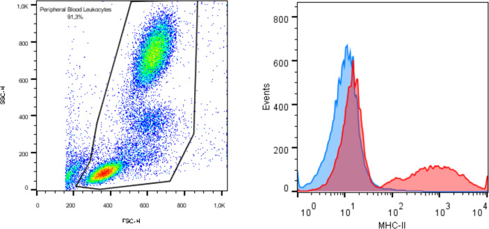
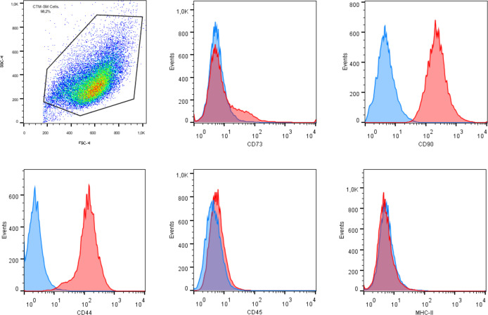
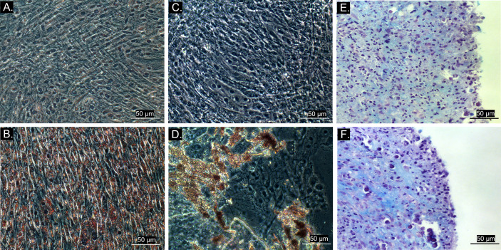
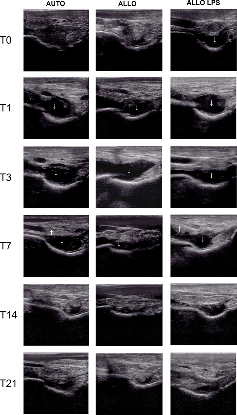
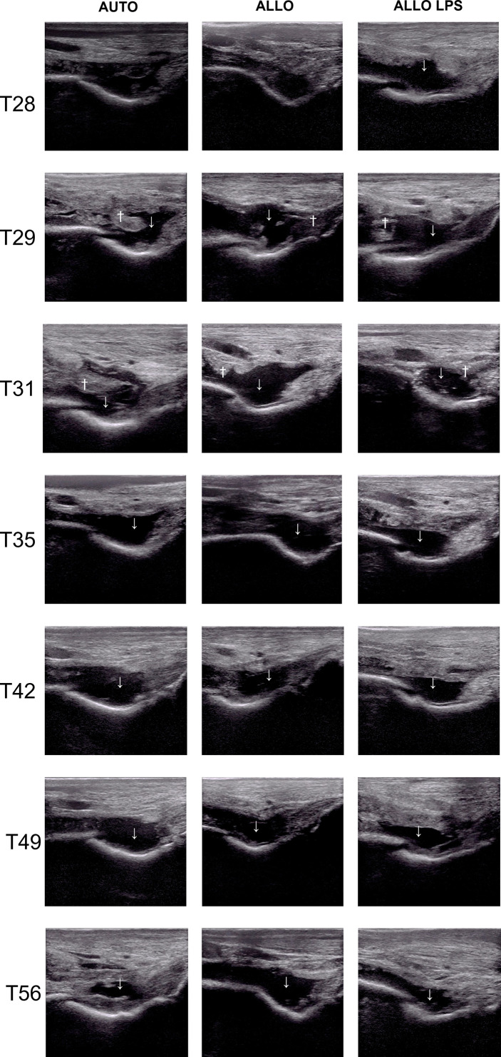
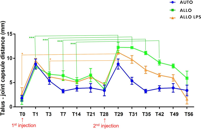
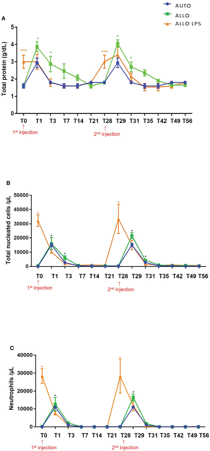
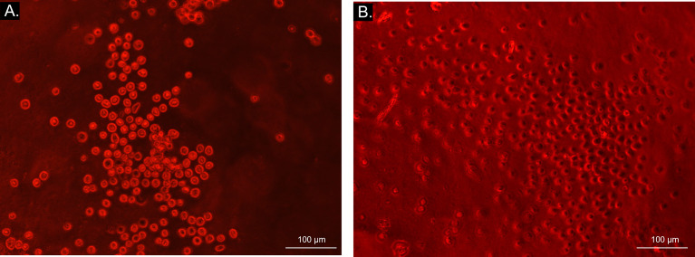
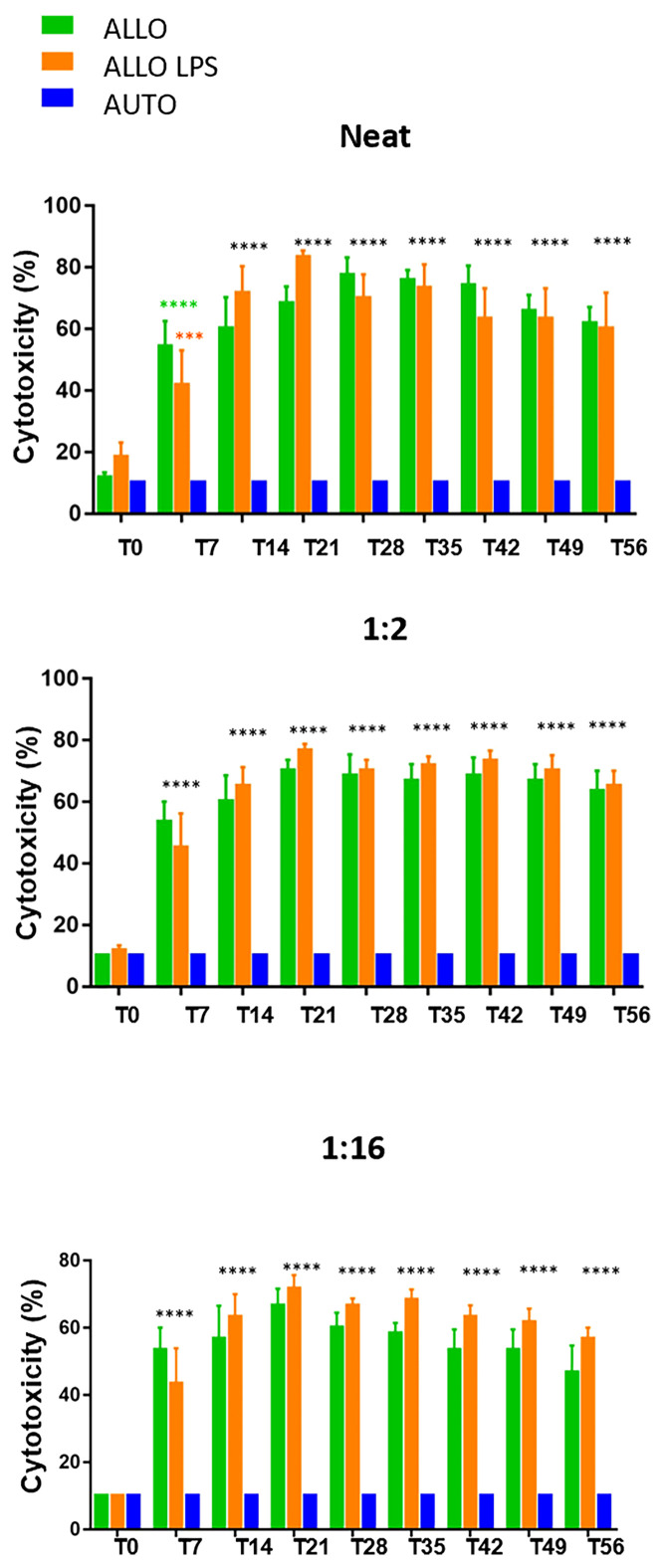
Allogeneic mesenchymal stem cells (MSC) are widely used in clinical routine due to the shorter expansion time and reliability of its quality. However, some recipients can produce alloantibodies that recognize MSCs and activate the immune system, resulting in cell death. Although antibody production was already described after MSC injection, no previous studies described the immune response after intra-articular MSC injection in acute synovitis. This study aimed to evaluate the influence of inflammation on immune response after single and repeated intra-articular injections of synovial membrane MSC (SMMSC). Horses were divided in three groups: control group (AUTO) received autologous synovial membrane MSCs; whereas group two (ALLO) received allogeneic SMMSCs and group three (ALLO LPS) was submitted to acute experimental synovitis 8 h before SMMSCs injection. The procedure was repeated for all groups for 28 days. Physical and lameness evaluations and synovial fluid analysis were performed. Sera from all animals were obtained before and every 7 days after each injection up to 4 weeks, to perform microcytotoxicity assays incubating donor SMMSCs with recipients' sera. The first injection caused a mild and transient synovitis in all groups, becoming more evident and longer in ALLO and ALLO LPS groups after the second injection. Microcytotoxicity assays revealed significant antibody production as soon as 7 days after SMMSC injection in ALLO and ALLO LPS groups, and cytotoxicity scores of both groups showed no differences at any time point, being equally different from AUTO group. Although inflammation is capable of inducing MHC expression in MSCs, which enhances immune recognition, cytotoxicity scores were equally high in ALLO and ALLO LPS groups, making it difficult to determine the potentiation effect of inflammation on antibody production. Our findings suggest that inflammation does not display a pivotal role in immune recognition on first allogeneic MSC injection. In a translational way, since specific antibodies were produced against MSCs, patients that need more than one MSC injection may benefit from a first allogeneic injection followed by subsequent autologous injections.

Keywords: MHC; cell rejection reactions; horses; humoral immune response; intraarticular injection; mesenchymal stromal cells; microcytotoxicity.


Conflict of interest statement

The authors declare that the research was conducted in the absence of any commercial or financial relationships that could be construed as a potential conflict of interest.

Figures

Figure 1
Figure 1
Immunophenotyping by flow cytometry exhibiting expression of MHC class II in equine peripheral blood as positive control.
Figure 2
Figure 2
Flow cytometry analysis of mesenchymal stem cell surface markers in synovial membrane derived MSCs revealing expression of CD90, CD44, and CD73, and lack of expression of CD45 and MHC class II.
Figure 3
Figure 3
Bright field microscopy (20x objective) of MSC trilineage differentiation. (A) Control group of adipogenic differentiation; (B) Differentiated cell culture stained with Oil Red O, showing intracellular lipid droplets in red; (C) Control group of osteogenic differentiation; (D) Differentiated cell culture stained with Alizarin Red S, exhibiting extracellular hydroxyapatite crystals stained in red; (E) Control group of chondrogenic differentiation; (F) Differentiated tridimensional cell culture stained with Alcian Blue after 21 days, revealing chondral matrix sulphated proteoglycans stained in blue.
Figure 4
Figure 4
Ultrasonography of tibiotarsal joints before and after the first MSC injection, showing synovial effusion (↓) and synovial membrane hyperplasia (†). Synovial effusion was observed at T0 in ALLO LPS, and at T1 in AUTO and ALLO, and decreased at T14 in all groups. Synovial membrane proliferation occurred at T7 in all groups, although it was more evident in ALLO and ALLO LPS.
Figure 5
Figure 5
Ultrasonographic aspect of tibiotarsal joints before and after the second MSC injection. Extended synovial effusion (↓) and synovial membrane hyperplasia (†) can be noticed in all groups.
Figure 6
Figure 6
Mean ± SD values of synovial effusion, measured by the distance between the talus and the joint capsule. ALLO group presented statistical differences between first and second injection at T0, T1, T3, T7 and T14 (***P<0.005), whereas ALLO LPS group showed differences between first and second injection comparing T0 to T28 and T3 to T31 (*P<0.05).
Figure 7
Figure 7
(A) Mean ± SD values of synovial fluid total protein (TP). Higher TP values were observed in ALLO LPS group at T0 and T28 (****P<0.0001). Statistical differences were observed between ALLO and AUTO groups at T1, T3, T29 and T31 (*P<0.05). (B) Mean ± SD values of synovial fluid total nucleated cell count (TNCC), showing absence of statistical differences between groups in first and second injections. Higher TNCC was observed in ALLO LPS group at T0 and T28 (*). Compared to the initial time point (T0 or T28), there was a significant increase in TNCC of all groups 1 and 3 days after SMMSC injection (T1 and T3; T29 and T31, respectively) (*P<0.05). (C) Mean ± SD values of synovial fluid neutrophil count, showing a higher neutrophil count in ALLO LPS group at T0 and T28 (*P<0.05), whereas in ALLO and AUTO the peak occurred at T1 and T29(*).
Figure 8
Figure 8
Bright field microscopy (10x objective) visualization of microcytotoxicity assay. Bright light and round cells represent live cells in AUTO group (A), while dark-centered flat cells correspond to dead cells (B) in ALLO LPS group.
Figure 9
Figure 9
Microcytotoxicity assay before and after synovial membrane mesenchymal stem cell injections in neat, 1:2 and 1:16 sera dilutions. Initial time point did not reveal antibody production. However, comparison between T0 and T7 revealed P<0.0001(****) in neat serum of ALLO group and P=0.0038 (***) in ALLO LPS group. Subsequent time points showed significant antibody production in ALLO and ALLO LPS groups, from T14 to T56 (****P<0.0001) in all sera dilutions. No differences were observed between ALLO and ALLO LPS groups.

References

    1. Ilic D, Polak J. Stem Cell-Based Therapy–Where are We Going? Lancet (2012) 379(9819):877–8. doi: 10.1016/S0140-6736(12)60155-X - DOI - PubMed
    1. Bartholomew A, Sturgeon C, Siatskas M, Ferrer K, McIntosh K, Patil S, et al. Mesenchymal Stem Cells Suppress Lymphocyte Proliferation In Vitro and Prolong Skin Graft Survival In Vivo . Exp Hematol (2012) 30:42–8. doi: 10.1016/S0301-472X(01)00769-X - DOI - PubMed
    1. Ankrum JA, Ong JF, Karp JM. Mesenchymal Stem Cells: Immune Evasive, Not Immune Privileged. Nat Biotechnol (2014) 32:252–60. doi: 10.1038/nbt.2816 - DOI - PMC - PubMed
    1. Joswig AJ, Mitchell A, Cummings KJ, Levine GJ, Gregory CA, Smith R 3rd, et al. Repeated Intra-Articular Injection of Allogeneic Mesenchymal Stem Cells Causes an Adverse Response Compared to Autologous Cells in the Equine Model. Stem Cell Res Ther (2017) 8:42. doi: 10.1186/s13287-017-0503-8 - DOI - PMC - PubMed
    1. Owens SD, Kol A, Walker NJ, Borjesson DL. Allogeneic Mesenchymal Stem Cell Treatment Induces Specific Alloantibodies in Horses. Hindawi Publ Corp Stem Cells Int (2016) 8:8. doi: 10.1155/2016/5830103 - DOI - PMC - PubMed

Publication types

Substances