

Risk factors for cardiac rupture after acute ST-segment elevation myocardial infarction during the percutaneous coronary intervention era: a retrospective case-control study
- PMID: 35572908
- PMCID: PMC9096283
- DOI: 10.21037/jtd-22-394
Risk factors for cardiac rupture after acute ST-segment elevation myocardial infarction during the percutaneous coronary intervention era: a retrospective case-control study
Abstract
Background: In percutaneous coronary intervention (PCI) era, more clinically valuable risk factors are still needed to determine the occurrence of cardiac rupture (CR). Therefore, we aimed to provide evidence for the early identification of CR in ST-segment elevation myocardial infarction (STEMI).
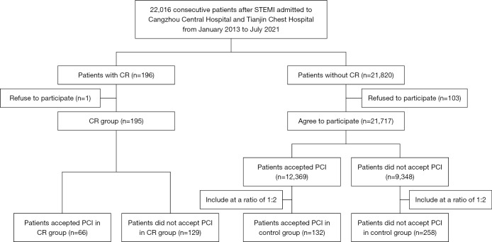
Methods: A total of 22,016 consecutive patients with STEMI admitted to Cangzhou Central Hospital and Tianjin Chest Hospital from January 2013 to July 2021 were retrospectively included, among which 195 patients with CR were included as CR group. From the rest 21,820 STEMI patients without CR, 390 patients at a ratio of 1:2 were included as the control group. A total of 66 patients accepted PCI in the CR group, and 132 patients who accepted PCI in the control group at a ratio of 1:2 were included. The status of first medical contact, laboratory examinations, and PCI characteristics were recorded. Multivariate logistic regression analysis was used to investigate the risk factors related to CR.
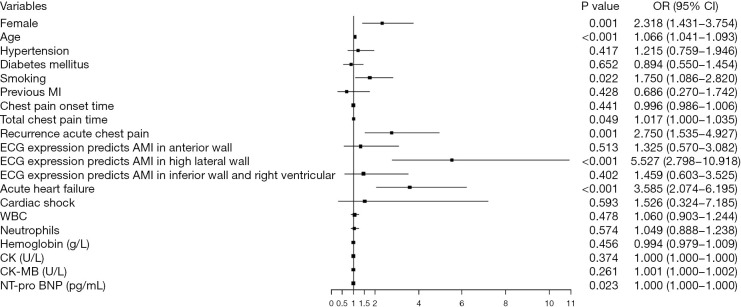
Results: There was a higher proportion of patients with myocardial infarction (MI) in the high lateral wall in the CR group (23.6% vs. 8.2%, P<0.001). The proportion of single lesions was lower in the CR group (24.2% vs. 45.5%, P=0.004). Female (OR =2.318, 95% CI: 1.431-3.754, P=0.001), age (OR =1.066, 95% CI: 1.041-1.093, P<0.001), smoking (OR =1.750, 95% CI: 1.086-2.820, P=0.022), total chest pain time (OR =1.017, 95% CI: 1.000-1.035, P=0.049), recurrent acute chest pain (OR =2.750, 95% CI: 1.535-4.927, P=0.001), acute myocardial infarction (AMI) in the high lateral wall indicated by ECG (OR =5.527, 95% CI: 2.798-10.918, P<0.001), acute heart failure (OR =3.585, 95% CI: 2.074-6.195, P<0.001), and NT-proBNP level (OR =1.000, 95% CI: 1.000-1.000, P=0.023) were risk factors for CR in all patients. In patients who accepted PCI, single lesion (OR =0.421, 95% CI: 0.204-0.867, P=0.019), preoperative thrombolysis in myocardial infarction (TIMI) grade (OR =0.358, 95% CI: 0.169-0.760, P=0.007), and postoperative TIMI grade (OR =0.222, 95% CI: 0.090-0.546, P=0.001) were risk factors for CR.
Conclusions: Non-single lesions and preoperative and postoperative TIMI grades were risk factors for CR in patients who accepted PCI. In addition to previously reported indicators, we found that AMI in the high lateral wall maybe helpful in early and accurate identification and prevention of possible CR.
Keywords: Cardiac rupture (CR); ST-segment elevation myocardial infarction; acute myocardial infarction in high lateral wall; percutaneous coronary intervention; thrombolysis in myocardial infarction grades.
2022 Journal of Thoracic Disease. All rights reserved.
Conflict of interest statement
Conflicts of Interest: All authors have completed the ICMJE uniform disclosure form (available at https://jtd.amegroups.com/article/view/10.21037/jtd-22-394/coif). The authors have no conflicts of interest to declare.
Figures


Comment in
-
Can we predict cardiac rupture in patients with ST-segment elevation myocardial infarction?J Thorac Dis. 2022 Jul;14(7):2451-2453. doi: 10.21037/jtd-22-655. J Thorac Dis. 2022. PMID: 35928604 Free PMC article. No abstract available.
-
Mechanical complications of ST segment elevation myocardial infarction: are they tangible?J Thorac Dis. 2022 Jul;14(7):2458-2460. doi: 10.21037/jtd-22-705. J Thorac Dis. 2022. PMID: 35928618 Free PMC article. No abstract available.
-
Cardiac rupture after ST-elevation myocardial infarction (STEMI): a 'Stitch' in time?J Thorac Dis. 2022 Jul;14(7):2454-2457. doi: 10.21037/jtd-22-720. J Thorac Dis. 2022. PMID: 35928626 Free PMC article. No abstract available.
-
Mechanical complications of acute myocardial infarction: a not "mechanical" preventive and therapeutic strategy.J Thorac Dis. 2022 Aug;14(8):2732-2734. doi: 10.21037/jtd-2022-09. J Thorac Dis. 2022. PMID: 36071781 Free PMC article. No abstract available.
References
LinkOut - more resources
Full Text Sources
Research Materials
Miscellaneous