

Toward Precise Localization of Abnormal Brain Activity: 1D CNN on Single Voxel fMRI Time-Series
- PMID: 35573265
- PMCID: PMC9094401
- DOI: 10.3389/fncom.2022.822237
Toward Precise Localization of Abnormal Brain Activity: 1D CNN on Single Voxel fMRI Time-Series
Abstract
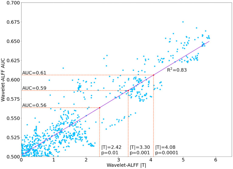
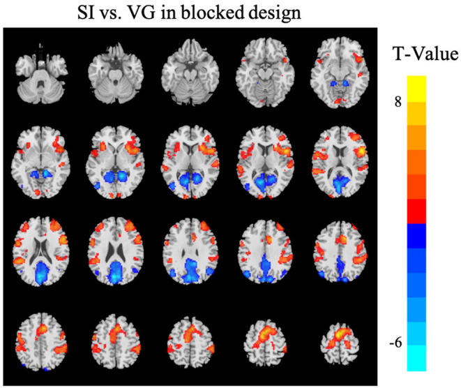
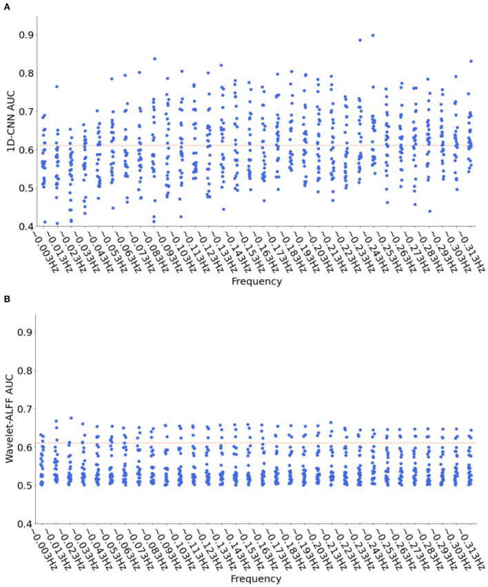
Functional magnetic resonance imaging (fMRI) is one of the best techniques for precise localization of abnormal brain activity non-invasively. Machine-learning approaches have been widely used in neuroimaging studies; however, few studies have investigated the single-voxel modeling of fMRI data under cognitive tasks. We proposed a hybrid one-dimensional (1D) convolutional neural network (1D-CNN) based on the temporal dynamics of single-voxel fMRI time-series and successfully differentiated two continuous task states, namely, self-initiated (SI) and visually guided (VG) motor tasks. First, 25 activation peaks were identified from the contrast maps of SI and VG tasks in a blocked design. Then, the fMRI time-series of each peak voxel was transformed into a temporal-frequency domain by using continuous wavelet transform across a broader frequency range (0.003-0.313 Hz, with a step of 0.01 Hz). The transformed time-series was inputted into a 1D-CNN model for the binary classification of SI and VG continuous tasks. Compared with the univariate analysis, e.g., amplitude of low-frequency fluctuation (ALFF) at each frequency band, including, wavelet-ALFF, the 1D-CNN model highly outperformed wavelet-ALFF, with more efficient decoding models [46% of 800 models showing area under the curve (AUC) > 0.61] and higher decoding accuracies (94% of the efficient models), especially on the high-frequency bands (>0.1 Hz). Moreover, our results also demonstrated the advantages of wavelet decompositions over the original fMRI series by showing higher decoding performance on all peak voxels. Overall, this study suggests a great potential of single-voxel analysis using 1D-CNN and wavelet transformation of fMRI series with continuous, naturalistic, steady-state task design or resting-state design. It opens new avenues to precise localization of abnormal brain activity and fMRI-guided precision brain stimulation therapy.
Keywords: 1D-CNN; continuous task states; fMRI; single-voxel analysis; wavelet transformation.
Copyright © 2022 Wu, Hu, Wang, Zang and Zhang.
Conflict of interest statement
The authors declare that the research was conducted in the absence of any commercial or financial relationships that could be construed as a potential conflict of interest.
Figures


Similar articles
-
fMRI volume classification using a 3D convolutional neural network robust to shifted and scaled neuronal activations.Neuroimage. 2020 Dec;223:117328. doi: 10.1016/j.neuroimage.2020.117328. Epub 2020 Sep 5. Neuroimage. 2020. PMID: 32896633
-
Higher reliability and validity of Wavelet-ALFF of resting-state fMRI: From multicenter database and application to rTMS modulation.Hum Brain Mapp. 2023 Feb 15;44(3):1105-1117. doi: 10.1002/hbm.26142. Epub 2022 Nov 17. Hum Brain Mapp. 2023. PMID: 36394386 Free PMC article.
-
Higher Sensitivity and Reproducibility of Wavelet-Based Amplitude of Resting-State fMRI.Front Neurosci. 2020 Mar 31;14:224. doi: 10.3389/fnins.2020.00224. eCollection 2020. Front Neurosci. 2020. PMID: 32300288 Free PMC article.
-
A hybrid learning framework for fine-grained interpretation of brain spatiotemporal patterns during naturalistic functional magnetic resonance imaging.Front Hum Neurosci. 2022 Sep 30;16:944543. doi: 10.3389/fnhum.2022.944543. eCollection 2022. Front Hum Neurosci. 2022. PMID: 36248685 Free PMC article.
-
Value of Frequency Domain Resting-State Functional Magnetic Resonance Imaging Metrics Amplitude of Low-Frequency Fluctuation and Fractional Amplitude of Low-Frequency Fluctuation in the Assessment of Brain Tumor-Induced Neurovascular Uncoupling.Brain Connect. 2017 Aug;7(6):382-389. doi: 10.1089/brain.2016.0480. Brain Connect. 2017. PMID: 28657344 Free PMC article.
Cited by
-
Language task-based fMRI analysis using machine learning and deep learning.Front Radiol. 2024 Nov 27;4:1495181. doi: 10.3389/fradi.2024.1495181. eCollection 2024. Front Radiol. 2024. PMID: 39664795 Free PMC article.
References
LinkOut - more resources
Full Text Sources