. 2022 Feb 1:33:100415.
doi: 10.1016/j.jbo.2022.100415. eCollection 2022 Apr.

Baicalin induces apoptosis and autophagy in human osteosarcoma cells by increasing ROS to inhibit PI3K/Akt/mTOR, ERK1/2 and β-catenin signaling pathways

Affiliations

Baicalin induces apoptosis and autophagy in human osteosarcoma cells by increasing ROS to inhibit PI3K/Akt/mTOR, ERK1/2 and β-catenin signaling pathways

He Pang et al. J Bone Oncol. .

Abstract

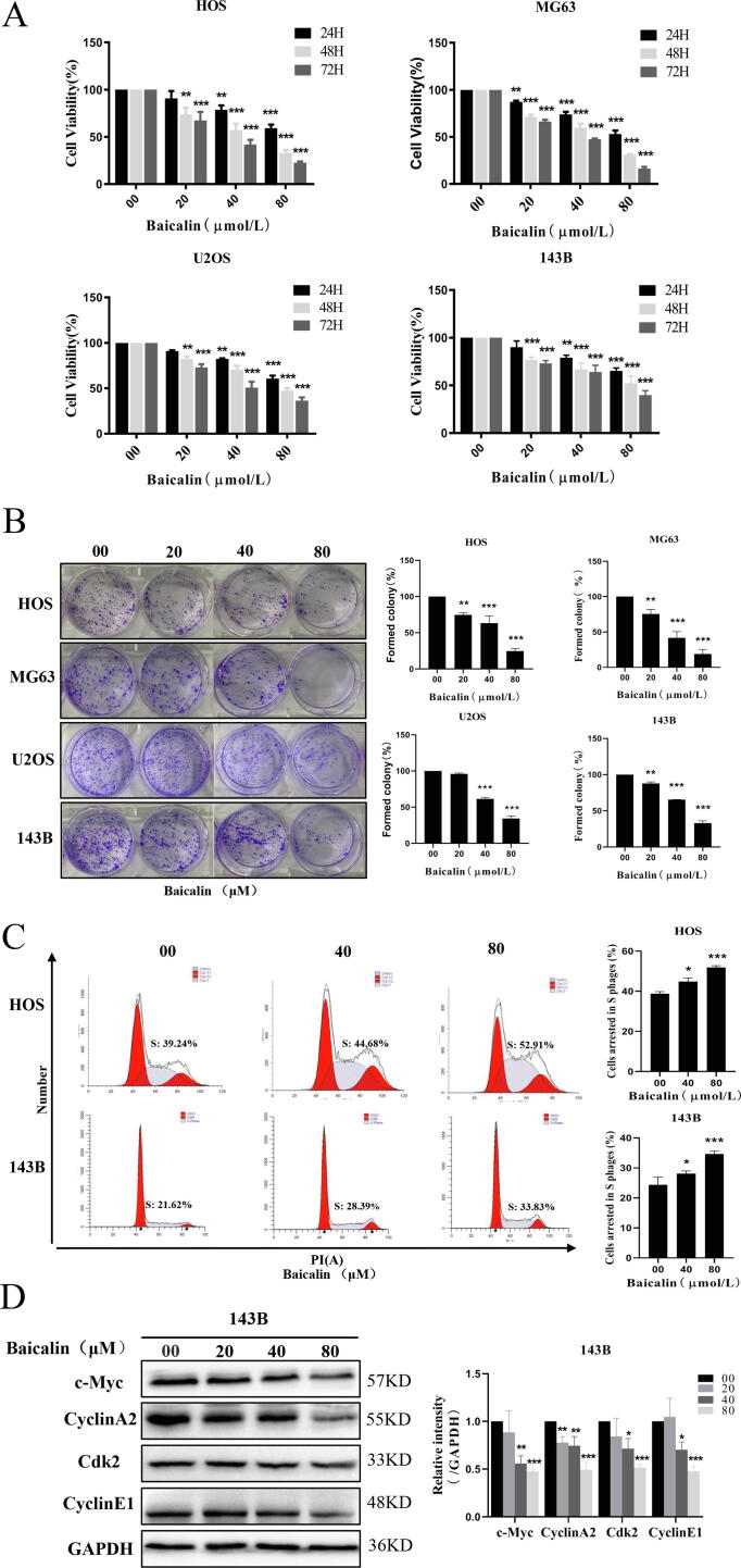
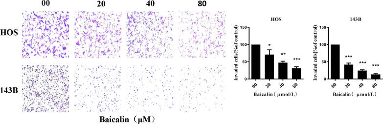
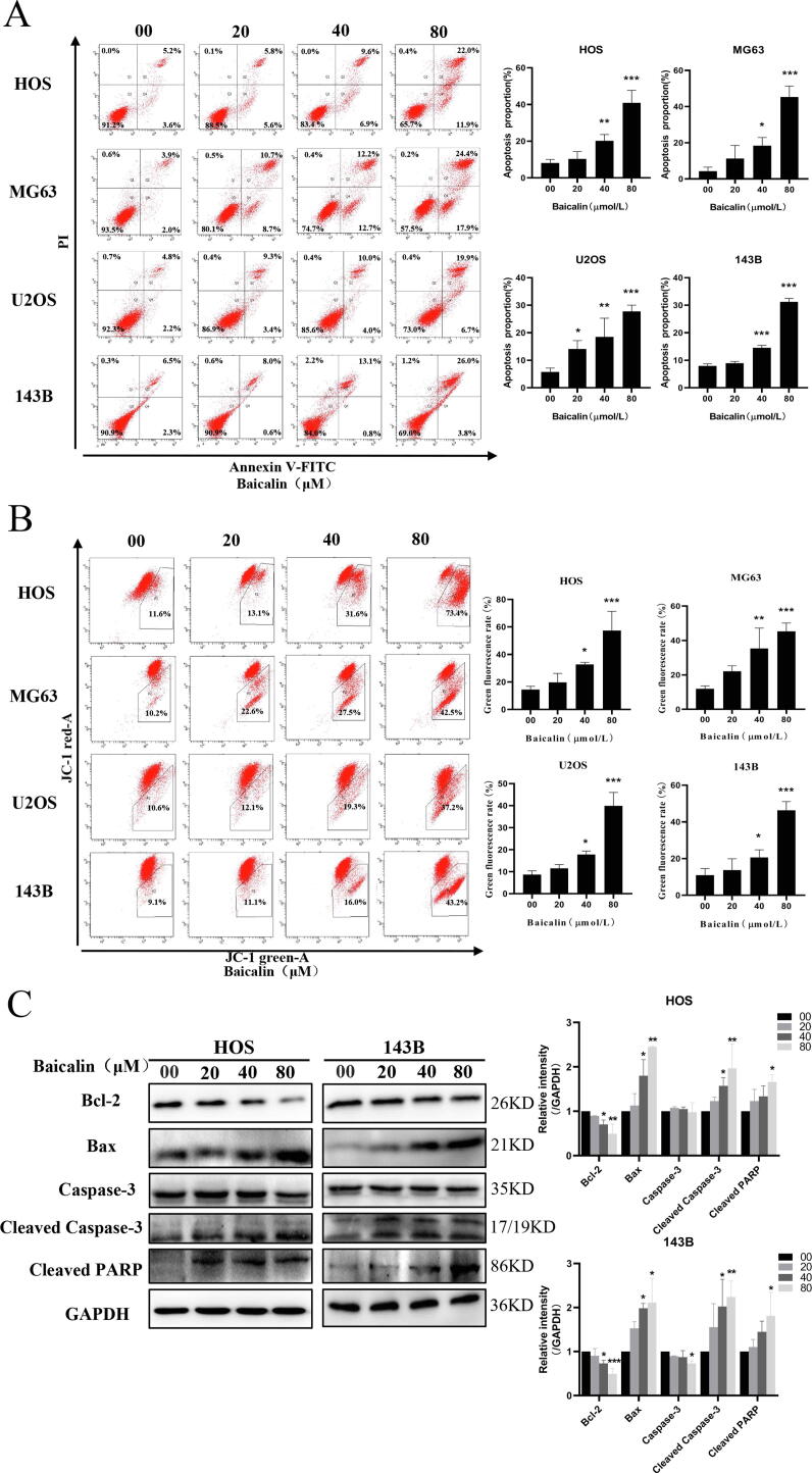
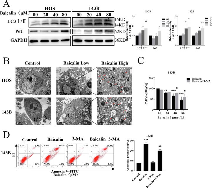
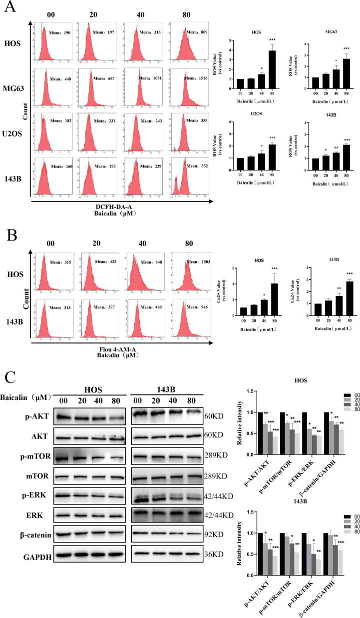
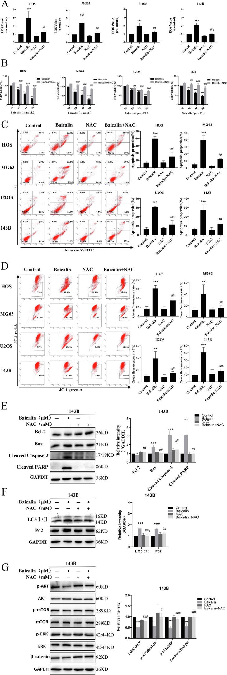
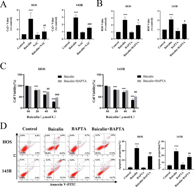
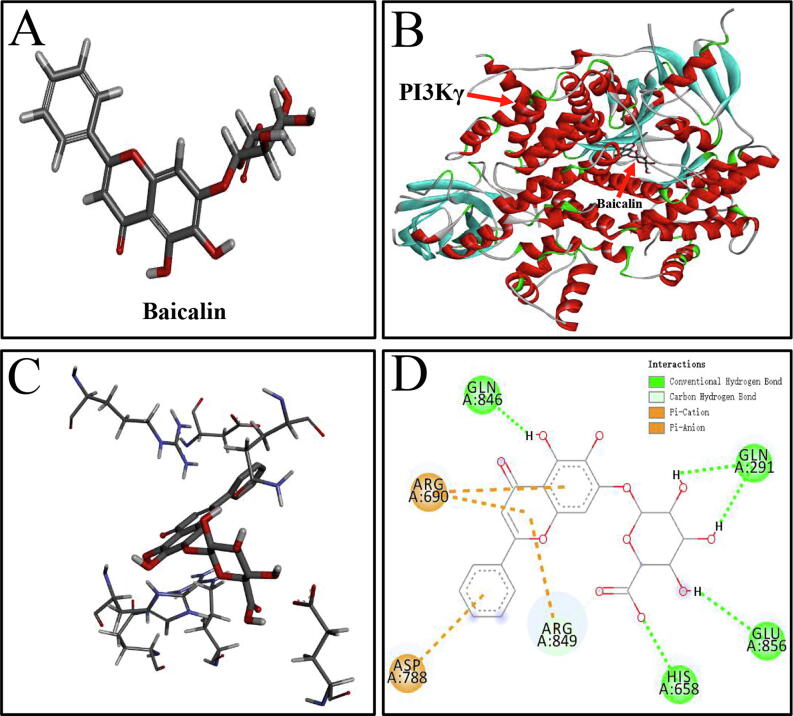
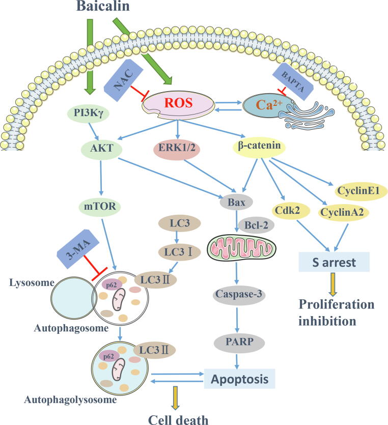
Baicalin, a flavonoid derivative, exerts antitumor activity in a variety of neoplasms. However, whether baicalin exerts antitumor effects on osteosarcoma cells remains to be elucidated. In this study, treatment with baicalin reduced the proliferation and invasive potential of osteosarcoma cells and reduced the mitochondrial membrane potential, which eventually caused mitochondrial apoptosis. In addition, baicalin increased intercellular Ca2+ and ROS concentrations. Baicalin-induced apoptosis was confirmed by enhanced Bax, cleaved caspase-3, and cleaved PARP levels and decreased Bcl-2 levels. The increase in LC3-II and p62 suggested that baicalin induced autophagosome formation but ultimately inhibited downstream autophagy. Moreover, apoptosis induced by baicalin was attenuated by the addition of 3-MA. Furthermore, we found that baicalin inhibited the PI3K/Akt/mTOR, ERK1/2 and β-catenin signaling pathways. Chelation of free Ca2+ by BAPTA-AM also inhibited both apoptosis induction and ROS concentration changes. Finally, NAC pretreatment reversed baicalin treatment outcomes, including the increase in Ca2+ concentration, induction of apoptosis and autophagy, and inhibition of the pathways. Molecular docking results indicated that baicalin might interact with the structural domain of PI3Kγ. Thus, baicalin may be considered a potential candidate for osteosarcoma treatment.

Keywords: Apoptosis; Autophagy; Baicalin; Osteosarcoma; PI3Kγ; ROS.


Conflict of interest statement

The authors declare that they have no known competing financial interests or personal relationships that could have appeared to influence the work reported in this paper.

Figures

Fig. 1
Fig. 1
Baicalin represses proliferation of OS cells. (A) HOS, MG63, U2OS and 143B cells were incubated with different concentrations of baicalin for 24–72 h. Cell viability was quantified using a CCK-8 assay. (B) Colony formation after baicalin treatment was evaluated as determined by the colony formation assay. (C) Baicalin-induced S-phase cell cycle arrest was detected by flow cytometry. (D) Expression of c-Myc, cyclin A2, Cdk2 and cyclinE1was analyzed by western blotting. *p < 0.05, **p < 0.01, ***p < 0.001 vs. control group.
Fig. 2
Fig. 2
Baicalin suppresses invasion in OS cells. Invasion assays assessed the invasion ability of osteosarcoma cells. The number of invasive cells in five independent microscopic fields was quantified using ImageJ. *p < 0.05, **p < 0.01, ***p < 0.001 vs. control group.
Fig. 3
Fig. 3
Baicalin induces apoptosis in osteosarcoma cells. (A) The percentage of OS cells undergoing apoptosis was evaluated by flow cytometry analysis. (B) MMP was subjected to flow cytometry analysis using the fluorescent mitochondrial probe JC-1. (C) Bcl-2, Bax, caspase-3, cleaved caspase-3 and cleaved PARP expression was examined by western blotting. *p < 0.05, **p < 0.01, ***p < 0.001 vs. control group.
Fig. 4
Fig. 4
Baicalin triggers autophagy in osteosarcoma cells. (A) Western blot analysis showing LC3-I/II and p62 expression in 143B and HOS cells after baicalin treatment. (B) Detection of autophagosomes in osteosarcoma cells by transmission electron microscopy. Low: 8000X; High: 30,000X. 143B cells were pretreated with 3-MA (3 mM) for 2 h followed by treatment with baicalin for 48 h. (C) Cell proliferation was analyzed by cell viability assay. (D) The apoptosis rate was detected by flow cytometry analysis. *p < 0.05, **p < 0.01, ***p < 0.001 vs. control group. #p < 0.05 vs. baicalin-treated group.
Fig. 5
Fig. 5
Baicalin induces ROS and Ca2+ generation and inhibits the PI3K/Akt/mTOR, ERK1/2 and β-catenin pathways in OS cells. Different concentrations of baicalin were used to treat OS cell for 48 h. (A) Flow cytometry analysis was used to assess ROS levels. (B) Intercellular Ca2+ was measured by determining Fluo-4 fluorescence using flow cytometry. (C) The signaling pathway protein levels of AKT, p-AKT, mTOR, p-mTOR, p-ERK, ERK, and β-catenin were measured by western blotting. *p < 0.05, **p < 0.01, ***p < 0.001 vs. control group.
Fig. 6
Fig. 6
Baicalin induces apoptosis and autophagy by accumulating ROS to suppress the PI3K/Akt/mTOR, ERK1/2 and β-catenin pathways in OS cells. Osteosarcoma cells were pretreated with NAC for 1 h and then treated with baicalin for 48 h. (A) ROS content was assessed by flow cytometry analysis. (B) A CCK-8 assay was conducted to evaluate cell viability. (C) The percentage of apoptotic cells was evaluated by flow cytometry. (D) MMP was measured using flow cytometry. (E, F) Apoptosis-related protein and autophagy-related protein levels were measured by western blot analysis. (G) The signaling pathway protein levels of p-mTOR, mTOR, p-AKT, AKT, p-ERK, ERK, and β-catenin in 143B OS cells were measured by western blotting. *p < 0.05, **p < 0.01, ***p < 0.001 vs. control group. #p < 0.05, ##p < 0.01, ###p < 0.001 vs. the baicalin-treated group.
Fig. 7
Fig. 7
Baicalin-induced ROS and Ca2+ interactions induce apoptosis. Osteosarcoma cells were pretreated with BAPTA-AM or NAC for 1 h followed by treatment with baicalin for 48 h. (A) Fluo-4 AM fluorescence was used to assess the intercellular Ca2+ concentration using flow cytometry. (B) Flow cytometry was used to measure the content of ROS. (C) The CCK-8 assay was conducted to evaluate cell viability. (D) The apoptotic rate was evaluated by flow cytometry. *p < 0.05, **p < 0.01, ***p < 0.001 vs. control group. #p < 0.05, ##p < 0.01, ###p < 0.001 vs. the baicalin-treated group.
Fig. 8
Fig. 8
The intermolecular interactions between PI3Kγ and baicalin. (A) The 3D structure of baicalin. (B) Interaction sites between PI3Kγ and baicalin in the 3D diagram. (C) Specific interactions between baicalin and PI3K γ-acid residues in the 3D structural diagram. (D) Hydrogen-bonding interactions between PI3Kγ and baicalin in the 2D diagram. Conventional hydrogen bonds are represented by green dashes. Pi-Cation and Pi-Anion are represented by brown dashes. (For interpretation of the references to color in this figure legend, the reader is referred to the web version of this article.)
Fig. 9
Fig. 9
Mechanism of baicalin-induced apoptosis and autophagy in osteosarcoma cells.

References

    1. Lilienthal I., Herold N. Targeting molecular mechanisms underlying treatment efficacy and resistance in osteosarcoma: a review of current and future strategies. Int. J. Mol. Sci. 2020;21(18):6885. doi: 10.3390/ijms21186885. - DOI - PMC - PubMed
    1. Raymond A., Jaffe N. Osteosarcoma multidisciplinary approach to the management from the pathologist's perspective. Cancer Treat. Res. 2009;152:63–84. - PubMed
    1. Gelberg K., Fitzgerald E., Hwang S., Dubrow R. Growth and development and other risk factors for osteosarcoma in children and young adults. Int. J. Epidemiol. 1997;26(2):272–278. - PubMed
    1. Mirabello L., Pfeiffer R., Murphy G., Daw N., Patiño-Garcia A., Troisi R., Hoover R., Douglass C., Schüz J., Craft A., Savage S. Height at diagnosis and birth-weight as risk factors for osteosarcoma. Cancer Causes & Control. 2011;22(6):899–908. - PMC - PubMed
    1. Berner K., Johannesen T.B., Berner A., Haugland H.K., Bjerkehagen B., Bøhler P.J., Bruland S.Ø. Time-trends on incidence and survival in a nationwide and unselected cohort of patients with skeletal osteosarcoma. Acta Oncol. 2015;54(1):25–33. - PMC - PubMed