. 2022 Apr 27:10:888279.
doi: 10.3389/fcell.2022.888279. eCollection 2022.

Improved Methods for Deamination-Based m6A Detection

Affiliations

Improved Methods for Deamination-Based m6A Detection

Huanyu Zhu et al. Front Cell Dev Biol. .

Abstract

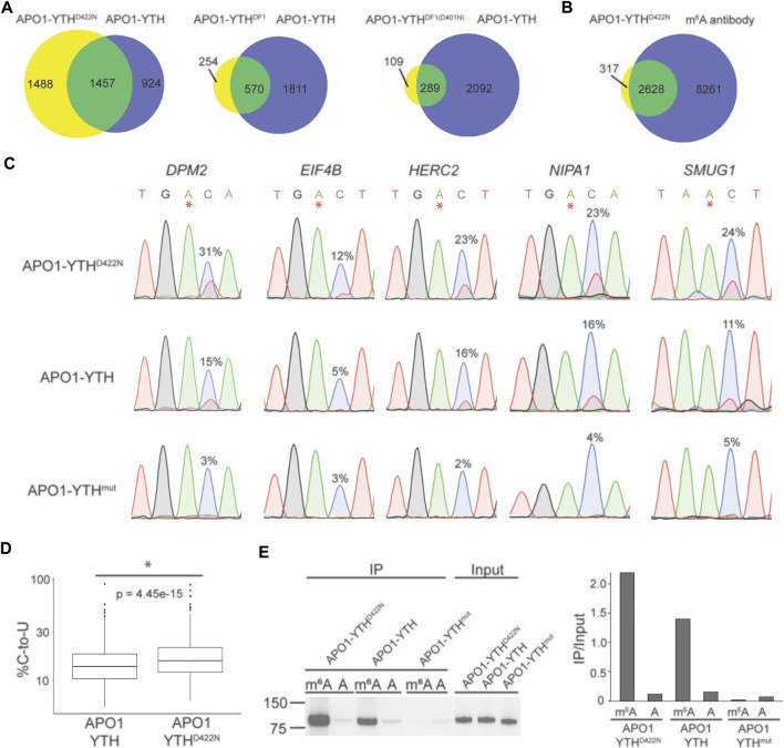
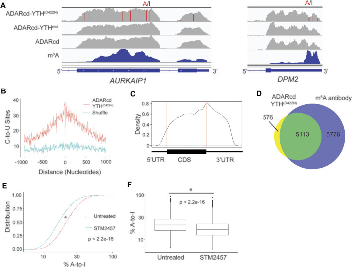
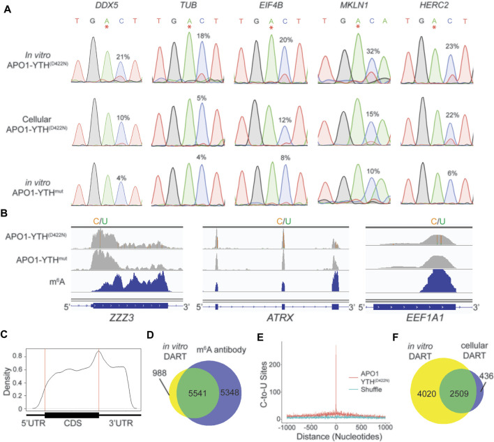
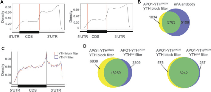
N 6-methyladenosine (m6A) is a critical regulator of gene expression and cellular function. Much of our knowledge of m6A has been enabled by the identification of m6A sites transcriptome-wide. However, global m6A profiling methods require high amounts of input RNA to accurately identify methylated RNAs, making m6A profiling from rare cell types or scarce tissue samples infeasible. To overcome this issue, we previously developed DART-seq, which relies on the expression of a fusion protein consisting of the APOBEC1 cytidine deaminase tethered to the m6A-binding YTH domain. APOBEC1-YTH directs C-to-U mutations adjacent to m6A sites, therefore enabling single nucleotide-resolution m6A mapping. Here, we present an improved version of DART-seq which utilizes a variant of the YTH domain engineered to achieve enhanced m6A recognition. In addition, we develop in vitro DART-seq and show that it performs similarly to cellular DART-seq and can map m6A in any sample of interest using nanogram amounts of total RNA. Altogether, these improvements to the DART-seq approach will enable better m6A detection and will facilitate the mapping of m6A in samples not previously amenable to global m6A profiling.

Keywords: DART-seq; RNA biology; RNA modification; epitranscriptome; m6A.


Conflict of interest statement

KM has filed a patent application for the DART-seq technology through Duke University. The remaining authors declare that the research was conducted in the absence of any commercial or financial relationships that could be construed as a potential conflict of interest.

Figures

FIGURE 1
FIGURE 1
Identification of an improved variant of the DART fusion protein. (A) Comparison of methylated RNAs identified by cellular DART-seq using expression of either APO1-YTHD422N, APO1-YTHDF1, or APO1-YTHDF1(D401N) in HEK293T cells. Venn diagrams compare each DART protein variant to the original APO1-YTH protein. (B) Overlap of methylated RNAs identified by cellular DART-seq using the APO1-YTHD422N protein with those identified by antibody-based methods. (C) Sanger sequencing traces showing C-to-U editing adjacent to m6A sites in cells expressing APO1-YTHD422N, APO1-YTH, and APO1-YTHmut for five mRNAs previously shown to contain m6A: DPM2, EIF4B, HERC2, NIPA1, and SMUG1. m6A sites are indicated by asterisks. C-to-U editing rate (%U) is indicated above the adjacent cytidine. Data are representative of three biological replicates. (D) Box plot showing the global C-to-U editing percentage of all sites common to HEK293T cells expressing APO1-YTHD422N or APO1-YTH. (E) Western blot following RNA pulldown assays using purified DART proteins and bait RNAs. APO1-YTHD422N exhibits improved binding to m6A compared to APO1-YTH.
FIGURE 2
FIGURE 2
ADARcd can be used as an alternative to APO1 to identify methylated RNAs with DART-seq. (A) Genome browser tracks showing two methylated mRNAs, AURKAIP1 and DPM2, in HEK293T cells expressing ADARcd-YTHD422N, ADARcd-YTHmut, or ADARcd alone. A-to-I editing found in at least 10% of the reads are indicated by red/blue coloring. m6A peaks identified by MeRIP (Meyer et al., 2012) is indicated in the bottom blue track. (B) Absolute distance plot showing the distance between A-to-I edit sites identified by ADARcd-YTHD422N and m6A sites identified by miCLIP (Linder et al., 2015). (C) Metagene plot showing the distribution of A-to-I edit sites found in cells expressing ADARcd-YTHD422N. (D) Venn diagram showing overlap between methylated RNAs identified by cellular DART-seq from HEK293T cells expressing ADARcd-YTHD422N and methylated RNAs identified by antibody-based profiling (Meyer et al., 2012; Schwartz et al., 2014; Lichinchi et al., 2016). (E) Cumulative distribution plot (left) of %A-to-I for sites identified by ADARcd-YTHD422N in untreated and STM2457 treated HEK293T cells. (F) Box plot showing the global A-to-I editing percentage of all sites common to both untreated and STEM2457 treated HEK293T cells expressing ADARcd-YTHD422N. A Wilcoxon Rank-Sum test was conducted to access statistical significance.
FIGURE 3
FIGURE 3
In vitro DART-seq identifies m6A transcriptome-wide. (A) Comparison of C-to-U editing rates in methylated mRNAs obtained by in vitro DART-seq and cellular DART-seq. Sanger sequencing traces show C-to-U editing adjacent to m6A sites in a panel of five methylated mRNAs: DDX5, TUB, EIF4B, MKLN1, and HERC2. m6A sites are indicated by asterisks. C-to-U editing rate (%U) is indicated above the adjacent cytidine. Data Representative of three biological replicates. (B) Genome browser tracks of in vitro DART-seq data showing C-to-U editing in three representative mRNAs: ZZZ3, ATRX, and EEF1A1. C-to-U editing found in at least 10% of the reads is indicated by green/yellow coloring. m6A peaks identified by MeRIP (Meyer et al., 2012) is indicated in the bottom blue track. (C) Metagene analysis of m6A sites identified by in vitro DART-seq using the APO1-YTHD422N protein. (D) Venn diagram showing the overlap between methylated RNAs identified by in vitro DART-seq filtered against the APO1-YTHmut negative control and methylated RNAs identified by antibody-based methods (Meyer et al., 2012; Schwartz et al., 2014; Lichinchi et al., 2016). (E) Absolute distance plot showing the distance of C-to-U editing sites identified by in vitro DART-seq relative to m6A sites identified by miCLIP (Linder et al., 2015). m6A sites are centered at position 0. (F) Venn diagram showing the overlap between methylated RNAs identified by in vitro DART-seq compared to methylated RNAs found by cellular DART-seq.
FIGURE 4
FIGURE 4
Blocking with the YTH domain minimizes false positives with in vitro DART-seq. (A) Metagene analysis of C-to-U editing sites in mRNAs identified with in vitro DART-seq using the APO1-YTHD422N protein after pre-incubation with the YTH domain (left) or the APO1-YTHmut protein (right). (B) Venn diagram showing the overlap between methylated RNAs identified by in vitro DART-seq with APO1-YTHD422N filtered by YTH blocking and methylated RNAs identified by antibody-based methods (Meyer et al., 2012; Schwartz et al., 2014; Lichinchi et al., 2016). (C) Metagene analysis showing the distribution of C-to-U editing sites in mRNAs after filtering of in vitro DART-seq data against by YTH blocking (blue) or by APO1-YTHmut (red). (D) Venn diagram of C-to-U edit sites induced by in vitro DART-seq with APO1-YTHD422N, filtered by use of either YTH blocking or APO1-YTHmut (left). Venn diagram of methylated RNA identified by in vitro DART-seq with APO1-YTHD422N, filtered by use of either YTH blocking or APO1-YTHmut (right).

References

    1. Chen X.-Y., Zhang J., Zhu J.-S. (2019). The Role of m6A RNA Methylation in Human Cancer. Mol. Cancer 18, 103. 10.1186/s12943-019-1033-z - DOI - PMC - PubMed
    1. Dominissini D., Moshitch-Moshkovitz S., Schwartz S., Salmon-Divon M., Ungar L., Osenberg S., et al. (2012). Topology of the Human and Mouse m6A RNA Methylomes Revealed by m6A-Seq. Nature 485, 201–206. 10.1038/nature11112 - DOI - PubMed
    1. Eggington J. M., Greene T., Bass B. L. (2011). Predicting Sites of ADAR Editing in Double-Stranded RNA. Nat. Commun. 2, 319. 10.1038/ncomms1324 - DOI - PMC - PubMed
    1. Gu C., Shi X., Dai C., Shen F., Rocco G., Chen J., et al. (2020). RNA m6A Modification in Cancers: Molecular Mechanisms and Potential Clinical Applications. The Innovation 1, 100066. 10.1016/j.xinn.2020.100066 - DOI - PMC - PubMed
    1. Hafner M., Landthaler M., Burger L., Khorshid M., Hausser J., Berninger P., et al. (2010). Transcriptome-wide Identification of RNA-Binding Protein and microRNA Target Sites by PAR-CLIP. Cell 141, 129–141. 10.1016/j.cell.2010.03.009 - DOI - PMC - PubMed