. 2022 Apr 28:10:868352.
doi: 10.3389/fcell.2022.868352. eCollection 2022.

Refined Interpretation of the Pistillate Flower in Ceratophyllum Sheds Fresh Light on Gynoecium Evolution in Angiosperms

Affiliations

Refined Interpretation of the Pistillate Flower in Ceratophyllum Sheds Fresh Light on Gynoecium Evolution in Angiosperms

Dmitry D Sokoloff et al. Front Cell Dev Biol. .

Abstract

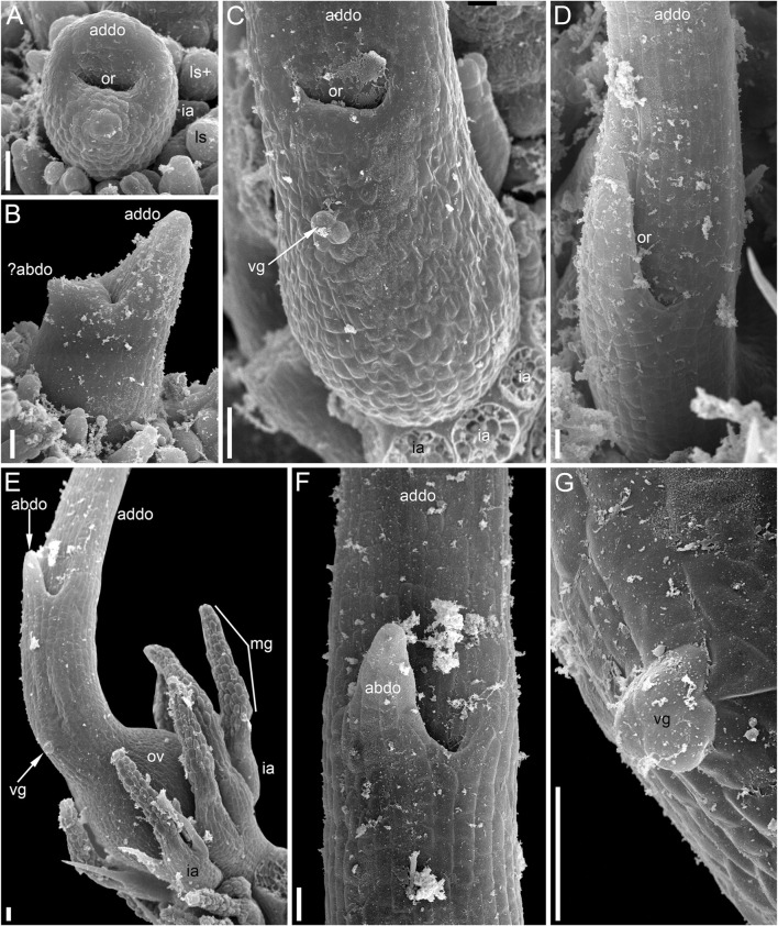
Molecular phylogenetic analyses have revealed a superclade of mesangiosperms with five extant lineages: monocots, eudicots, magnoliids, Ceratophyllum and Chloranthaceae. Both Ceratophyllum and Chloranthaceae are ancient lineages with a long fossil record; their precise placement within mesangiosperms is uncertain. Morphological studies have suggested that they form a clade together with some Cretaceous fossils, including Canrightia, Montsechia and Pseudoasterophyllites. Apart from Canrightia, members of this clade share unilocular gynoecia commonly interpreted as monomerous with ascidiate carpels. Alternatively, the gynoecium of Ceratophyllum has also been interpreted as syncarpous with a single fertile carpel (pseudomonomerous). We investigate patterns of morphological, anatomical and developmental variation in gynoecia of three Ceratophyllum species to explore the controversial interpretation of its gynoecium as either monomerous or pseudomonomerous. We use an angiosperm-wide morphological data set and contrasting tree topologies to estimate the ancestral gynoecium type in both Ceratophyllum and mesangiosperms. Gynoecia of all three Ceratophyllum species possess a small (sometimes vestigial) glandular appendage on the abaxial side and an occasionally bifurcating apex. The ovary is usually unilocular with two procambium strands, but sometimes bilocular and/or with three strands in C. demersum. None of the possible phylogenetic placements strongly suggest apocarpy in the stem lineage of Ceratophyllum. Rescoring Ceratophyllum as having two united carpels affects broader-scale reconstructions of the ancestral gynoecium in mesangiosperms. Our interpretation of the glandular appendage as a tepal or staminode homologue makes the Ceratophyllum ovary inferior, thus resembling (semi)inferior ovaries of most Chloranthaceae and potentially related fossils Canrightia and Zlatkocarpus. The entire structure of the flower of Ceratophyllum suggests strong reduction following a long and complex evolutionary history. The widely accepted notion that apocarpy is ancestral in mesangiosperms (and angiosperms) lacks robust support, regardless of which modes of carpel fusion are considered. Our study highlights the crucial importance of incorporating fossils into large-scale analyses to understand character evolution.

Keywords: Ceratophyllales; Chloranthales; apocarpy; congenital fusion; fossils; mesangiosperms; pseudomonomery; syncarpy.


Conflict of interest statement

The authors declare that the research was conducted in the absence of any commercial or financial relationships that could be construed as a potential conflict of interest.

Figures

FIGURE 1
FIGURE 1
Living plants of Ceratophyllum spp. in Khoper State Nature Reserve, Russia (photos: E.V. Pechenyuk). (A) Ceratophyllum submersum, portion of plant with two fruits, both sessile and smooth. (B) Terrestrial plant of C. tanaiticum with a stalked, spinose fruit (arrowhead). (C) Aquatic plant of C. tanaiticum with three fruits at successive developmental stages (arrowheads). Fruit spines appear late in development. (D) Postanthetic pistillate flower of C. tanaiticum.
FIGURE 2
FIGURE 2
Ambiguities among various phylogenetic trees of angiosperms considered in the present study. (A) Simplified diagrams of three major hypotheses of relationships among clades of mesangiosperms with the position of Ceratophyllum omitted. See text for details on naming these topologies. (B) simplified ‘main’ tree topology (JM tree) explored in an important recent study of character evolution in angiosperms (Sauquet et al., 2017). Dotted lines indicate contrasting positions for Ceratophyllum and three other problematic taxa. Abbreviations after taxon names indicate types of fusion between carpels in a rough sequence of their frequency/closeness to the root in each terminal group: Fr, carpels free; Cf, congenital fusion: Pf, postgenital fusion: C/Pf, both types of fusion in the same gynoecium: ? gynoecium monomerous or reportedly monomerous and thus the fusion character is apparently not applicable.
FIGURE 3
FIGURE 3
Pistillate flowers of Ceratophyllum demersum (SEM). (A) Very young flower with involucral appendages of the reproductive unit and any gynoecium appendages still inconspicuous. Adaxial side up. Small organs near the flower (asterisks) are leafy segments of a vegetative branch situated below the flower. (B,C) Young flowers, adaxial side up. The glandular gynoecial appendage is already well-developed, but distal outgrowth(s) are still very short. (B) Gynoecium developing a bilobed distal outgrowth or two distal outgrowths, a wider one adaxial and a narrower one left transversal; the gland is abaxial. (C) The gland and a young distal outgrowth are at the opposite radii, which are oblique relative to the median plane of the flower. Note that the flower is extra-axillary, located between two vegetative leafy segments of a node. (D) Abaxial view of a flower with two equal distal gynoecial outgrowths. The flower is attached to a stem node between two leafy segments that are excised. Sections of this flower are shown in Figure 7. (E) Abaxial view of flower with involucral appendages removed. The two distal appendages are of unequal size. (F) Side view of flower with unequally bilobed distal appendage. Scale bars = 30 μm in (A–C), 300 μm in (D–F).
FIGURE 4
FIGURE 4
Pistillate flowers of Ceratophyllum demersum (SEM). (A) Abaxial view of flower with single, unlobed distal gynoecial outgrowth. Some involucral appendages are removed to show the ovary. The glandular appendage of the gynoecium is abscised, but its scar is visible. (B) Detail of (A) showing the gynoecial orifice and a scar of the ascised glandular appendage. (C) Dissected flower with unequally bilobed distal gynoecial outgrowth. The ovary is unilocular, with a single pendent ovule. (D) Abaxial view of flower with bilobed distal gynoecial outgrowth. (E) Detail of (D) showing the two lobes of the distal outgrowth. (F) Detail of (D) showing glandular gynoecial appendage. Scale bars = 300 μm in (A,C,D), 100 μm in (B,E,F).
FIGURE 5
FIGURE 5
Ascending series of anatomical sections of pistillate flower of Ceratophyllum demersum with single, abaxial glandular gynoecial appendage (LM). The flower is similar to the one illustrated in Figure 4A. (A) Below the ovary locule, note two vascular bundles. (B) Close to the bottom of the ovule. (C) Middle part of the ovule. (D) Level of the ovule attachment. (E–H) Sections between the ovary locule and the gynoecial orifice (arrowheads, stylar canal). (F,G) Sections showing the glandular appendage. (I,J) Distal gynoecial outgrowth. Scale bars = 100 μm in (A–J).
FIGURE 6
FIGURE 6
Anatomy of pistillate flowers of Ceratophyllum demersum, ascending series of transverse anatomical sections (LM). (A–E) Flower with single, abaxial glandular gynoecial appendage and single horizontally directed ovule (involucral appendages not shown). Ovule insertion is nonmedian as evidenced by its position relative to the two procambial strands and the glandular appendage. (A) Below the ovary locule. (B–D) Ovary locule and ovule. (D) Detail of section just above (C); note the clearly visible boundary between the nucellus and the integument. (E) Level of glandular appendage. (F–Q) Flower with two glandular gynoecial appendages and a single horizontally directed ovule. (F) Level of attachment of involucral appendages. (G) Trilobed bundle in the gynoecium stalk. (H) Three distinct bundles in the gynoecium stalk. (I–K) The ovule is attached along almost the entire length of the ovary locule. (L–N) Sections between the ovary locule and the gynoecial orifice (arrowheads, stylar canal). (M, N) Sections showing the two glandular appendages. Each of these two sections shows only one glandular appendage, but these are two different appendages. Asterisk in (M) indicates the radius of the appendage visible in (N). (O) Gynoecium orifice. (P, Q) Distal outgrowths of the gynoecium. Scale bars = 100 μm in (A–Q).
FIGURE 7
FIGURE 7
Ascending series of transverse anatomical sections of pistillate flower of Ceratophyllum demersum with two glandular gynoecial appendages and basally bilocular gynoecium (LM). A general view of this flower is shown in Figure 3D. The flower has been serially sectioned after taking SEM images. (A) Level of attachment of involucral appendages. (B,C) Bilocular region of the gynoecium. Each locule has an ovule. (D–F) Unilocular region of the gynoecium with the third ovule. Arrowheads in D,E show radii of attachment of the two long distal outgrowths (see Figure 3D). Asterisks in D,E show radii of the two glandular appendages that are visible in (F). (F) Level of attachment of the two glandular appendages. Scale bars = 300 μm in (A,B,F), 100 μm in (C–E).
FIGURE 8
FIGURE 8
Pistillate flower development in Ceratophyllum tanaiticum (SEM). (A) Young apparently pistillate reproductive unit inserted between two leafy segments of a node. The bilobed leafy segment of the subsequent node is visible. Involucral appendages of the reproductive unit are not yet initiated. (B) Early stage of gynoecium development. The adaxial side of the gynoecium is indicated by asterisk. Involucral appendages are initiated. (C) Side view of a gynoecium with adaxial distal outgrowth initiated. Involucral appendages are still very short. (D) Pistillate flower (gynoecium) with vestigial abaxial glandular appendage. (E) Pistillate flower with well-developed abaxial glandular appendage. (F–I) Successive developmental stages of pistillate flowers lacking glandular appendage. Scale bars = 30 μm in (A–I).
FIGURE 9
FIGURE 9
Pistillate flower and fruit morphology of Ceratophyllum tanaiticum (SEM). (A) Abaxial view of flower with long adaxial and short abaxial distal gynoecial outgrowth. (B) Detail of (A) showing abaxial outgrowth. The gynoecium orifice is hidden by the abaxial outgrowth. (C) Side view of flower with a single (adaxial) distal gynoecial outgrowth; two involucral appendages removed, but their scars are visible in the bottom-right part of the image. (D) Detail of abaxial view of gynoecium similar to that in (C), but with a vestigial gland below the orifice. (E) Young fruit; note the ab initio absence of involucral appendages on one side of the fruit base (asterisk). Scale bars = 100 μm in (A–E).
FIGURE 10
FIGURE 10
Pistillate flower anatomy of Ceratophyllum tanaiticum (A–L) and C. submersum (M–Q), transverse sections, LM. Adaxial side up in all images. (A,B) Midsection of anthetic ovary (A) and detail of its adaxial side showing poorly recognizable procambium strand (B). (C–L) Ascending serial sections of postanthetic flower similar to that in Figure 9E; involucral appendages removed before sectioning. (C) Stalk of pistillate reproductive unit. (D) Below the ovary locule and above the insertion of involucral appendages. (E–H) Ovary locule. Note that note that the epidermis is biseriate in (D–H). (E) Bottom of the ovule showing globular proembryo (red cells in the centre). (F) Detail of adaxial part of the ovary wall from (E) showing vascular bundle. (G) Middle part of the ovule with a large endosperm cell in the centre. (H) Level of ovule attachment. (I–L) Above the ovary locule (arrowheads, stylar canal). (J,K) Level of vestigial gland. (M–Q) Ascending serial sections of anthetic flower above the ovary locule (arrowheads, stylar canal). The flower is similar to that in Figures 11D, 12D. (M) Level of vestigial gland (only two gland cells visible). (N) Above the gland. (O) Asymmetric gynoecium orifice. (P) Asymmetric proximal part of adaxial distal outgrowth of gynoecium. (Q) Symmetrical distal part of the outgrowth. Scale bars = 50 μm in (A–Q).
FIGURE 11
FIGURE 11
Reproductive structures of Ceratophyllum submersum (SEM). (A,B) Two views of pistillate reproductive unit. (C,D) Two views of shoot apex with anthetic pistillate reproductive unit and pre-anthetic staminate reproductive unit located a node below. There is also a vegetative branch located right below the staminate reproductive unit in yet another node. The staminate reproductive unit has numerous stamens and a peripheral whorl of involucral appendages. Scale bars = 300 μm in (A–D).
FIGURE 12
FIGURE 12
Pistillate reproductive units of Ceratophyllum submersum (SEM). (A–C) Successive developmental stages. (A,C) Adaxial side up; (B) adaxial side right. Each reproductive unit has a pistillate flower surrounded by a whorl of involucral appendages. The involucral appendages are removed in (C), but their scars are visible. In (A,B), the involucral appendages and leafy segments of stem nodes are yet small and densely crowded, so that it is difficult to distinguish them from each other without further dissection in (B). (D) Detail of anthetic gynoecium seen from the abaxial side; the gynoecial orifice is asymmetrically spaced. (E–G) Anthetic gynoecium with two distal outgrowths of unequal size. (E) General view from lateral side. (F) View from abaxial side; the gynoecial orifice is behind the short abaxial outgrowth. (G) Detail of (E) with vestigial gland. Scale bars = 300 μm in (A–G).
FIGURE 13
FIGURE 13
Summary of ancestral state reconstructions with parsimony of fusion between carpels in mesangiosperms on different tree topologies and with two types of coding for the state ‘syncarpy’ (any fusion = carpel fusion may be congenital or postgenital; congenital fusion = carpel fusion concerns only congenital fusion) in angiosperms. For more detail, see Supplementary File 2. Each pie chart shows the ratio between inferred ancestral mesangiosperm conditions among 32 tree topologies that differ in placement of Ceratophyllum, Nuphar, Euptelea and Tofieldiaceae. Each diagram shows what pattern of relationships between monocots, eudicots, magnoliids and Chloranthaceae was fixed in each set of 32 tree topologies.
FIGURE 14
FIGURE 14
Diagrams of pistillate or bisexual flower morphology in Ceratophyllum and some of its possible phylogenetic relatives. In each case, longitudinal and cross-sections are provided, with their positions reciprocally indicated by arrows. (A) One of the rare conditions observed here in Ceratophyllum demersum (Figures 3D, 7). (B) The most common type of pistillate flower in Ceratophyllum. (C) Cretaceous fossil Canrightia (modified from Friis and Pedersen, 2011). (D,E) Extant members of Chloranthaceae. (D) Hedyosmum (modified from Endress, 1971). (E) Chloranthus (modified from Endress, 1987b).

References

    1. Aboy H. E. (1936). A Study of the Anatomy and Morphology of Ceratophyllum demersum . MS Thesis. Ithaca: Cornell University.
    1. Albert V. A., Renner T. (2020). Aquatic Angiosperm Ambiguities Answered. Nat. Plants 6, 181–183. 10.1038/s41477-020-0607-5 - DOI - PubMed
    1. Antonov A. S., Troitsky A. V., Samigullin T. K., Bobrova V. K., Valiejo-Roman K. M., Martin W. (2000). “Early Events in the Evolution of Angiosperms Deduced from Cp rDNA ITS 2–4 Sequence Comparisons,” in Proceedings of the International Symposium on the Family Magnoliaceae. Editors Liu Y.-H., Fan H.-M., Chen Z.-Y., Wu Q.-G., Zeng Q.-W. (Beijing: Science Press; ), 210–214.
    1. Arber A. (1920). Water Plants: A Study of Aquatic Angiosperms. Cambridge: Cambridge University Press.
    1. Armbruster W. S., Debevec E. M., Willson M. F. (2002). Evolution of Syncarpy in Angiosperms: Theoretical and Phylogenetic Analyses of the Effects of Carpel Fusion on Offspring Quantity and Quality. J. Evol. Biol. 15, 657–672. 10.1046/j.1420-9101.2002.00414.x - DOI