. 2022 Apr 28:10:866491.
doi: 10.3389/fcell.2022.866491. eCollection 2022.

Dissection of the microRNA Network Regulating Hedgehog Signaling in Drosophila

Affiliations

Dissection of the microRNA Network Regulating Hedgehog Signaling in Drosophila

Tao He et al. Front Cell Dev Biol. .

Abstract

The evolutionarily conserved Hedgehog (Hh) signaling plays a critical role in embryogenesis and adult tissue homeostasis. Aberrant Hh signaling often leads to various forms of developmental anomalies and cancer. Since altered microRNA (miRNA) expression is associated with developmental defects and tumorigenesis, it is not surprising that several miRNAs have been found to regulate Hh signaling. However, these miRNAs are mainly identified through small-scale in vivo screening or in vitro assays. As miRNAs preferentially reduce target gene expression via the 3' untranslated region, we analyzed the effect of reduced expression of core components of the Hh signaling cascade on downstream signaling activity, and generated a transgenic Drosophila toolbox of in vivo miRNA sensors for core components of Hh signaling, including hh, patched (ptc), smoothened (smo), costal 2 (cos2), fused (fu), Suppressor of fused (Su(fu)), and cubitus interruptus (ci). With these tools in hand, we performed a genome-wide in vivo miRNA overexpression screen in the developing Drosophila wing imaginal disc. Of the twelve miRNAs identified, seven were not previously reported in the in vivo Hh regulatory network. Moreover, these miRNAs may act as general regulators of Hh signaling, as their overexpression disrupts Hh signaling-mediated cyst stem cell maintenance during spermatogenesis. To identify direct targets of these newly discovered miRNAs, we used the miRNA sensor toolbox to show that miR-10 and miR-958 directly target fu and smo, respectively, while the other five miRNAs act through yet-to-be-identified targets other than the seven core components of Hh signaling described above. Importantly, through loss-of-function analysis, we found that endogenous miR-10 and miR-958 target fu and smo, respectively, whereas deletion of the other five miRNAs leads to altered expression of Hh signaling components, suggesting that these seven newly discovered miRNAs regulate Hh signaling in vivo. Given the powerful effects of these miRNAs on Hh signaling, we believe that identifying their bona fide targets of the other five miRNAs will help reveal important new players in the Hh regulatory network.

Keywords: Drosophila wing; hedgehog signaling; in vivo miRNA sensor toolbox; miR-10; miR-958.


Conflict of interest statement

The authors declare that the research was conducted in the absence of any commercial or financial relationships that could be construed as a potential conflict of interest.

Figures

FIGURE 1
FIGURE 1
The developing Drosophila wing is an ideal model system for studying Hh signaling. (A) Shown are a wild-type third instar larval wing imaginal disc and an adult wing blade. Expression of Hh-GFP (green), Col (blue), and Ci (red) is shown. Hh protein is produced in the posterior compartment and diffuses into the anterior compartment to stabilize CiFL protein and activate col expression. The wing pouch (outlined by a yellow dashed circle) eventually metamorphose into the adult wing blade. The area between the longitudinal L3 and L4 veins of the adult wing is predetermined in the area between the two blue dashed lines in the larval wing disc, where Col is expressed. (B) A diagram showing ap-Gal4 expression area. Cells in the ventral compartment were used as an internal control. Scale bar, 100 μm.
FIGURE 2
FIGURE 2
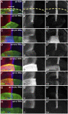
Knockdown of core Hh pathway components alters CiFL levels and Col expression. CiFL levels and Col expression were visualized by immunostaining in third instar larval wing discs of the indicated genotypes. GFP marks dsRNA-expressing cells in the dorsal compartment. (A–B’’,D–D’’,H–H’’) ap-Gal4-driven RNAi of the positive regulators of Hh signaling (i.e. hh, smo, and ci) resulted in significantly reduced levels of CiFL (B’,D’) and loss of Col expression (B”,D”,H”) in dorsal compartment cells. (C–C’’,E–E’’) Conversely, knockdown of ptc caused expansion of the labile CiA region (C’) and expansion and increase in Col expression (C”). Significant increases in CiFL levels (E’) and expanded Col expression (E”) were observed when cos2 was knocked down. (F–G’’) Decreased expression of fu resulted in elevated levels of CiFL (F’) and downregulation of Col (F”), whereas knockdown of Su(fu) led to significantly decreased levels of CiFL (G’) but had no effects on Col expression (G”). The dorsal-ventral boundary of the wing disc is marked with a yellow dashed line. Scale bar, 50 μm.
FIGURE 3
FIGURE 3
Overexpression of previously known Hh signaling-regulating miRNAs alters CiFL levels and kn-lacZ reporter activity. CiFL levels and kn-lacZ activity were monitored by immunostaining in third instar larval wing discs of the indicated genotypes. (A–B’’,D–D’’) ap-Gal4-driven overexpression of miR-7 or miR-14 resulted in decreased levels of CiFL (B’,D’) and loss of kn-lacZ expression (B”,D”) in the dorsal compartment. (C–C’’,F–F’’) Increased expression of miR-12 or miR-960 led to markedly elevated levels of CiFL (C’,F’), but little change in kn-lacZ activity (C”,F”). (E–E’’) A mild increase in CiFL levels (E’) and a weak downregulation of kn-lacZ activity (E”) were observed in GFP-positive cells overexpressing miR-932. Scale bar, 50 μm.
FIGURE 4
FIGURE 4
miR-10 negatively regulates Hh signaling by targeting fu. (A–B”) Overexpression of miR-10 by ap-Gal4 in the dorsal compartment of the wing disc resulted in an obvious increase in CiFL levels (B’) and a significant decrease in kn-lacZ activity (B”). Smo protein levels were significantly reduced in both anterior and posterior compartment cells (D’), and the expression region of Col was narrowed (D”). (E) Schematic showing the composition of a miRNA sensor containing the αTub84B promoter, the egfp coding sequence, and the 3′ UTR of individual core Hh pathway genes. (F) Two predicted miR-10 binding sites are present in the 3′ UTR of fu. Mutated binding sites are shown in red. (G–H”) GFP expression of gfp: 3′UTR fu and gfp: 3′UTRmut fu sensor lines in dpp-Gal4-driven miR-10 overexpression wing discs are shown. Increased miR-10 expression resulted in significantly reduced GFP expression (G′) and elevated CiFL levels (G”) along the A-P boundary in gfp: 3′UTR fu sensor wing discs. In contrast, overexpressed miR-10 did not affect GFP expression in gfp: 3′UTRmut fu sensor wing discs (H’), although CiFL was upregulated (H”). Scale bar, 50 μm.
FIGURE 5
FIGURE 5
miR-133, miR-375, and miR-927 are negative regulators of Hh signaling. Smo, CiFL, and Col levels and kn-lacZ activity were visualized by immunostaining in wing discs overexpressing miR-133 (A–B”), miR-375 (C–D”), or miR-927 (E–F”). GFP marks the expression region of ap-Gal4. Although overexpression of miR-133, miR-375, and miR-927 all resulted in significant decrease in CiFL levels (A’,C’,E’), kn-lacZ activity (A”,C”,E”), and Col expression (B”,D”,F”), different effects on Smo expression were observed. Increased miR-133 expression led to a marked increase in Smo protein levels in the posterior compartment, whereas the increase in the anterior compartment was very mild (B’). It is difficult to determine the effect of miR-375 overexpression on Smo expression because the formation of the dorsal compartment of the wing disc was disrupted (D’). Overexpression of miR-927 resulted in mildly elevated Smo levels, especially in the anterior compartment (F’). Scale bar, 50 μm.
FIGURE 6
FIGURE 6
miR-958 targets smo to regulate Hh signaling (A–B”) ap-Gal4-driven overexpression of miR-958 in the dorsal compartment resulted in a marked reduction in CiFL levels (A’), abrogation of Smo expression (B’) and complete loss of kn-lacZ activity (A”) and Col expression (B”). (C) The predicted miR-958 binding site is present in the 3′ UTR of smo, and the mutated binding site is shown in red (D–E”) GFP expression in wing discs of gfp: 3′UTR smo and gfp: 3′UTRmut smo sensor lines is shown when miR-958 was overexpressed. ptc-Gal4-driven overexpression of miR-958 resulted in obviously reduced GFP (D’) and Smo expression (D”) along the A-P boundary in the gfp:3’UTR fu sensor wing disc. In contrast, increased miR-958 expression had no effects on GFP expression in the gfp: 3′UTRmut fu sensor wing disc (E’), although Smo protein levels were significantly reduced (E”). Scale bar, 50 μm.
FIGURE 7
FIGURE 7
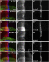
Effects of core Hh pathway gene knockdown on Smo protein levels. Smo expression in wing discs of the indicated genotypes is shown. (A–B’’) ap-Gal4-driven hh RNAi caused a slight decrease in Smo protein levels in both anterior and posterior compartment cells (B’). (C,C’,H,H’) Knockdown of either ptc or ci resulted in a similar increase in Smo protein levels in the cells of the anterior compartment adjacent to the A-P boundary (C’,H’). (D,D’) Knockdown of smo resulted in loss of Smo expression, as expected (D’). (E–G’) Reducing cos2 or Su(fu) expression had no apparent effects on Smo expression (E’,G’). In addition, knockdown of fu specifically reduced Smo protein levels in the posterior compartment (F’). Scale bar, 50 μm.
FIGURE 8
FIGURE 8
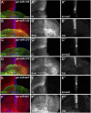
miR-190 and miR-964 act as negative regulators of Hh signaling. CiFL levels and expression of kn-lacZ, Smo, and Col in wing discs overexpressing either miR-190 (A–B”) or miR-964 (C–D”) were monitored by immunostaining. Overexpression of miR-190 or miR-964 led to obviously reduced levels of CiFL (A’,C’) and almost unchanged kn-lacZ activity (A”,C”). Increased miR-190 expression induced a significant increase in Smo protein levels in both anterior and posterior compartment cells (B’) and abrogation of Col expression (B”), whereas overexpressing miR-964 did not affect the expression of Smo and Col (D’,D”). Scale bar, 50 μm.
FIGURE 9
FIGURE 9
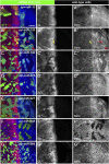
Newly discovered miRNAs regulate Hh signaling in a cell-autonomous manner. All miRNAs were overexpressed in GFP-marked clones in wing discs induced by the FLIPout technique. CiFL levels and Smo expression were visualized by immunostaining. (A–A’’) In miR-10 overexpressed cells, CiFL levels were slightly increased (A’), while Smo protein levels were reduced in clones located in the anterior compartment (A”). (B–B’’) CiFL levels were only decreased in miR-133-expressing clones adjacent to the A-P boundary (yellow arrow in B’), and Smo expression was increased in clones located in the posterior compartment (red arrows in B”), but not anterior compartment (yellow arrows in B”). (C–E’’,G–G’’) Overexpression of miR-190, miR-375, miR-927, and miR-964 all resulted in a significant decrease in CiFL levels (C’,D’,E’,G’). However, they had different effects on Smo expression. Overexpression of miR-190 led to increased Smo levels in anterior and posterior clones (C”), whereas increased expression of miR-375 resulted in decreased Smo levels, especially in anterior clones (D”). Overexpression of miR-927 or miR-964 had no apparent effects on Smo expression (E”,G”). (F–F’’) Increased expression of miR-958 led to a mild decrease in CiFL levels in clones located in the anterior compartment, whereas Smo expression was completely lost in clones located in both anterior and posterior compartments (F”). Scale bar, 50 μm.
FIGURE 10
FIGURE 10
Newly discovered miRNAs, except miR-964, are involved in Hh signaling-controlled CySC maintenance during spermatogenesis. (A–H”) All newly discovered miRNAs were specifically overexpressed by tj-Gal4 in early cyst cells. CiFL levels and expression of Smo and Tj were monitored by immunostaining. Overexpression of miR-10, miR-133, miR-190, miR-375, miR-927, and miR-958, but not miR-964, obviously reduced the number of Tj-positive cells in the testis. (I) Shown is statistical analysis of the number of Tj-positive cells per testis. Data are presented as mean ± S.D. (n tj-Gal4 = 13, n tj>miR−10 = 14, n tj>miR−133 = 12, n tj>miR−190 = 14, n tj>miR−375 = 19, n tj>miR−927 = 15, n tj>miR−958 = 13, n tj>miR−964 = 15). One-way ANOVA followed by Dunnett’s tests was used. NS, not significant. *p < 0.05. **p < 0.01. ***p < 0.001. Scale bar, 25 μm.
FIGURE 11
FIGURE 11
Loss-of-function analysis of the seven newly discovered miRNAs. (A) Quantification of mRNA expression of hh, ptc, smo, cos2, fu, Su(fu), ci, ihog, boi, dally, dlp, col, and dpp in seven miRNA knockout mutant larvae by qPCR. Bar plots represent relative mRNA levels of indicated genotypes (n = 3); error bars represent standard deviation (S.D.). (B–E) The GFP expression of the gfp: 3’UTR fu sensor was significantly increased in miR-10 KO homozygotes (C) compared with miR-10 KO heterozygotes (B). The same goes for the gfp:3’UTR smo sensor in miR-958 KO homozygotes (D,E). (F–L) Adult wings of the indicated genotypes are shown. The distance between L3-L4 veins (green line in F), the distance between L2-L5 veins (blue line in F), and the size of adult wings (yellow area in F) were measured, (M,N) Shown is statistical analysis of the ratio of L3-L4 distance to L2-L5 distance (M), and wing size (N) for the indicated genotypes. Data are presented as mean ± S.D. (n = 20). One-way ANOVA followed by Dunnett’s tests was used. NS, not significant. *p < 0.05. **p < 0.01. ***p < 0.001. Scale bar, (B–E), 50 μm; F-L, 100 μm.
FIGURE 12
FIGURE 12
CiFL and Smo levels and Col expression in loss-of-function mutants of newly discovered miRNAs. CiFL and Smo protein levels and Col expression were visualized by immunostaining in third instar larval wing discs of the indicated genotypes. Loss of miR-10 or miR-927 had no apparent effects on CiFL and Smo levels and Col expression (B–B’’’, F–F’’’). CiFL levels and Col expression were significantly reduced in miR-133 KO mutants (C’,C’’’), while Smo levels remained unchanged (C”). CiFL and Smo levels were unaffected in miR-190 KO clones negatively marked by GFP (D–D’’’). Col expression was significantly decreased in miR-375 KO mutants (E’’’), whereas CiFL and Smo levels were unchanged (E’,E”). Deletion of miR-958 or miR-964 significantly elevated Smo levels in the posterior compartment (G”,H”), but hardly changed CiFL levels and Col expression (G’,G’’’,H’,H’’’). Scale bar, 50 μm.

Similar articles

Cited by

References

    1. Agirre X., Jiménez-Velasco A., San José-Enériz E., Garate L., Bandrés E., Cordeu L., et al. (2008). Down-regulation of Hsa-miR-10a in Chronic Myeloid Leukemia CD34+ Cells Increases USF2-Mediated Cell Growth. Mol. Cancer Res. 6, 1830–1840. 10.1158/1541-7786.mcr-08-0167 - DOI - PubMed
    1. Alcedo J., Zou Y., Noll M. (2000). Posttranscriptional Regulation of Smoothened Is Part of a Self-Correcting Mechanism in the Hedgehog Signaling System. Mol. Cell 6, 457–465. 10.1016/s1097-2765(00)00044-7 - DOI - PubMed
    1. Alexandre C., Jacinto A., Ingham P. W. (1996). Transcriptional Activation of Hedgehog Target Genes in Drosophila Is Mediated Directly by the Cubitus Interruptus Protein, a Member of the GLI Family of Zinc finger DNA-Binding Proteins. Genes Dev. 10, 2003–2013. 10.1101/gad.10.16.2003 - DOI - PubMed
    1. Alves G., Limbourg-Bouchon B., Tricoire H., Brissard-Zahraoui J., Lamour-Isnard C., Busson D. (1998). Modulation of Hedgehog Target Gene Expression by the Fused Serine-Threonine Kinase in Wing Imaginal Discs. Mech. Dev. 78, 17–31. 10.1016/s0925-4773(98)00130-0 - DOI - PubMed
    1. Amoyel M., Sanny J., Burel M., Bach E. A. (2013). Hedgehog Is Required for CySC Self-Renewal but Does Not Contribute to the GSC Niche in the Drosophila Testis. Development 140, 56–65. 10.1242/dev.086413 - DOI - PMC - PubMed

LinkOut - more resources