

Case Series of Percutaneous Mechanical Aspiration of Mitral Valve Endocarditis
- PMID: 35573849
- PMCID: PMC9091537
- DOI: 10.1016/j.jaccas.2022.02.019
Case Series of Percutaneous Mechanical Aspiration of Mitral Valve Endocarditis
Abstract
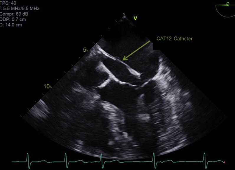
Infective endocarditis of the mitral valve that is refractory to medical therapy requires surgical debridement. However, patients who are high risk for surgery have limited options. We report 3 cases of refractory infective endocarditis involving the mitral valve that were treated with percutaneous mechanical aspiration with an embolic protection system. (Level of Difficulty: Intermediate.).
Keywords: CT, computed tomography; IE, infective endocarditis; IVDU, intravenous drug use; MRSA, methicillin-resistant Staphylococcus aureus; STS, Society of Thoracic Surgery; TEE, transesophageal echocardiogram; drug abuse; endocarditis; mitral valve.
© 2022 The Authors.
Conflict of interest statement
The authors have reported that they have no relationships relevant to the contents of this paper to disclose.
Figures


References
-
- Otto C.M., Nishimura R.A., Bonow R.O., et al. 2020 ACC/AHA guideline for the management of patients with valvular heart disease: executive summary: a report of the American College of Cardiology/American Heart Association Joint Committee on Clinical Practice Guidelines. J Am Coll Cardiol. 2021;77:450–500. - PubMed
-
- Mylonakis E., Calderwood S.B. Infective endocarditis in adults. N Engl J Med. 2001;345:1318. - PubMed
-
- Veve M.P., Akhtar Y., McKeown P.P., et al. Percutaneous mechanical aspiration vs. valve surgery for tricuspid-valve endocarditis in drug users. Ann Thorac Surg. 2021;111:1451–1457. - PubMed
-
- Kang D.H., Kim Y.J., Kim S.H., et al. Early surgery versus conventional treatment for infective endocarditis. N Engl J Med. 2012;366:2466. - PubMed
-
- Akhtar Y.N., Walker W.A., Shakur U., et al. Clinical outcomes of percutaneous debulking of tricuspid valve endocarditis in intravenous drug users. Catheter Cardiovasc Interv. 2021;97:1290–1295. - PubMed
Publication types
LinkOut - more resources
Full Text Sources
