. 2022 May 11;8(1):veac021.
doi: 10.1093/ve/veac021. eCollection 2022.

An antibody-escape estimator for mutations to the SARS-CoV-2 receptor-binding domain

Affiliations

An antibody-escape estimator for mutations to the SARS-CoV-2 receptor-binding domain

Allison J Greaney et al. Virus Evol. .

Abstract

A key goal of severe acute respiratory syndrome coronavirus 2 (SARS-CoV-2) surveillance is to rapidly identify viral variants with mutations that reduce neutralization by polyclonal antibodies elicited by vaccination or infection. Unfortunately, direct experimental characterization of new viral variants lags their sequence-based identification. Here we help address this challenge by aggregating deep mutational scanning data into an 'escape estimator' that estimates the antigenic effects of arbitrary combinations of mutations to the virus's spike receptor-binding domain. The estimator can be used to intuitively visualize how mutations impact polyclonal antibody recognition and score the expected antigenic effect of combinations of mutations. These scores correlate with neutralization assays performed on SARS-CoV-2 variants and emphasize the ominous antigenic properties of the recently described Omicron variant. An interactive version of the estimator is at https://jbloomlab.github.io/SARS2_RBD_Ab_escape_maps/escape-calc/ (last accessed 11 March 2022), and we provide a Python module for batch processing. Currently the calculator uses primarily data for antibodies elicited by Wuhan-Hu-1-like vaccination or infection and so is expected to work best for calculating escape from such immunity for mutations relative to early SARS-CoV-2 strains.

Keywords: Omicron; SARS-CoV-2 variants; antibody escape; deep mutational scanning; epitope.


Figures

Figure 1.
Figure 1.
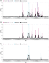
Escape map for a hypothetical polyclonal mix consisting of an equipotent mixture of three monoclonal antibodies targeting distinct epitopes on the SARS-CoV-2 RBD. (A) Experimentally measured escape maps for three antibodies, and the mean of these maps (thick black line). Each point on the x-axis represents a site in the RBD, and the y-axis represents the total measured escape by all mutations at that site scaled so the maximum for each antibody is one. (B) Escape map if the contribution of antibody LY-CoV555 is ablated. (C) Escape map if the contributions of antibodies LY-CoV555 and LY-CoV016 are ablated. An interactive version of this figure is at https://jbloomlab.github.io/SARS2_RBD_Ab_escape_maps/mini-example-escape-calc/ (last accessed 11 March 2022).
Figure 2.
Figure 2.
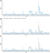
An escape estimator generated by aggregating deep mutational scanning for thirty-three neutralizing antibodies targeting the SARS-CoV-2 RBD. (A) The blue line shows the extent of escape mediated by mutations at each site, as estimated by simply averaging the data for all the individual antibodies. (B) The blue line shows escape map after a mutation to Site 484 (red point) ablates recognition by antibodies strongly targeting that site, while the gray line shows the original escape map in the absence of any mutations. (C) The escape map after mutating Sites 417, 484, and 501 (the three RBD sites mutated in the Beta variant). An interactive version of this figure is at https://jbloomlab.github.io/SARS2_RBD_Ab_escape_maps/escape-calc/ (last accessed 11 March 2022).
Figure 3.
Figure 3.
Correlation of calculated binding scores with experimentally measured fold changes in neutralization for SARS-CoV-2 variants and mutants (smaller values indicate worse neutralization). The data of Lucas et al. (2021) were generated using authentic SARS-CoV-2 and sera from vaccinated individuals who were (top) or were not (bottom) previously infected with SARS-CoV-2. The data of Uriu et al. (2021) and Wang et al. (2021) were generated using pseudovirus against convalescent (top) or vaccine (bottom) sera, with vaccine sera from Pfizer BNT162b2 or Moderna mRNA-1273 vaccines, respectively. The fold changes are geometric means over all subjects in each cohort. Labels at the top of each plot show the Pearson’s R and associated P-value. An interactive version of this figure that allows mousing over points to see details is at https://jbloomlab.github.io/RBD_escape_calculator_paper/neut_studies.html (last accessed 11 March 2022). Supplementary Fig. S1 shows almost identical results are obtained if the binding scores are computed using the mean of mutations at a site rather than the sum.
Figure 4.
Figure 4.
Escape calculations for the Omicron variant. (A) The calculated binding scores for SARS-CoV-2 variants and the artificial polymutant spike (PMS20) generated by Schmidt et al. (2021). Scores of one indicate no mutations affect binding, and scores of zero indicate no antibody binding remains. An interactive version of this plot that allows mousing over points to see details is at https://jbloomlab.github.io/RBD_escape_calculator_paper/variants.html (last accessed March-11-2022). (B) The calculated escape map for the Omicron variant’s RBD (blue) compared to an unmutated RBD (gray), with sites of mutations in the Omicron variant in red. The mutated RBD sites for each variant are in Table 1.

Update of

References

    1. Barnes C. O. et al. (2020) ‘SARS-CoV-2 Neutralizing Antibody Structures Inform Therapeutic Strategies’, Nature, 588: 682–687. - PMC - PubMed
    1. Chen E. C. et al. (2021) ‘Convergent Antibody Responses to the SARS-CoV-2 Spike Protein in Convalescent and Vaccinated Individuals’, Cell Reports, 36: 109604. doi: 10.1016/j.celrep.2021.109604 - DOI - PMC - PubMed
    1. Cobey S. and Hensley S. E. (2017) ‘Immune History and Influenza Virus Susceptibility’, Current Opinion in Virology, 22: 105–111. doi: 10.1016/j.coviro.2016.12.004 - DOI - PMC - PubMed
    1. de Oliveira T. (2021) <https://twitter.com/Tuliodna/status/1463911554538160130>.
    1. Dong J. et al. (2021) ‘Genetic and Structural Basis for SARS-CoV-2 Variant Neutralization by a Two-Antibody Cocktail’, Nature Microbiology, 6: 1233–1244. doi: 10.1038/s41564-021-00972-2 - DOI - PMC - PubMed