Review
. 2022 Apr 28:13:886533.
doi: 10.3389/fendo.2022.886533. eCollection 2022.

The Transcoelomic Ecosystem and Epithelial Ovarian Cancer Dissemination

Affiliations
Review

The Transcoelomic Ecosystem and Epithelial Ovarian Cancer Dissemination

Sabrina J Ritch et al. Front Endocrinol (Lausanne). .

Abstract

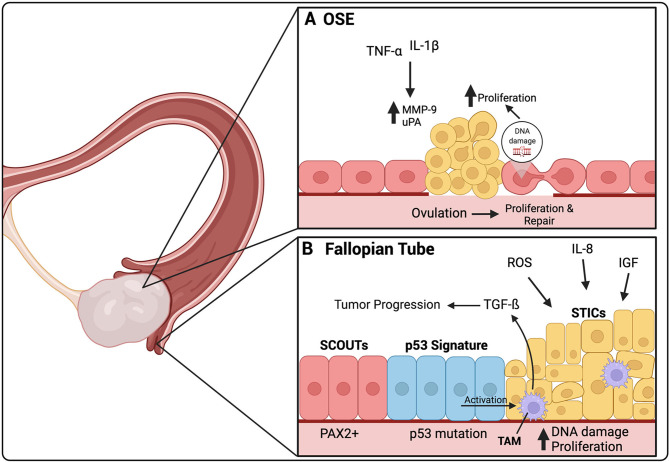
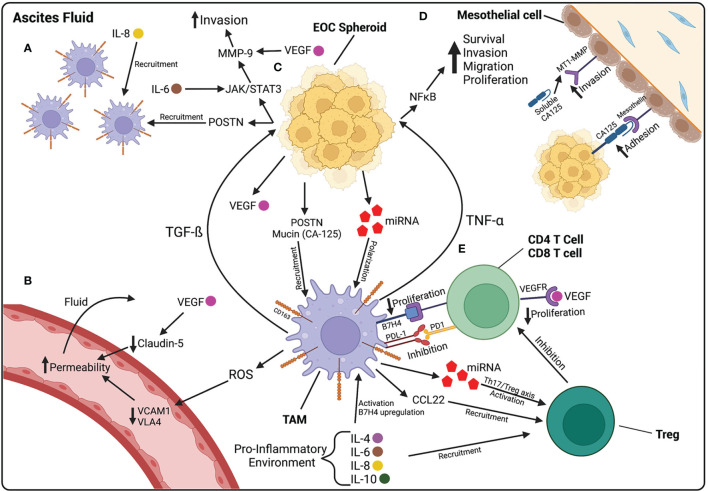
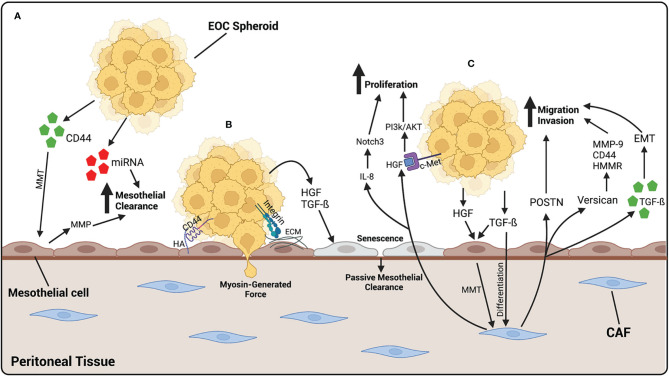
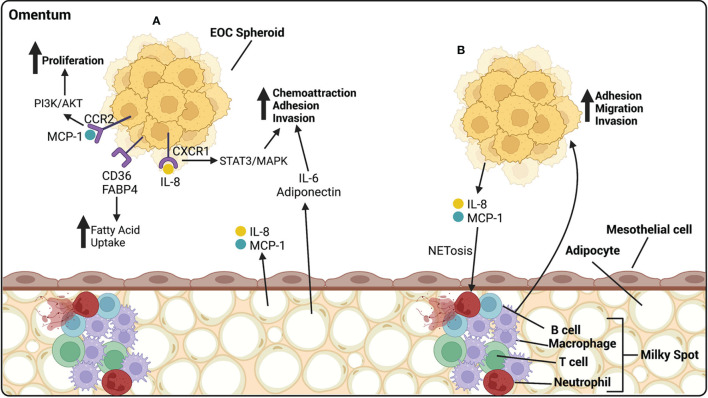
Epithelial ovarian cancer (EOC) is considered the deadliest gynecological disease and is normally diagnosed at late stages, at which point metastasis has already occurred. Throughout disease progression, EOC will encounter various ecosystems and the communication between cancer cells and these microenvironments will promote the survival and dissemination of EOC. The primary tumor is thought to develop within the ovaries or the fallopian tubes, both of which provide a microenvironment with high risk of causing DNA damage and enhanced proliferation. EOC disseminates by direct extension from the primary tumors, as single cells or multicellular aggregates. Under the influence of cellular and non-cellular factors, EOC spheroids use the natural flow of peritoneal fluid to reach distant organs within the peritoneal cavity. These cells can then implant and seed distant organs or tissues, which develop rapidly into secondary tumor nodules. The peritoneal tissue and the omentum are two common sites of EOC metastasis, providing a microenvironment that supports EOC invasion and survival. Current treatment for EOC involves debulking surgery followed by platinum-taxane combination chemotherapy; however, most patients will relapse with a chemoresistant disease with tumors developed within the peritoneum. Therefore, understanding the role of the unique microenvironments that promote EOC transcoelomic dissemination is important in improving patient outcomes from this disease. In this review article, we address the process of ovarian cancer cellular fate at the site of its origin in the secretory cells of the fallopian tube or in the ovarian surface epithelial cells, their detachment process, how the cells survive in the peritoneal fluid avoiding cell death triggers, and how cancer- associated cells help them in the process. Finally, we report the mechanisms used by the ovarian cancer cells to adhere and migrate through the mesothelial monolayer lining the peritoneum. We also discuss the involvement of the transcoelomic ecosystem on the development of chemoresistance of EOC.

Keywords: ascites fluid; fallopian tube epithelium; high-grade serous ovarian cancer (HGSOC); omental metastasis; ovarian surface epithelium; peritoneal carcinomatosis; transcoelomic dissemination.


Conflict of interest statement

The authors declare that the research was conducted in the absence of any commercial or financial relationships that could be construed as a potential conflict of interest.

Figures

Figure 1
Figure 1
The origin of HGSOC is thought to be either in the secretory cells of the fallopian tube or the ovarian surface epithelium (OSE). (A) Ovulation induces a constant damage and repair cycle in the OSE. In certain cases, these constant DNA repair cycles can cause DNA damage, leading to uncontrolled proliferation of the OSE. TNF-α and IL-1β are present in the inflammatory environment created by ovulation. They increase the expression of MMP-9 and uPA, both involved in ECM repair after ovulation, but have also been linked to HGSOC premetastatic lesions. (B) SCOUTs, an extended area of secretory cells, are thought to be the initial precursor lesion for HGSOC. SCOUTs then evolve into p53 signature, secretory cells that have developed p53 mutations. STICs are thought to be the immediate precursor lesion of HGSOC. ROS, IL-8 and IGF released during ovulation, are thought to cause increases in DNA damage leading to increased proliferation of STIC and development into HGSOC. Activated TAMs can be found in STIC lesions, secreting TGF-β, which contributes to tumor progression. TAMs are activated by p53 mutant cells within STIC lesions. Created with BioRender.com.
Figure 2
Figure 2
The ascites fluid is composed of cellular and non-cellular components that contribute to the progression of EOC. (A) Macrophages are recruited to the peritoneal cavity by IL-8, found at high levels in ascites fluid, and POSTN, released by EOC cells. (B) Increased vascular permeability contributes to the accumulation of fluid within the peritoneal cavity. ROS released by macrophages decreases VCAM1 and VLA4, leading to increased permeability. VEGF, released by HGSOC cells, also contributes to increased permeability by decreasing the junctional protein Claudin-5 between endothelial cells. (C) EOC spheroids floating in ascites release POSTN and mucins, involved in macrophage recruitment, and miRNA-containing exosomes that polarize macrophages towards a TAM phenotype. TAMs then release TGF-β which activates further POSTN release and the JAK/STAT3 pathway in HGSOC cells, involved in increasing invasion capabilities through MMP-9 activation. TAMs also release TNF-α which activates the NF-κB pathway, involved in increasing the survival, proliferation, migration, and invasion of EOC cells. (D) CA125 on the surface of EOC cells plays a role in the adhesion of cancer cells to the mesothelium by binding to mesothelin on the surface of mesothelial cells. CA125 is also found in a soluble form in ascites fluid and increases the expression of MT1-MMP on mesothelial cells, leading to increased invasion of EOC cells. (E) TAMs play a role in suppressing the immune system in ascites fluid. Pro-inflammatory cytokines in ascites upregulate B7H4 receptor on TAMs. B7H4 binds to T cells and decreases their proliferation. TAMs also release CCL22, that recruit Tregs, and miRNA containing exosomes, that activate Tregs. Created with BioRender.com.
Figure 3
Figure 3
EOC metastasizes to the peritoneal tissue, where it binds and invades through mesothelial cells. (A) EOC cells release CD44 which cause MMT of mesothelial cells, giving them a more fibroblast-like function. MMP, released by mesothelial cells, and miRNA-containing exosomes, released by EOC cells, increase mesothelial clearance of cancer cells. (B) EOC spheroids adhere to the mesothelium by the binding of CD44 on EOC cells to HA on mesothelial cells or integrins binding to ECM components. Once adhered, EOC cells undergo mesothelial clearance, either by using myosin-generated force, or in a more passive way by generating cellular senescence. HGF and TGF-β released by EOC cells initiates senescence of mesothelial cells, leading to an increase in passive mesothelial clearance. (C) Fibroblasts differentiate into CAFs by TGF-β released by EOC cells. HGF and TGF-β released by EOC also contributes to the activation of CAFs, by initiating MMT in mesothelial cells. Once activated, CAFs release POSTN and versican which contribute to the migratory and invasive abilities of EOC cells. CAFs also release exosomes containing TGF-β which increase the ability of cancer cells to undergo EMT, contributing to an increased ability to invade. CAFs also release HGF which binds to c-Met on EOC cells, activating the PI3k/AKT pathway leading to an increase in proliferation. IL-8 released by CAFs activates the Notch3 pathway also leading to an increase in EOC cell proliferation. Created with BioRender.com.
Figure 4
Figure 4
EOC metastasize preferentially to the omentum, composed of mesothelial cells atop of adipocytes. (A) Adipocytes release cytokines and chemokines that influence the progression of EOC. IL-6 and adiponectin act as chemoattractants, homing HGSOC spheroids to the omentum, and increasing their ability to adhere and invade. IL-8, released by adipocytes, binds CXCR1 on EOC cells, activating the STAT3/MAPK pathway and increasing adhesion and invasion of EOC cells. MCP-1, released by adipocytes, binds CCR2 on EOC cells, activating the PI3k/AKT pathway, increasing proliferation. CD36 and FABP4 receptors on EOC cells are involved in the uptake of fatty acids that can be utilized by the cancer cells as a source of energy. (B) Milky spots are highly vascularized areas, rich in immune cells. They are primarily composed of macrophages with a lesser proportion of T cells, B cells and neutrophils. EOC cells release IL-8 and MCP-1 that cause NETosis of neutrophils, to which cancer cells preferentially bind. Macrophages release many cytokines aiding in the adhesion, migration and invasion of HGSOC spheroids. Created with BioRender.com.

References

    1. Lisio MA, Fu L, Goyeneche A, Gao ZH, Telleria C. High-Grade Serous Ovarian Cancer: Basic Sciences, Clinical and Therapeutic Standpoints. Int J Mol Sci (2019) 20(4):952. doi: 10.3390/ijms20040952 - DOI - PMC - PubMed
    1. Tan DS, Agarwal R, Kaye SB. Mechanisms of Transcoelomic Metastasis in Ovarian Cancer. Lancet Oncol (2006) 7(11):925–34. doi: 10.1016/S1470-2045(06)70939-1 - DOI - PubMed
    1. Mutsaers SE, Prele CM, Pengelly S, Herrick SE. Mesothelial Cells and Peritoneal Homeostasis. Fertil Steril (2016) 106(5):1018–24. doi: 10.1016/j.fertnstert.2016.09.005 - DOI - PubMed
    1. van Baal JO, Van de Vijver KK, Nieuwland R, van Noorden CJ, van Driel WJ, Sturk A, et al. . The Histophysiology and Pathophysiology of the Peritoneum. Tissue Cell (2017) 49(1):95–105. doi: 10.1016/j.tice.2016.11.004 - DOI - PubMed
    1. Auersperg N, Wong AS, Choi KC, Kang SK, Leung PC. Ovarian Surface Epithelium: Biology, Endocrinology, and Pathology. Endocr Rev (2001) 22(2):255–88. doi: 10.1210/edrv.22.2.0422 - DOI - PubMed

Publication types