Review
. 2022 Dec;16(1):128-151.
doi: 10.1080/19336934.2022.2058309.

A standardized nomenclature and atlas of the female terminalia of Drosophila melanogaster

Affiliations
Review

A standardized nomenclature and atlas of the female terminalia of Drosophila melanogaster

Eden W McQueen et al. Fly (Austin). 2022 Dec.

Abstract

The model organism Drosophila melanogaster has become a focal system for investigations of rapidly evolving genital morphology as well as the development and functions of insect reproductive structures. To follow up on a previous paper outlining unifying terminology for the structures of the male terminalia in this species, we offer here a detailed description of the female terminalia of D. melanogaster. Informative diagrams and micrographs are presented to provide a comprehensive overview of the external and internal reproductive structures of females. We propose a collection of terms and definitions to standardize the terminology associated with the female terminalia in D. melanogaster and we provide a correspondence table with the terms previously used. Unifying terminology for both males and females in this species will help to facilitate communication between various disciplines, as well as aid in synthesizing research across publications within a discipline that has historically focused principally on male features. Our efforts to refine and standardize the terminology should expand the utility of this important model system for addressing questions related to the development and evolution of animal genitalia, and morphology in general.

Keywords: Drosophila melanogaster; Genitalia; anatomy; nomenclature; terminalia.


Conflict of interest statement

No potential conflict of interest was reported by the authors.

Figures
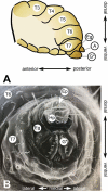
Figure 1.
Figure 1.
The terminalia of female D. melanogaster. [A] Model diagram of posterior female abdomen of D. melanogaster, lateral view. [B] Scanning electron micrograph of D. melanogaster female terminalia, posterior view. T3-T7 = female abdominal tergites 3–7. G* = female genitalia, A = female analia, Eg = epigynium (T8), Hp = hypoproct, Ep = epiproct. The hypoproct and the epiproct together form the female analia. *Note that the female genitalia includes the epigynium, which is indicated separately in this figure.
Figure 2.
Figure 2.
Visual atlas of the external female terminalia. Light microscopy images showing the whole external terminalia in lateral view (panel A) and the genitalia in posteroventral view (panel B). Individual structures are highlighted below each image, with line drawings to aid identification. Previous FlyBase terms are listed in the left column and revised terms are given in the right column. Panel C is a detail of a lateral view with internal structures extruded (as during egg laying), to highlight interior membranous structures.
Figure 3.
Figure 3.
Hypogynial sensilla. [A] Light microscopy image of hypogynial lobes. Inset is a closeup of the posterior tip of one lobe. [B] Line tracing of [A], showing locations of bristle types. Hypogynial short sensilla are barely visible from this angle, but one is shown in the inset (arrow). [C] Scanning electron micrograph of female genitalia, posterior view. Colour-coding of sensilla types is as follows: Red, hypogynial tooth; Green, hypogynial long sensillum; Blue, hypogynial short sensillum. [D] Scanning electron micrograph of female genitalia, posterior view. The region covered with oviprovector scales is indicated with a dashed purple line.
Figure 4.
Figure 4.
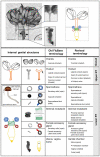
Visual atlas of internal female genitalia and reproductive structures. Confocal bright-field images and schematic of Drosophila melanogaster female (Canton S strain) reproductive system. Scale bar is 500 µm. The upper box shows the upper reproductive tract (upper RT) and the ovaries, the lower box is the lower reproductive tract (Lower RT). The lower panel displays individual structures with line drawings to aid identification. The internal structures and substructures include the gonad (ovaries), the upper RT (oviduct) and the lower RT (seminal receptacle, spermatheca, female accessory glands, genital chamber). Inset is a detail of the spermatheca to highlight substructures. Previous FlyBase terms are on the left and revised terms are on the right.
Figure 5.
Figure 5.
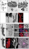
Internal genital structures of the female reproductive tract. [A] The Drosophila female reproductive tract consists of a pair of ovaries (OV) connected to a median common oviduct (CO) by two lateral oviducts (LO), and a uterus (UT) that leads to the vagina, which opens to the exterior through the vulva. The reproductive tract also includes specialized organs: a pair of spermathecae (Spt), seminal receptacle (SR), and a pair of female accessory glands (AG); drawing by Zohar Nir-Amitin. [B, C] The whole system with fat body [C] or without the fat body [B] that covers the spermatheca (Spt-FB) and the female accessory glands (AG-FB) (scale bar is 500 µm). [D-F] Upper RT that includes the lateral and common oviducts (scale bar is 100 µm), [D]. Toluidine blue stained 1 µm thick section of the oviduct that highlight the luminal space (l) and the epithelial cells (Epi) [E]. Upper RT stained with Alexa Fluor 594-phalloidin (red) showing the muscle fibres in different regions of oviduct (scale bar is 50 µm), [F]. [G, H] Lower reproductive tract, including the spermatheca (Spt), seminal receptacle (SR), and female accessory glands (AG). Note the red and blue arrowheads that mark the connection of the Spt and AG stalks to the uterus (scale bar is 50 µm). The panel also presents bright-field, phalloidin and DAPI images: SR showing the proximal (Pro) and distal (Dis) regions and the surrounding layer of visceral muscle (scale bar is 50 µm); Spt showing the spermathecal secretory cells (SSC), the lumen where sperm is stored (L), the stalks (St) (scale bar is 20 µm), the end apparatus (EA), and the fat body (FB, stained with DAPI) that surrounds the Spt; the female accessory glands (AG) showing the secretory cells (SC) (scale bar is 20 µm). [I-L] Zoom-in image of the uterus: [J] layers of circular muscle fibres (UTm) (scale bar is 50 µm), [K] micro-CT of the uterus highlighting the structure of the uterine lumen (L) (scale bar is 50 µm), [L] DsRed expression (magenta) showing the location of the fertilization chamber (FC), a structure to which the stalks of the SR, Spt and AG enter.
Figure 6.
Figure 6.
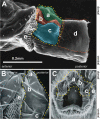
Scanning electron micrographs of the furca and furcal folds. [A] Lateral view with internal structures extruded, [B] lateral view, unextended, [C] posterior view. In each image, the vaginal furca is indicated by the yellow dashed line. a. vaginal furcal dorsal fold, b. vaginal furcal dorsolateral fold, c. vaginal furcal lateral fold, d. uterine furca. Not shown in the figure is the portion of the uterine furca that extends internally until the entry point of the spermathecal and accessory gland ducts into the genital chamber. e. oviprovector scales.

References

    1. Eberhard WG. Sexual selection and animal genitalia. Cambridge Massachusetts USA: Harvard University Press; 1985.
    1. Hosken DJ, Stockley P. Sexual selection and genital evolution. Trends Ecol Evol. 2004;19(2):87–93. - PubMed
    1. Simmons LW. Sexual selection and genital evolution. Austral Entomol. 2014;53(1):1–17.
    1. Eberhard WG. Evolution of genitalia: theories, evidence, and new directions. Genetica. 2010;138(1):5–18. - PubMed
    1. Ah-King M, Barron AB, Herberstein ME. Genital evolution: why are females still understudied? PLoS Biol. 2014;12(5):e1001851. - PMC - PubMed

Publication types

LinkOut - more resources