. 2022 May 16;132(10):e154997.
doi: 10.1172/JCI154997.

UNC45A deficiency causes microvillus inclusion disease-like phenotype by impairing myosin VB-dependent apical trafficking

Affiliations

UNC45A deficiency causes microvillus inclusion disease-like phenotype by impairing myosin VB-dependent apical trafficking

Rémi Duclaux-Loras et al. J Clin Invest. .

Abstract

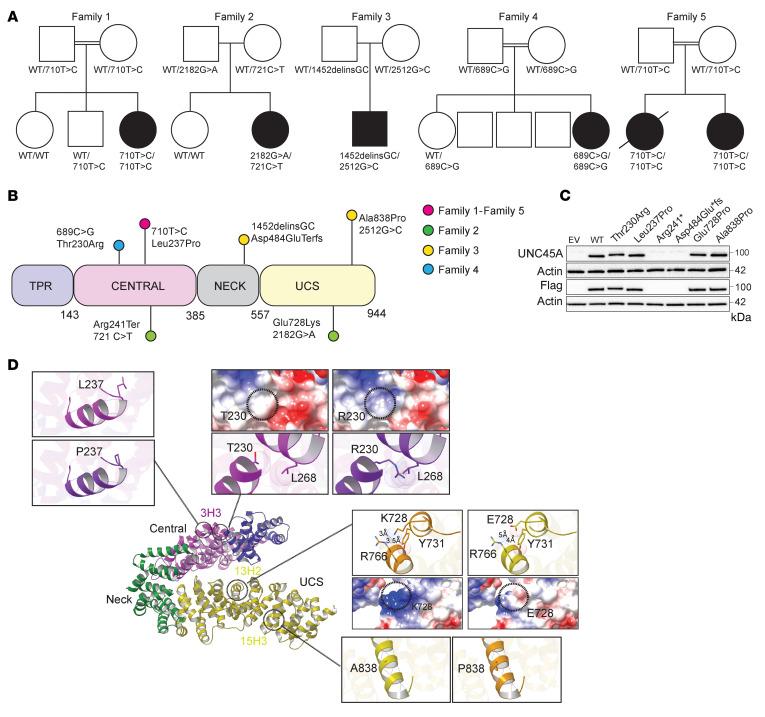
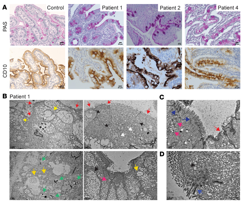
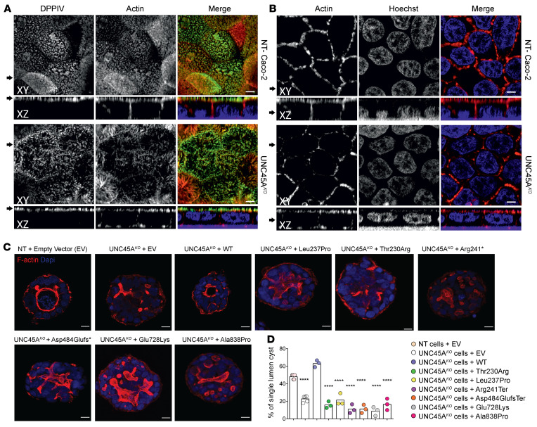
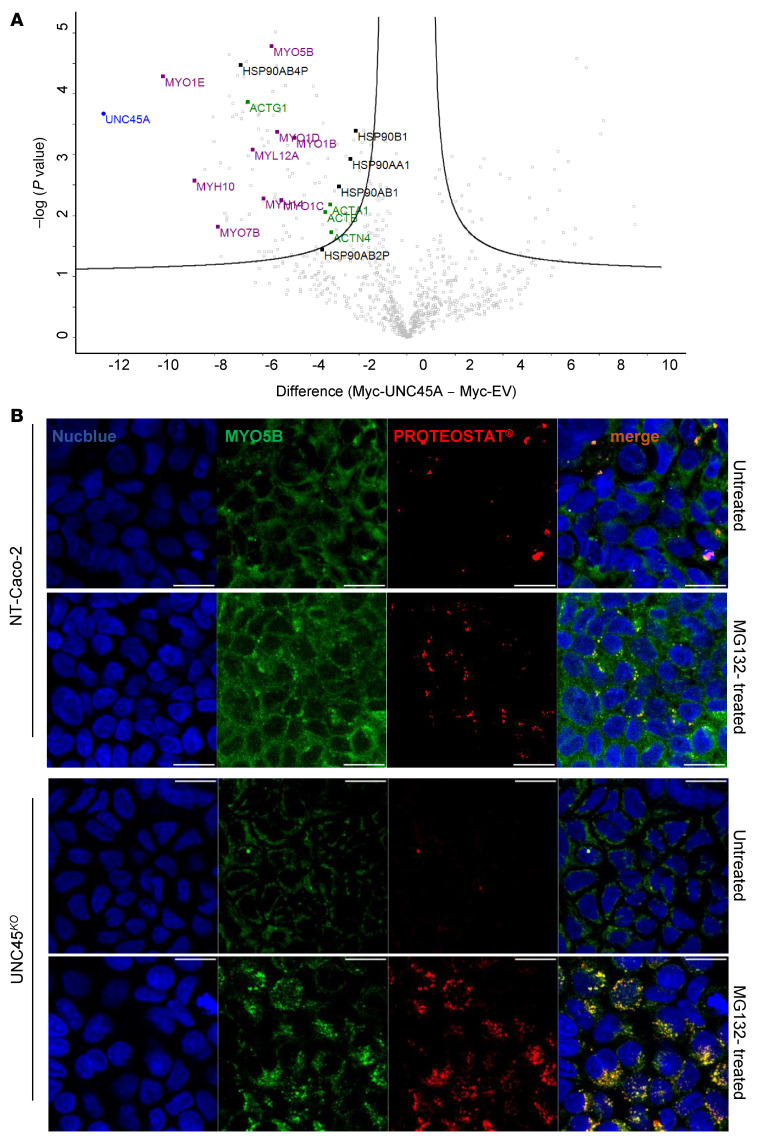
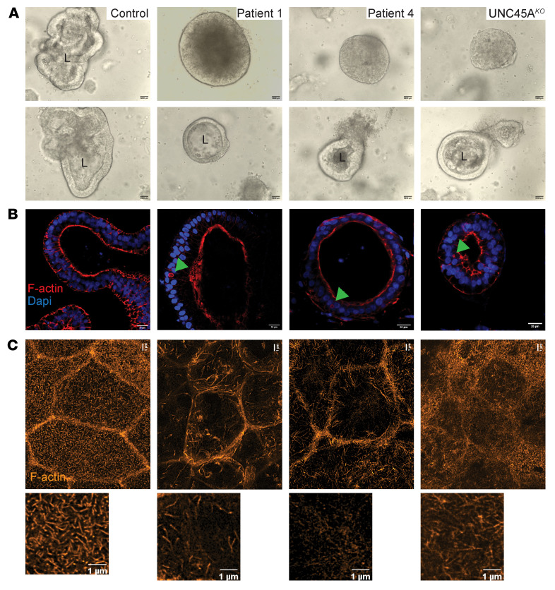
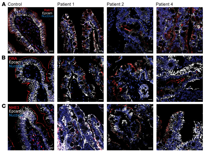
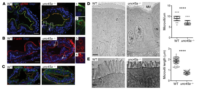
Variants in the UNC45A cochaperone have been recently associated with a syndrome combining diarrhea, cholestasis, deafness, and bone fragility. Yet the mechanism underlying intestinal failure in UNC45A deficiency remains unclear. Here, biallelic variants in UNC45A were identified by next-generation sequencing in 6 patients with congenital diarrhea. Corroborating in silico prediction, variants either abolished UNC45A expression or altered protein conformation. Myosin VB was identified by mass spectrometry as client of the UNC45A chaperone and was found misfolded in UNC45AKO Caco-2 cells. In keeping with impaired myosin VB function, UNC45AKO Caco-2 cells showed abnormal epithelial morphogenesis that was restored by full-length UNC45A, but not by mutant alleles. Patients and UNC45AKO 3D organoids displayed altered luminal development and microvillus inclusions, while 2D cultures revealed Rab11 and apical transporter mislocalization as well as sparse and disorganized microvilli. All those features resembled the subcellular abnormalities observed in duodenal biopsies from patients with microvillus inclusion disease. Finally, microvillus inclusions and shortened microvilli were evidenced in enterocytes from unc45a-deficient zebrafish. Taken together, our results provide evidence that UNC45A plays an essential role in epithelial morphogenesis through its cochaperone function of myosin VB and that UNC45A loss causes a variant of microvillus inclusion disease.

Keywords: Epithelial transport of ions and water; Gastroenterology.


Figures

Figure 1
Figure 1. Genetics of 5 families with UNC45A deficiency and molecular characterization of UNC45A variants.
(A) Pedigree of families with AR UNC45A deficiency (filled shapes indicate affected individuals). (B) Schematic representation of UNC45A protein showing location of the variants identified in this study. (C) Western blot analysis of UNC45A protein in HEK293T cells transfected with EV, WT, and mutant alleles (n = 2). (D) Ribbon representation of the human UNC45a model generated by MODELLER (based on the D. melanogaster and C. elegans UNC45 protein structures) showing its domain architecture. The variants were modeled in silico using Chimera. The location and a close-up view of each variant are shown. For the L237P and A838P mutations, hydrogen bonds are depicted by dark-blue dashed lines. For the T230R and E728K mutations, Van der Waals radii are represented for each atom and steric clashes occurring in the structure are shown with brown dashed lines. Coulombic surface representations are also shown. Red surface indicates the lowest electrostatic potential energy and blue the highest. Locations of the mutated residue are highlighted by a black circle. Distances are indicated using black dotted lines.
Figure 2
Figure 2. Histopathological and ultrastructural MVID features in the intestinal epithelium of patient.
(A) PAS and CD10 staining in control, P1, P2, and P4 duodenal biopsies. Scale bars: 20 μm. (BD) TEM of P1’s duodenal enterocytes showing ultrastructural features of MVID: regions containing vesicles and tubulovesicular structures (black arrowheads), enlarged lysosomes (green arrowheads), and swollen (i.e., stressed) endoplasmic reticulum (white arrowheads) as well as more or less complex or partially degraded MVIs (yellow arrowheads) and basolateral microvilli (pink arrowheads). Brush border showed defective microvilli anchored deep into the cytoplasm and thickened terminal web (blue arrowheads) or partially depleted microvilli (red arrowheads). Scale bars: 2 μm (B); 1 μm (C); 0.5 μm (D).
Figure 3
Figure 3. Disrupted enterocyte architecture in UNC45A deficiency.
(A) Confocal images of polarized NT control and Unc45AKO Caco-2 cells grown on a filter and stained for an apical brush border marker DPPIV and actin. (B) Actin staining in polarized Caco-2 cells. Nuclei were visualized with HOECHST. Arrows on the left mark the corresponding XY and XZ planes. Scale bars: 20 μm. Panels A and B were from the same experiment. (C) NT control and UNC45AKO Caco-2 cells complemented or not with WT or mutant alleles were cultured in 3D for 5 days to form cysts. Nuclei are stained with Nucblue (blue); actin is stained with phalloidin AF 455 (red). Single confocal sections through the middle of the cyst are shown. Scale bars: 10 μm. (D) Single-lumen cysts were counted in each experiment. Results from 3 independent experiments (35 cysts each) are shown, 1-way ANOVA. ****P < 0.0001.
Figure 4
Figure 4. MYO5B as a client of the HSP90-UNC45A chaperone complex.
(A) Volcano plot showing proteins enriched in the myc-tagged UNC45A WT immunoprecipitates over the myc-tagged EV control identified by mass spectrometry in 3 independent pull-down assays. The difference of the average of the logarithm of Label Free Quantification (LQF) intensities (WT vs. EV) is plotted against negative logarithmic P values of a 2-sided, 2-sample Welsh t test. The hyperbolic curve delimitates significantly enriched proteins from common hits. High LFQ hits of interest are indicated in black (HSP90 family), in purple (Myosin protein family), and in green (Actin protein family). (B) Accumulation of myosin VB aggregates (aggresomes) in UNC45AKO cells treated with MG-132 (10 μM) overnight. Scale bars: 10 μm. n = 3. Nuclei are stained with Nucblue (blue).
Figure 5
Figure 5. UNC45A-deficient organoids recapitulate MVID features.
(A) Bright-field images of control, P1, P4, and UNC45AKO1 3D organoids. Scale bar: 5000 μm. (B) F-actin staining revealing MVIs in the small subset of P1, P4, and UNC45AKO 3D organoids that display a central lumen. Scale bar: 20 μm. (C) STED image of F-actin at the apical pole of 2D enterocyte cultures derived from control, P1, P4, and UNC45AKO1 organoids after a 21-day culture on filters. Scale bars: 1 μm. L, lumen.
Figure 6
Figure 6. Impaired apical targeting in UNC45A-deficient patients.
(AC) Confocal imaging showing localization of Rab11 (A) and of apical transporters DRA (B) and NHE3 (C) in duodenum of control and patients. Scale bars: 20 μm.
Figure 7
Figure 7. MVID features in enterocytes of unc45a zebrafish mutants.
(AC) Confocal microscopy analysis of the intestinal bulb of WT and unc45a–/– mutant larvae stained for villin and pERM (A), F-actin (phalloidin) (B), and Rab11 (C) at 5 dpf. Scale bars: 20 μm. Boxes showing microvillus-like inclusions are enlarged ×4. (D and E) TEMs of thin sections of intestinal bulb of 5 dpf WT and unc45a–/– mutant larvae showing defects in the organization of the brush border. Scale bars: 2 μm (D); 0.5 μm (E). Quantitative analysis showing decrease in microvillus length and density in unc45a–/– enterocytes as compared with WT enterocytes. Data are represented as mean + SD. ****P < 0.001, t test. N nucleus; MV microvilli.

Comment in

References

    1. Canani RB, et al. Congenital diarrhoeal disorders: advances in this evolving web of inherited enteropathies. Nat Rev Gastroenterol Hepatol. 2015;12(5):293–302. doi: 10.1038/nrgastro.2015.44. - DOI - PMC - PubMed
    1. Charbit-Henrion F, et al. Intestinal immunoregulation: lessons from human mendelian diseases. Mucosal Immunol. 2021;14(5):1017–1037. doi: 10.1038/s41385-021-00398-3. - DOI - PubMed
    1. Esteve C, et al. Loss-of-function mutations in UNC45A cause a syndrome associating cholestasis, diarrhea, impaired hearing, and bone fragility. Am J Hum Genet. 2018;102(3):364–374. doi: 10.1016/j.ajhg.2018.01.009. - DOI - PMC - PubMed
    1. Lee CF, et al. The UNC-45 myosin chaperone: from worms to flies to vertebrates. Int Rev Cell Mol Biol. 2014;313:103–144. - PMC - PubMed
    1. Gazda L, et al. The myosin chaperone UNC-45 is organized in tandem modules to support myofilament formation in C. elegans. Cell. 2013;152(1–2):183–195. - PMC - PubMed

Publication types

MeSH terms

Substances

Supplementary concepts