. 2022 May 16;132(10):e148333.
doi: 10.1172/JCI148333.

HDAC6 modulates myofibril stiffness and diastolic function of the heart

Affiliations

HDAC6 modulates myofibril stiffness and diastolic function of the heart

Ying-Hsi Lin et al. J Clin Invest. .

Abstract

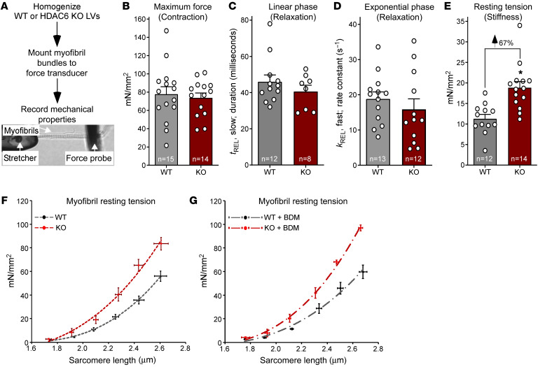
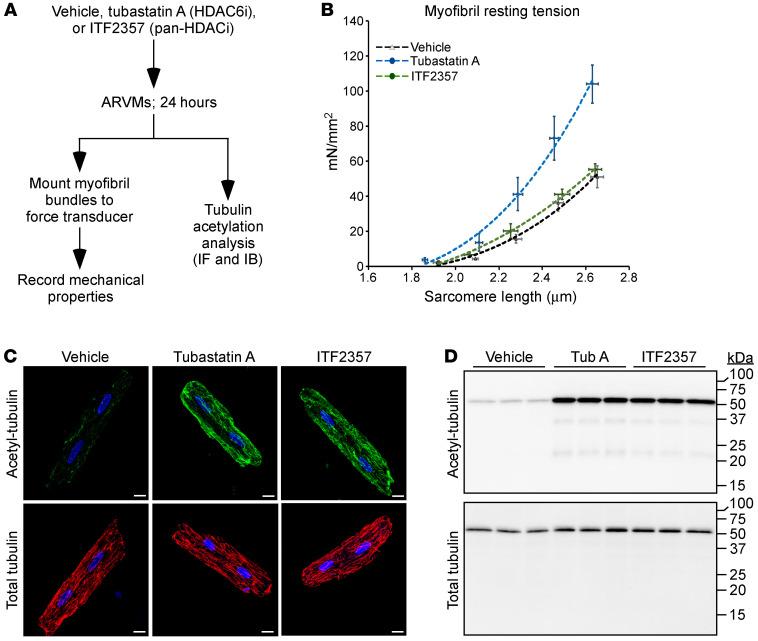
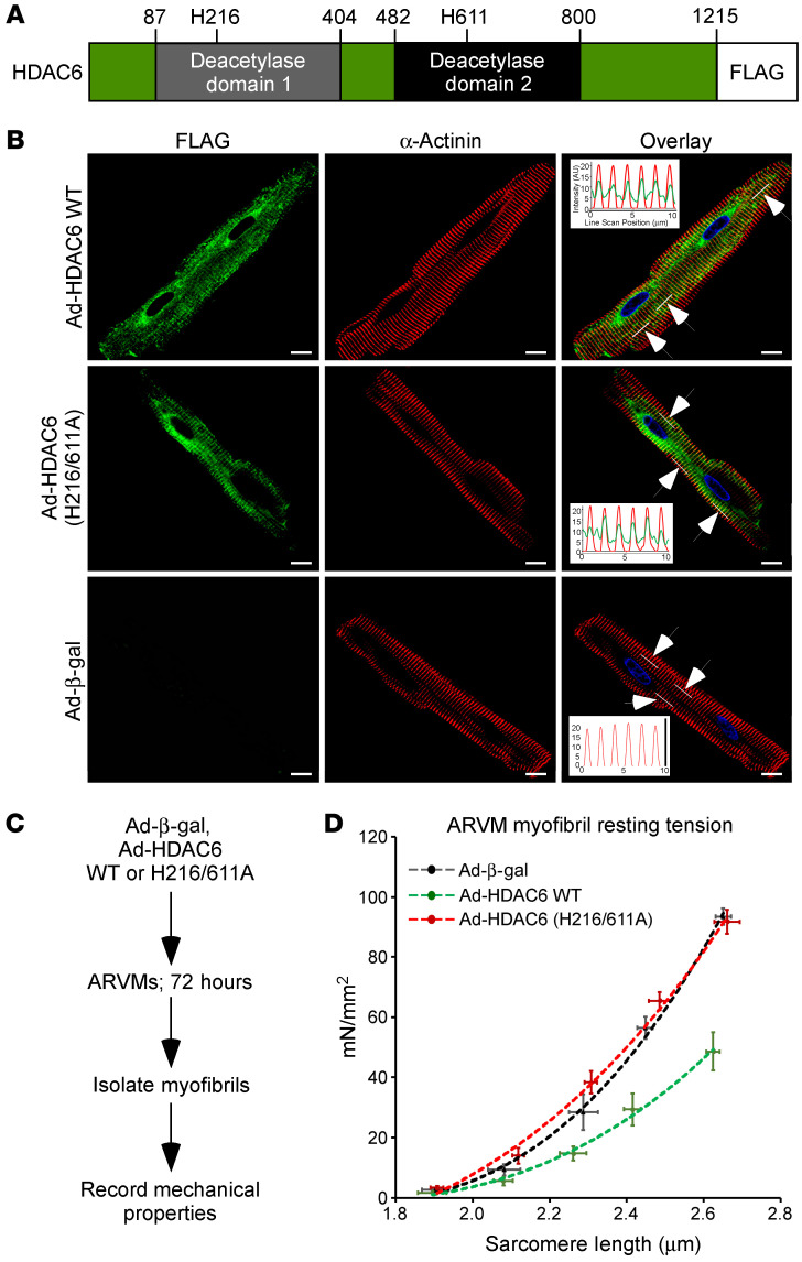
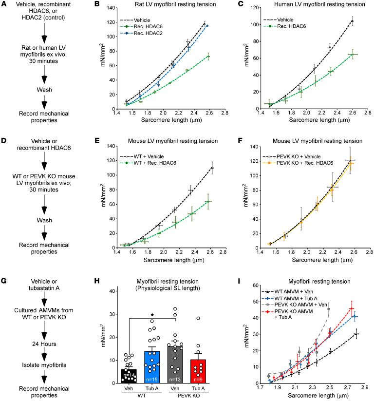
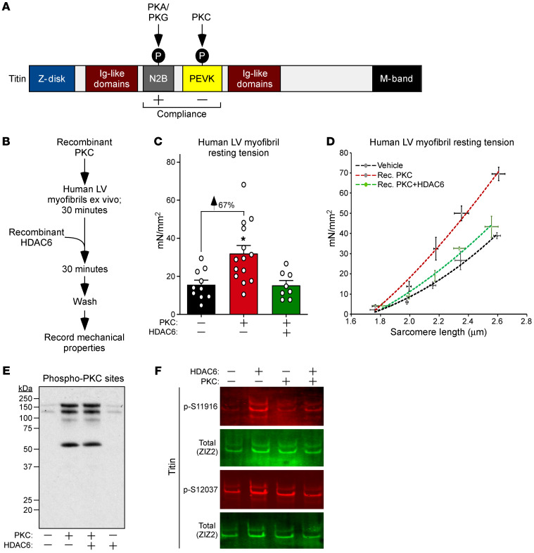
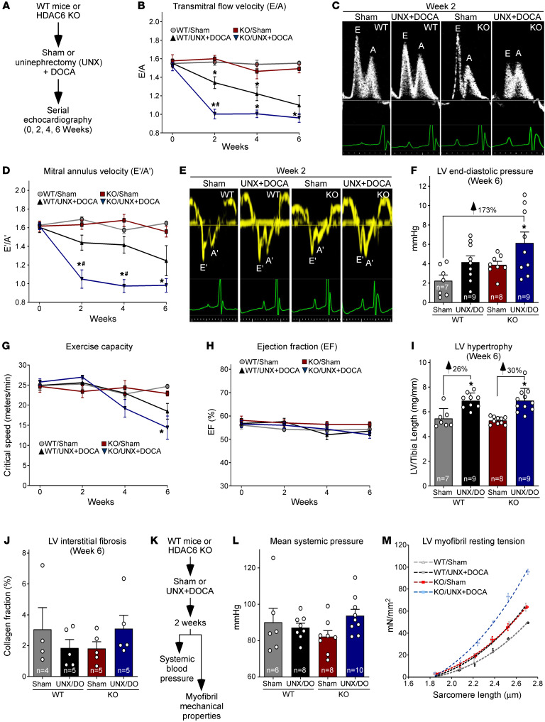
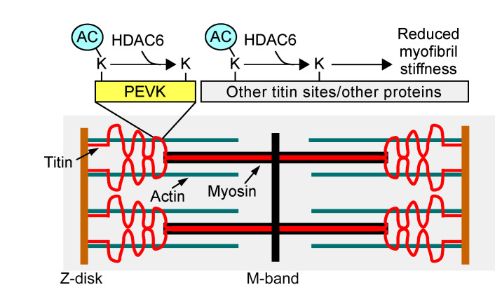
Passive stiffness of the heart is determined largely by extracellular matrix and titin, which functions as a molecular spring within sarcomeres. Titin stiffening is associated with the development of diastolic dysfunction (DD), while augmented titin compliance appears to impair systolic performance in dilated cardiomyopathy. We found that myofibril stiffness was elevated in mice lacking histone deacetylase 6 (HDAC6). Cultured adult murine ventricular myocytes treated with a selective HDAC6 inhibitor also exhibited increased myofibril stiffness. Conversely, HDAC6 overexpression in cardiomyocytes led to decreased myofibril stiffness, as did ex vivo treatment of mouse, rat, and human myofibrils with recombinant HDAC6. Modulation of myofibril stiffness by HDAC6 was dependent on 282 amino acids encompassing a portion of the PEVK element of titin. HDAC6 colocalized with Z-disks, and proteomics analysis suggested that HDAC6 functions as a sarcomeric protein deacetylase. Finally, increased myofibril stiffness in HDAC6-deficient mice was associated with exacerbated DD in response to hypertension or aging. These findings define a role for a deacetylase in the control of myofibril function and myocardial passive stiffness, suggest that reversible acetylation alters titin compliance, and reveal the potential of targeting HDAC6 to manipulate the elastic properties of the heart to treat cardiac diseases.

Keywords: Cardiology; Heart failure.


Conflict of interest statement

Conflict of interest: TAM is on the scientific advisory boards of Artemes Bio and Eikonizo Therapeutics, received funding from Italfarmaco for an unrelated project, and has a subcontract from Eikonizo Therapeutics related to a Small Business Innovation Research grant from the National Institutes of Health (HL154959).

Figures

Figure 1
Figure 1. HDAC6 KO increases cardiac myofibril stiffness.
(A) Schematic representation of ex vivo myofibril mechanics system. Myofibrils from left ventricles (LVs) of 6-month-old male mice were evaluated. (B) Myofibril tension (mN/mm2) generation in response to maximal calcium (pCa 4.5). (C and D) Linear (tREL, slow) and exponential (kREL, fast) myofibril relaxation upon removal of calcium (pCa 9.0). (E) Myofibril resting tension (mN/mm2) at a sarcomere length of 2.0–2.2 μm. For BE, dots represent data from individual myofibrils. Mean + SEM is shown; *P < 0.05 vs. WT based on unpaired, 2-tailed t test. (F) Myofibril resting tension–to–sarcomere length curves. (G) Myofibrils were treated with the myosin ATPase inhibitor butanedione monoxime (BDM; 50 mM) before assessment of resting tension at the given sarcomere lengths. For F and G, data are presented as mean ± SEM, fitted by third-order polynomials, from 4 animals per group, with 6–8 myofibrils per mouse analyzed.
Figure 2
Figure 2. Selective pharmacological inhibition of HDAC6 increases cardiac myofibril stiffness.
(A) Schematic representation of the adult rat ventricular myocyte (ARVM) experiment. (B) Resting tension–to–sarcomere length curves obtained with myofibrils isolated from ARVMs treated as indicated. Data are presented as mean ± SEM, fitted by third-order polynomials, from 4 separate ARVM preparations per group, with 6–8 myofibrils per preparation analyzed. (C) Indirect immunofluorescence analysis of acetyl-tubulin and total tubulin in ARVMs; scale bars: 10 μm. (D) Immunoblot analysis of acetyl-tubulin and total tubulin in whole-cell homogenates from treated ARVMs. See complete unedited blots in the supplemental material.
Figure 3
Figure 3. Ectopic HDAC6 colocalizes with cardiomyocyte sarcomeres and reduces myofibril stiffness.
(A) Schematic representation of HDAC6 with amino acid numbers indicated. (B) Indirect immunofluorescence of ARVMs infected with adenoviruses encoding FLAG-tagged WT HDAC6, HDAC6 harboring 2 amino acid substitutions that abolish enzymatic activity (H216/611A), and β-galactosidase (β-gal) as a negative control. Insets in the overlay are line scans of fluorescence intensity in the 488 nm and 568 nm channels within the regions of the cells indicated by the white lines (arrows point to white lines). Averages from the 3 regions are shown, and overlapping peaks of fluorescence reveal colocalization of HDAC6 and sarcomeric α-actinin. DAPI fluorescence of nuclei is also shown. Scale bars: 10 μm. (C) Schematic representation of the ARVM myofibril experiment using adenoviruses. (D) Myofibril resting tension–to–sarcomere length curves. Data are presented as mean ± SEM, fitted by third-order polynomials, from 4 animals per group, with 6–8 myofibrils per mouse analyzed.
Figure 4
Figure 4. Recombinant HDAC6 regulates myofibril stiffness in a manner that is dependent on a region of titin encompassing a portion of the PEVK element and an adjacent Ig-like domain.
(A) Schematic representation of the ex vivo assay using recombinant HDACs and myofibrils isolated from rat or human left ventricles (LVs). (B and C) Myofibril resting tension–to–sarcomere length curves. (D) Schematic representation of the ex vivo assay using myofibrils from WT and PEVK/Ig-like domain–KO mouse LVs. (E and F) Myofibril resting tension–to–sarcomere length curves. (G) Schematic representation of the experiment using cultured adult mouse ventricular myocytes (AMVMs) from WT and PEVK-KO mice. (H) Myofibril resting tension (mN/mm2) at a sarcomere length of 2.0–2.2 μm. (I) Myofibril resting tension–to–sarcomere length curves. For B, C, E, F, and I, data are presented as mean ± SEM, fitted by third-order polynomials. For E, F, H, and I, 6–8 myofibrils from 3 mice per group (WT and KO) were analyzed. For H, mean + SEM is shown; *P < 0.05 vs. WT + vehicle based on 1-way ANOVA with Tukey’s multiple-comparison test.
Figure 5
Figure 5. HDAC6 reverses PKC-mediated stiffening of human myofibrils.
(A) Schematic representation of titin, with the impact of phosphorylation of the N2B and PEVK regions indicated. (B) Schematic representation of the ex vivo assay using human myofibrils and recombinant forms of PKCα and HDAC6. (C) Myofibril resting tension measurements at physiological sarcomere length (~2.19 μm). Mean + SEM is shown; *P < 0.05 vs. untreated myofibrils based on 1-way ANOVA with Tukey’s multiple-comparison test. (D) Myofibril resting tension–to–sarcomere length curves. Data are presented as mean ± SEM, fitted by third-order polynomials. Myofibrils from 3 nonfailing human hearts were each treated with vehicle, recombinant PKCα, or recombinant PKCα followed by recombinant HDAC6; 6–8 myofibrils per treatment were analyzed and averaged per heart. (E) Immunoblotting was performed with an anti–PKC substrates antibody and solubilized proteins from myofibrils treated as indicated. (F) Titin was immunoblotted with antibodies specific for phospho-S11916 (S11878 in human) and phospho-S12037 (S12022 in human) in the PEVK domain, as well as an antibody against the titin Z1Z2 element to assess total titin levels.
Figure 6
Figure 6. HDAC6 deletion exacerbates DD and cardiac myofibril stiffening in the UNX/DOCA model.
(A) Schematic representation of mouse model of DD with preserved ejection fraction. (B) Serial Doppler echocardiographic measurements of mitral inflow velocity (E/A), a parameter of diastolic cardiac function. (C) Representative E/A images. (D) Serial echocardiographic measurements of septal mitral annulus velocity (E′/A′), another measure of diastolic function. (E) Representative E′/A′ images. (F) Invasive, catheter-based measurements of LV end-diastolic pressure at study endpoint (6 weeks). Mean + SEM values are shown and were compared by 2-way ANOVA with Tukey’s multiple-comparison test; *P < 0.05 vs. WT/sham. (G) Critical speed was determined as a measure of exercise capacity. (H) Echocardiographic assessment of systolic function as determined by ejection fraction. For B, D, G, and H, mean ± SEM values are shown and were compared by a mixed-effects model for repeated measures with Tukey’s multiple-comparison test; *P < 0.05 vs. WT/sham, #P < 0.05 vs. WT/UNX + DOCA; animal numbers for each time point are provided in Supplemental Table 1. Echocardiographic data are summarized in Supplemental Table 2. (I) LV–to–tibia length assessment of cardiac hypertrophy upon necropsy. Mean + SEM values are shown and were compared by 1-way ANOVA with Tukey’s multiple-comparison test; *P < 0.05 vs. corresponding sham control. (J) LV sections were stained with Picrosirius red dye, and the ratio of positively stained (red) pixels to the total pixel number of each section (collagen fraction %) was calculated. Mean values are shown (+ SEM). Two-way ANOVA revealed no significant difference between groups. (K) Schematic representation of the 2-week study to assess the impact of HDAC6 deletion on blood pressure and myofibril stiffness in the mouse model of DD with preserved ejection fraction. (L) Tail cuff measurements of mean systemic pressure (mmHg). Data are presented as mean + SEM. (M) Myofibril resting tension–to–sarcomere length curves. Data are presented as mean ± SEM, fitted by third-order polynomials, from 4 animals per group, with 6–8 myofibrils per mouse analyzed.
Figure 7
Figure 7. A model for the regulation of titin stiffness by HDAC6.
HDAC6-mediated deacetylation reduces myofibril stiffness, whereas inhibition of HDAC6-mediated deacetylation leads to myofibril stiffening. We propose that HDAC6 regulates myofibril compliance, at least in part, through deacetylation of lysines embedded within, or adjacent to, the PEVK element of titin, and possibly through deacetylation of other regions of titin and/or other sarcomeric proteins.

References

    1. Granzier HL, Irving TC. Passive tension in cardiac muscle: contribution of collagen, titin, microtubules, and intermediate filaments. Biophys J. 1995;68(3):1027–1044. doi: 10.1016/S0006-3495(95)80278-X. - DOI - PMC - PubMed
    1. Linke WA. Titin gene and protein functions in passive and active muscle. Annu Rev Physiol. 2018;80:389–411. doi: 10.1146/annurev-physiol-021317-121234. - DOI - PubMed
    1. Borbely A, et al. Hypophosphorylation of the stiff N2B titin isoform raises cardiomyocyte resting tension in failing human myocardium. Circ Res. 2009;104(6):780–786. doi: 10.1161/CIRCRESAHA.108.193326. - DOI - PubMed
    1. Makarenko I, et al. Passive stiffness changes caused by upregulation of compliant titin isoforms in human dilated cardiomyopathy hearts. Circ Res. 2004;95(7):708–716. doi: 10.1161/01.RES.0000143901.37063.2f. - DOI - PubMed
    1. Nagueh SF, et al. Altered titin expression, myocardial stiffness, and left ventricular function in patients with dilated cardiomyopathy. Circulation. 2004;110(2):155–162. doi: 10.1161/01.CIR.0000135591.37759.AF. - DOI - PubMed

Publication types