Review
. 2022 Oct 1;102(4):1669-1701.
doi: 10.1152/physrev.00045.2021. Epub 2022 May 16.

Circadian clocks of the kidney: function, mechanism, and regulation

Affiliations
Review

Circadian clocks of the kidney: function, mechanism, and regulation

Hannah M Costello et al. Physiol Rev. .

Abstract

An intrinsic cellular circadian clock is located in nearly every cell of the body. The peripheral circadian clocks within the cells of the kidney contribute to the regulation of a variety of renal processes. In this review, we summarize what is currently known regarding the function, mechanism, and regulation of kidney clocks. Additionally, the effect of extrarenal physiological processes, such as endocrine and neuronal signals, on kidney function is also reviewed. Circadian rhythms in renal function are an integral part of kidney physiology, underscoring the importance of considering time of day as a key biological variable. The field of circadian renal physiology is of tremendous relevance, but with limited physiological and mechanistic information on the kidney clocks this is an area in need of extensive investigation.

Keywords: chronic kidney disease; renal; rhythm.


Conflict of interest statement

No conflicts of interest, financial or otherwise, are declared by the authors.

Figures

None
Graphical abstract
FIGURE 1.
FIGURE 1.
Circadian physiology and molecular mechanism. The circadian system is organized as a hierarchical multioscillator network, with the central clock in the suprachiasmatic nucleus (SCN) of the hypothalamus. The SCN directly receives photic cues for entrainment to the light-dark cycle and synchronizing peripheral clocks. Although the SCN is responsive to photic cues, the peripheral oscillators are more responsive to nonphotic cues such as feeding. The mechanism of the molecular clock is a transcriptional/translational feedback loop. The core loop consists of a positive (BMAL1, CLOCK) and a negative (PER, CRY) arm, which act on the E-box response element. This clock mechanism regulates tissue-specific target genes to regulate rhythmic outputs in physiology and behavior, with these rhythmic outputs influencing the circadian system. Diagram created with BioRender.com, with permission.
FIGURE 2.
FIGURE 2.
Renal rhythms in electrolyte excretion persist in the absence of time cues. Cave studies involved human subjects underground with no knowledge of clock time. Rhythms in renal sodium, potassium, and chloride excretion, indicative of rhythms in renal function, remained intact after 8 wk underground in a male subject in complete isolation from any environmental time cues. By the end of the study (12 wk), rhythms had shifted but were still apparent. Reference data from Ref. . Diagram created with BioRender.com, with permission.
FIGURE 3.
FIGURE 3.
Circadian expression of endothelin axis genes in the kidney. Relative circadian expression of endothelin-1 (ET-1) and ET-1 receptors ETA (ETRA) and ETB (ETRB); data adapted with permission from CircaDB (http://circadb.hogeneschlab.org/) (104) and fit to cosine curves. Gray shading indicates the active phase, and white background indicates the inactive phase.
FIGURE 4.
FIGURE 4.
Circadian clocks regulate gene expression in a cell-type specific manner in the kidney. Blood enters the nephron and is filtered at the glomerulus. A: podocytes cover the outside of the glomerular capillary to support the structure and function of the glomerulus. BMAL1-target genes, G protein-coupled receptor class C group 5 member A (Gprc5a), cathepsin L (Ctsl), transcription factor 21 (Tcf21), N-ethylmaleimide sensitive factor (Nsf), and G protein subunit alpha 12 (Gna12), are rhythmic (depicted with clocks) and involved in podocyte development. From the glomerulus, filtrate moves to the proximal tubule. B: proximal tubule cells express the sodium/hydrogen exchanger isoform 3 (NHE3), sodium-phosphate cotransporter 2 (NPT2A), phosphate transporter (PIT1/2), and the sodium-glucose linked transporter isoform 1 (SGLT1), all of which are located on the apical membrane (facing the filtrate) and are regulated by the circadian clock. Sodium reabsorbed from the filtrate is pumped back into the blood by the basolateral Na+-K+ pump, found on each of the cell types illustrated here. Filtrate then moves to the loop of Henle. C: cells in the thick ascending limb contain the sodium-potassium-chloride cotransporter isoform 2 (NKCC2), which is positively regulated by the nuclear estrogen-related receptor ERRβ and with-no-lysine 1 (WNK1)-oxidative stress responsive kinase 1 (OSR1)/Ste20-related proline-alanine rich kinase (SPAK) pathway. NKCC2 is negatively regulated by Alström syndrome 1 protein (ALMS1). Expressions of these regulators, as well as NKCC2, have a circadian rhythm (depicted by clocks). Again, these cells have a basolateral Na+-K+ pump and are regulated by the clock. D: the distal convoluted tubule cells contain apically located sodium-chloride cotransporters (NCC), which reabsorb Na+ and Cl from the filtrate. This cotransporter is regulated by the circadian clock and WNK1-OSR1/SPAK pathway. The final segment of the nephron that the filtrate enters is the collecting duct. E: in principal cells, the epithelial sodium channel (ENaC) reabsorbs Na+ from the filtrate and is regulated by the circadian clock. WNK1 positively regulates ENaC, with endothelin-1 (ET-1) and serum and glucocorticoid-inducible kinase 1 (SGK1), via neuronal precursor cell-expressed developmentally downregulated 4-2 (Nedd4-2), which negatively regulates ENaC. Expressions of all of these regulators in the kidney, as well as ENaC but not Nedd4-2, are regulated by the circadian clock. FXYD5 is also regulated by the circadian clock and in turn positively regulates the Na+-K+ pump. Expression of vasopressin receptors (V1aR/V2R) and aquaporin receptors (AQP2/4), involved in water reabsorption in the collecting duct, has been shown to have a circadian rhythm. Together, this highlights the extensiveness of the circadian clocks’ role in the regulation of genes throughout the cells of the kidney and its potential implications on renal function. Adapted from Ref. . Diagram created with BioRender.com, with permission.
FIGURE 5.
FIGURE 5.
Circadian expression of renal Na+-phosphate cotransporters. Relative circadian expression of Na+-phosphate cotransporters Pit1, Pit2, and NPT2A. Data adapted with permission from CircaDB (http://circadb.hogeneschlab.org/) (104) and fit to cosine curves. Gray shading indicates the active phase, and the white background represents the inactive phase for mice.
FIGURE 6.
FIGURE 6.
Integration of glucocorticoid signaling and circadian clock-mediated transcriptional regulation. Glucocorticoids (GCs) bind to their receptor, the glucocorticoid receptor (GR), leading to translocation of GR into the nucleus. GR regulates the transcription/translation feedback loops of the circadian clock mechanism through glucocorticoid response elements (GREs) of Per1/2 and negative GREs (nGREs) of Reverbα and Ror. Both positive and negative arms of the circadian clock inhibit GR activity. Diagram created with BioRender.com, with permission.
FIGURE 7.
FIGURE 7.
Integrative physiology of the kidney clocks. The central clock in the suprachiasmatic nucleus (SCN) directly receives photic cues for entrainment to the light-dark cycle to synchronize peripheral clocks including the kidney clocks. The kidney clock is responsive to entrainment from nonphotic cues including feeding, core temperature, and stress. Hormone signaling, including adrenal hormones (aldosterone and cortisol), vasopressin (AVP), and endothelin-1, has been shown to regulate the circadian clock. With stress and glucocorticoids involved in entrainment of the kidney clock, the hypothalamic-pituitary-adrenal (HPA) axis plays a key role in control of the kidney clock, but the kidney clocks’ role in the regulation of the HPA axis remains unclear. The clocks in the kidney have been suggested to communicate with the central clock and adrenal clock, but the mechanisms are not fully understood. Diagram created with BioRender.com, with permission.
FIGURE 8.
FIGURE 8.
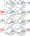
Blood pressure phenotypes in kidney-specific BMAL1 knockout (KO) mice. Blood pressure tracings over the course of 24 h in mice with various BMAL1 KOs compared with control mice. All panels are based on 12:12-h light-dark conditions while maintained on standard rodent diets. Red shaded areas of nephron demonstrate the location of knockout. A: male global BMAL1 KO mice were the first to be characterized, which lack BMAL1 expression in the entire body including the kidney. MAP, mean arterial pressure. Data from Ref. . B: male Nephrin-Cre BMAL1 KO is specific to podocytes in the glomerulus. SBP, systolic blood pressure. Data from Ref. . C: male inducible Pax8-Cre BMAL1 KO is specific to the entire nephron. Data from Ref. . D: male Renin-Cre BMAL1 KO mice. BMAL1 expression is lost in cells that express renin, which includes juxtaglomerular cells and collecting duct. Data from Ref. . E and F: female (E) and male (F) Aqp2-Cre BMAL1 KO (151) is specific to collecting duct cells. Data from Ref. . G and H: female (G) and male (H) Ksp-cadherin BMAL1 KO is specific to thick ascending limb, distal tubule, and collecting duct cells. Data from Ref. .
FIGURE 9.
FIGURE 9.
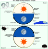
Time of day is a key biological variable. Humans are diurnal, with activity patterns and cardiovascular functions peaking during the daytime, whereas mice and rats are nocturnal, with activity patterns and cardiovascular functions peaking during the nighttime. Circadian rhythms in physiological function are very similar between rodents and humans, except that the peaks and troughs are antiphase to each other. For rodent studies to be as physiologically relevant to humans as possible, time of day should be considered a key biological variable. Diagram created with BioRender.com, with permission.

Similar articles

Cited by

References

    1. Pilorz V, Helfrich-Förster C, Oster H. The role of the circadian clock system in physiology. Pflugers Arch 470: 227–239, 2018. doi:10.1007/s00424-017-2103-y. - DOI - PubMed
    1. Dibner C, Schibler U, Albrecht U. The mammalian circadian timing system: organization and coordination of central and peripheral clocks. Annu Rev Physiol 72: 517–549, 2010. doi:10.1146/annurev-physiol-021909-135821. - DOI - PubMed
    1. Partch CL, Green CB, Takahashi JS. Molecular architecture of the mammalian circadian clock. Trends Cell Biol 24: 90–99, 2014. doi:10.1016/j.tcb.2013.07.002. - DOI - PMC - PubMed
    1. Healy KL, Morris AR, Liu AC. Circadian synchrony: sleep, nutrition, and physical activity. Front Netw Physiol 1: 732243, 2021. - PMC - PubMed
    1. Cederroth CR, Albrecht U, Bass J, Brown SA, Dyhrfjeld-Johnsen J, Gachon F, Green CB, Hastings MH, Helfrich-Förster C, Hogenesch JB, Lévi F, Loudon A, Lundkvist GB, Meijer JH, Rosbash M, Takahashi JS, Young M, Canlon B. Medicine in the fourth dimension. Cell Metab 30: 238–250, 2019. doi:10.1016/j.cmet.2019.06.019. - DOI - PMC - PubMed

Publication types

LinkOut - more resources