. 2022 Aug 3;35(3):ivac124.
doi: 10.1093/icvts/ivac124.

Effect of preservation solution and distension pressure on saphenous vein's endothelium

Affiliations

Effect of preservation solution and distension pressure on saphenous vein's endothelium

Matheus Duarte Pimentel et al. Interact Cardiovasc Thorac Surg. .

Abstract

Objectives: Approaches to improve saphenous vein (SV) patency in coronary artery bypass graft (CABG) surgery remain relevant. This study aimed to evaluate the effects of different preservation solutions and different pressures of intraluminal distention on the endothelium of SV segments in CABG.

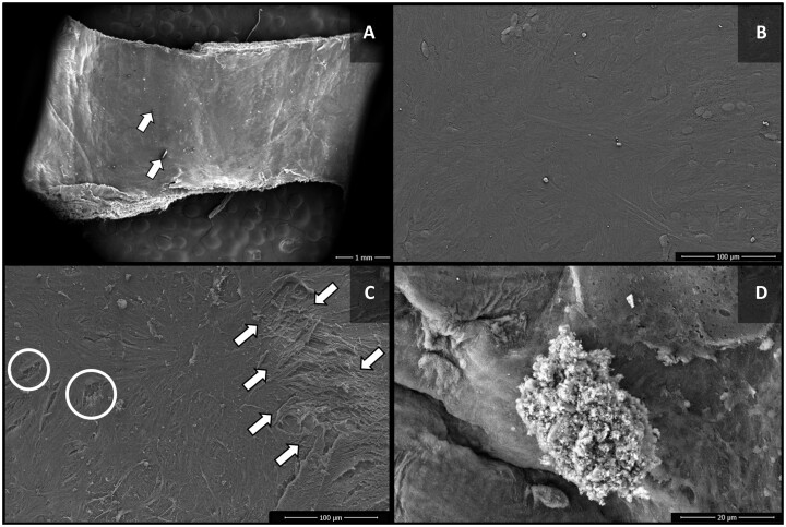
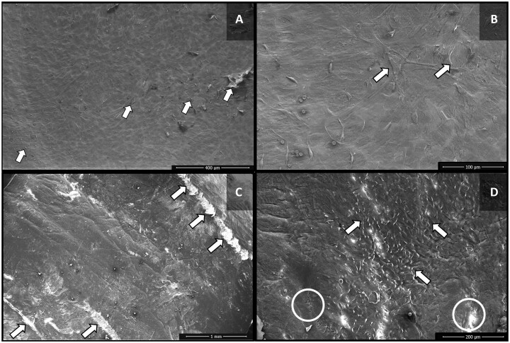
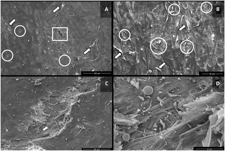
Methods: Forty-two SV segments obtained from 12 patients undergoing CABG were divided into 7 groups. Group 1 (control) was prepared without preservation or intraluminal distension, while the other 6 groups were preserved in autologous heparinized autologous arterial blood or normal saline (NS), with distention pressures 30, 100 and 300 mmHg. To assess the effects of using these solutions and pressures on the endothelium, the grafts were analysed by scanning electron microscopy, with the measurement of endothelial damage degree.

Results: Segments in group 1 showed minimal endothelial damage. SV grafts preserved with NS had significantly greater endothelial damage both compared to the control group and compared to groups preserved with autologous arterial blood (P < 0.001). Segments distended with pressures up to 100 mmHg showed less damage when compared to those distended at 300 mmHg, with the ones subjected to higher pressures presenting a maximum degree of damage, with considerable loss and separation of endothelial cells, extensive foci of exposure of the basement membrane and numerous fractures of the intimate layer, without differences regarding the solution used.

Conclusions: Preparation of SV using NS and with intraluminal distension pressures above 100 mmHg is factors related to increased damage to the venous endothelium.

Keywords: Coronary artery bypass grafting; Distension pressure; Preservation solution; Saphenous vein.


Figures

Figure 1:
Figure 1:
Scanning electron microscopy images of the luminal surface of the control group saphenous vein segments. (A) Venous endothelium with no signs of cell damage. (B) Intimal layer with preserved endothelial cells.
Figure 2:
Figure 2:
Scanning electron microscopy images of the luminal surface of saphenous vein segments of groups 2 (autologous arterial blood 30) and 3 (normal saline 30). (A) Group 2 (autologous arterial blood 30)—integrity of most of the endothelium, with discrete artefacts from the preparation for microscopic analysis (arrow). (B) Group 2 (autologous arterial blood 30)—intact endothelium. (C) Group 3 (normal saline 30)—marked separation of endothelial cells with exposure of subendothelial layers (arrow), in addition to fissures in the cellular intima layer (circle). (D) Group 3 (normal saline 30)—presence of a cluster of fibrin and platelets on the endothelial surface.
Figure 3:
Figure 3:
Scanning electron microscopy images of the luminal surface of saphenous vein segments of groups 4 (autologous arterial blood 100) and 5 (normal saline 100). (A) Group 4 (autologous arterial blood 100)—occasional separation of endothelial cells (arrow). (B) Group 4 (autologous arterial blood 100)—separation of endothelial cells, with exposure of the basement membrane (arrow). (C) Group 5 (normal saline 100)—areas with marked endothelial damage, including exposition of collagen fibres (arrows). (D) Group 5 (normal saline 100)—elongated pattern of the endothelial cells, with separation of these cells (arrows), in addition to fissures in the intima layer (circles).
Figure 4:
Figure 4:
Scanning electron microscopy images of the luminal surface of saphenous vein segments of groups 6 (autologous arterial blood 300) and 7 (normal saline 300). (A) Group 6 (autologous arterial blood 300)—Loss and separation of endothelial cells (arrows), with exposure of the basement membrane (circles), and of collagen fibres (square). (B) Group 6 (autologous arterial blood 300)—separation of endothelial cells (arrows), and exposure of basement membrane (circle). (C) Group 7 (normal saline 300)—fissure in the intima layer (arrow), with exposure of the subendothelial layers. (D) Group 7 (normal saline 300)—microenvironment of fissure of the endothelial layer, with disarrangement of collagen fibres, and presence of red blood cells, fibrin and platelet agglomerates.
Figure 5:
Figure 5:
Evaluation of the degree of endothelial damage in saphenous vein segments with different storage solutions and intraluminal distension pressures. The data correspond to the mean and standard deviation of measurements performed in each group. The symbols **(P <0.01) and ***(P <0.001) denote statistically significant differences in relation to the control group, while the symbol ###(P <0.001) means statistically significant differences in relation to the group stored with normal saline at the same distension pressure. The symbols +(P <0.05) and +++(P <0.001) indicate statistically significant differences in relation to the group submitted to distension pressure of 30 mmHg in the same storage solution, while the symbol xxx(P <0.001) corresponds to statistically significant differences in relation to the group submitted to distension pressure of 100 mmHg in the same storage solution.
None

Similar articles

Cited by

References

    1. Mack M, Gopal A.. Epidemiology, traditional and novel risk factors in coronary artery disease. Heart Fail Clin 2016;12:1–10. - PubMed
    1. Neumann F, Sousa-Uva M, Ahlsson A, Alfonso F, Banning AP, Benedetto U. et al.; ESC Scientific Document Group. 2018 ESC/EACTS Guidelines on myocardial revascularization. Eur Heart J 2019;40:87–165. - PubMed
    1. D'Agostino RS, Jacobs JP, Badhwar V, Fernandez FG, Paone G, Wormuth DW. et al. The Society of Thoracic Surgeons adult cardiac surgery database: 2018 update on outcomes and quality. Ann Thorac Surg 2018;105:15–23. - PubMed
    1. Ward AO, Caputo M, Angelini GD, George SJ, Zakkar M.. Activation and inflammation of the venous endothelium in vein graft disease. Atherosclerosis 2017;265:266–74. - PubMed
    1. de Vries MR, Simons KH, Jukema JW, Braun J, Quax PHA.. Vein graft failure: from pathophysiology to clinical outcomes. Nat Rev Cardiol 2016;13:451–70. - PubMed