. 2022 Jun 28;13(3):e0048022.
doi: 10.1128/mbio.00480-22. Epub 2022 May 16.

Adenosine Awakens Metabolism to Enhance Growth-Independent Killing of Tolerant and Persister Bacteria across Multiple Classes of Antibiotics

Affiliations

Adenosine Awakens Metabolism to Enhance Growth-Independent Killing of Tolerant and Persister Bacteria across Multiple Classes of Antibiotics

David A Kitzenberg et al. mBio. .

Abstract

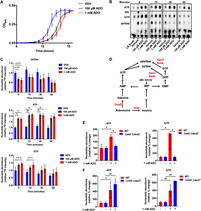
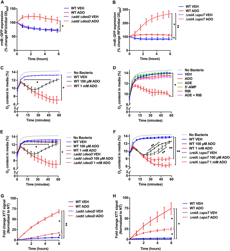
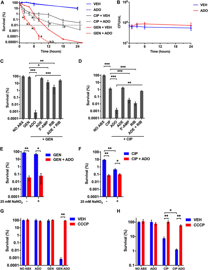
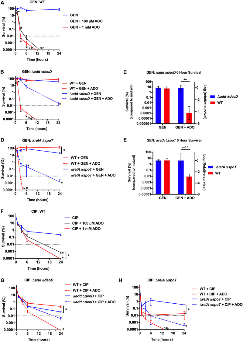
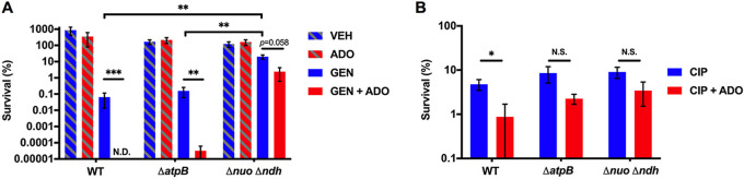
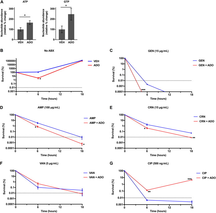
Metabolic and growth arrest are primary drivers of antibiotic tolerance and persistence in clinically diverse bacterial pathogens. We recently showed that adenosine (ADO) suppresses bacterial growth under nutrient-limiting conditions. In the current study, we show that despite the growth-suppressive effect of ADO, extracellular ADO enhances antibiotic killing in both Gram-negative and Gram-positive bacteria by up to 5 orders of magnitude. The ADO-potentiated antibiotic activity is dependent on purine salvage and is paralleled with a suppression of guanosine tetraphosphate synthesis and the massive accumulation of ATP and GTP. These changes in nucleoside phosphates coincide with transient increases in rRNA transcription and proton motive force. The potentiation of antibiotic killing by ADO is manifested against bacteria grown under both aerobic and anaerobic conditions, and it is exhibited even in the absence of alternative electron acceptors such as nitrate. ADO potentiates antibiotic killing by generating proton motive force and can occur independently of an ATP synthase. Bacteria treated with an uncoupler of oxidative phosphorylation and NADH dehydrogenase-deficient bacteria are refractory to the ADO-potentiated killing, suggesting that the metabolic awakening induced by this nucleoside is intrinsically dependent on an energized membrane. In conclusion, ADO represents a novel example of metabolite-driven but growth-independent means to reverse antibiotic tolerance. Our investigations identify the purine salvage pathway as a potential target for the development of therapeutics that may improve infection clearance while reducing the emergence of antibiotic resistance. IMPORTANCE Antibiotic tolerance, which is a hallmark of persister bacteria, contributes to treatment-refractory infections and the emergence of heritable antimicrobial resistance. Drugs that reverse tolerance and persistence may become part of the arsenal to combat antimicrobial resistance. Here, we demonstrate that salvage of extracellular ADO reduces antibiotic tolerance in nutritionally stressed Escherichia coli, Salmonella enterica, and Staphylococcus aureus. ADO potentiates bacterial killing under aerobic and anaerobic conditions and takes place in bacteria lacking the ATP synthase. However, the sensitization to antibiotic killing elicited by ADO requires an intact NADH dehydrogenase, suggesting a requirement for an energized electron transport chain. ADO antagonizes antibiotic tolerance by activating ATP and GTP synthesis, promoting proton motive force and cellular respiration while simultaneously suppressing the stringent response. These investigations reveal an unprecedented role for purine salvage stimulation as a countermeasure of antibiotic tolerance and the emergence of antimicrobial resistance.

Keywords: E. coli; Salmonella; antibiotic resistance; nucleosides; persistence; tolerance.


Conflict of interest statement

The authors declare no conflict of interest.

Figures

FIG 1
FIG 1
Extracellular ADO dysregulates nucleotide metabolism. (A) Growth of E. coli in M9 minimal medium supplemented with 0.4% glucose, treated at time zero with 100 μM or 1 mM ADO. (B) Representative thin-layer chromatography (TLC) autoradiogram of 32P-labeled nucleotide extracts from E. coli downshifted to M9 minimal medium supplemented with 0.4% glucose and treated at time zero with vehicle or ADO. Cells shifted to LB broth serve as a nutrient-rich control. (C) Relative changes in ATP, GTP, and ppGpp were determined by mean autoradiogram intensity of 3 replicate experiments from panel B. (D) Simplified purine salvage and (p)ppGpp synthesis pathways; enzymes of interest are shown in red. (E and F) HPLC analysis of intracellular nucleotide extracts 15 min after E. coli cells were downshifted from LB broth into PBS. Selected groups of cells were treated with ADO at time zero. Prior to treatment, experimental cultures were washed and diluted to equal starting CFU. Data are the mean of three biological replicates ± SEM. *, P < 0.05; **, P < 0.01, as assessed by two-way analysis of variance (ANOVA) with Tukey’s multiple-comparison and Mann-Whitney U tests. ADO, adenosine; VEH, vehicle control.
FIG 2
FIG 2
ADO hyperstimulates rRNA transcription, O2 respiration, and electron transport chain activity. (A and B) rrnB P1:GFP signal from wild-type and mutant E. coli following the downshift from LB broth to PBS. Selected samples were treated with 1 mM ADO at time zero. Fluorescent signal and OD600 were monitored over time at 37°C. Signal was normalized to initial inoculum. (C to F) O2 consumption was examined in PBS with E. coli at 37°C in a shaker incubator using PreSens OxoDish. Dotted gray line indicates O2 content in cell-free media. (G and H) Tetrazolium dye, XTT, in PBS; for each time point, the signal was normalized to the time zero of the wild-type vehicle control group. Readings were taken every 10 min at 37°C. No growth was observed under experimental conditions in panels A to H. Data are the mean of three biological replicates ± SEM. *P, < 0.05; **, P < 0.01, as estimated by two-way ANOVA with Tukey’s multiple comparison. ADE, adenine; ADO, adenosine; RIB, d-ribose; RFU, relative fluorescent unit; VEH, vehicle control.
FIG 3
FIG 3
ADO potentiates antibiotic lethality. (A) E. coli grown to stationary phase in LB broth was treated with very high concentrations of antibiotics for 0 to 24 h. Selected groups were treated with 80 μg/mL GEN, 3 μg/mL CIP, and/or 1 mM ADO. (B) E. coli downshifted from LB broth into PBS and treated at time zero with 1 mM ADO. Both control and treatment groups were established from one culture to ensure equal starting CFU for each group within an experiment. (C and D) Survival of E. coli after downshift from exponential-phase LB broth to PBS followed by an 18 h treatment with 5 μg/mL GEN or 500 ng/mL CIP. Selected samples were treated with 1 mM of the indicated nucleotides or metabolites. (E and F) Eighteen-hour survival experiment performed in an anaerobic chamber. Bacteria were passaged for at least 1 week to adapt to anaerobic conditions. Prior to treatment, bacteria were shifted from LB broth to PBS. Sodium nitrate (25 mM) was added at time of antibiotic treatment. Selected samples were treated with 5 μg/mL GEN, 500 ng/mL CIP, and/or 1 mM ADO. (G and H) Survival of aerobic E. coli after downshift from exponential-phase LB broth to PBS followed by a 3 h treatment with 5 μg/mL GEN or 500 ng/mL CIP. Selected samples were treated with 1 mM ADO and 50 μM CCCP at time of antibiotic treatment. Data are the mean of three biological replicates ± SEM. Dotted line indicates MDK99.99 threshold. *, P < 0.05; **, P < 0.01; ***, P < 0.001, as assessed by Student's t test, Mann-Whitney U test, or one-way ANOVA with Fisher’s least significant difference (LSD) multiple comparison at each time point. ADE, adenine; ADO, adenosine; RIB, d-ribose; VEH, vehicle control.
FIG 4
FIG 4
ADO shortens MDK99.99 duration. (A, B, D, and F to H) Survival of E. coli after downshift from exponential-phase LB broth to PBS followed by 0 to 24 h treatment to establish MDK values. Samples were treated with 5 μg/mL GEN or 500 ng/mL CIP. (C and E) Figures are data from MDK curves presented in panels B and D, respectively. Control strain survival values after 6 h of treatment were normalized to the mutant strain. For all experiments, ADO did not cause significant changes in growth or survival compared to VEH. Data are the mean of three biological replicates ± SEM. *, P < 0.05; **, P < 0.01; ***, P < 0.001, as assessed by one-way ANOVA with Fisher’s LSD multiple comparison at each time point or Student's t test. Dotted line indicates MDK99.99 threshold. Asterisk denotes significant difference between treatment groups within a strain unless indicated by a bracket for between-strain comparisons. ADO, adenosine; ND, no CFU detected; VEH, vehicle control.
FIG 5
FIG 5
ADO promotes antibiotic killing of S. Typhimurium. (A and B) Eighteen-hour survival of S. Typhimurium grown to stationary phase in LB broth followed by treatment with 50 μg/mL GEN or 5 μg/mL CIP. Selected samples were treated with 1 mM ADO. Data are the mean of three biological replicates ± SEM. *, P < 0.05; **, P < 0.01; ***, P < 0.001, as assessed by Student's t test. ADO, adenosine; ND, no CFU detected; NS, not statistically significant; VEH, vehicle control.
FIG 6
FIG 6
ADO sensitizes S. aureus to antibiotic killing. (A) S. aureus intracellular nucleotide concentrations 15 min after downshift from LB broth into M9 minimal medium. Selected samples were treated with vehicle or 1 mM ADO. (B to G) To establish MDK values, antibiotic killing was determined in S. aureus cultures after downshifting from exponential-phase LB broth to M9 medium supplemented with 0.2% Casamino Acids, 5 μg/mL thiamine, and 2 μg/mL nicotinic acid, without glucose. Selected samples were treated with vehicle or 1 mM ADO in the presence of 15 μg/mL GEN, 100 μg/mL AMP, 15 μg/mL CRN, 5 μg/mL VAN, 500 ng/mL CIP, or antibiotic vehicle control. Data are the mean of three biological replicates ± SEM. *, P < 0.05; **, P < 0.01; ***, P < 0.001, as assessed by Student's t test. Dotted line indicates MDK99.99 threshold. Asterisk denotes significant difference between treatment groups. ABX, antibiotics; ADO, adenosine; AMP, ampicillin; CIP, ciprofloxacin; CRN, ceftriaxone; GEN, gentamicin; VAN, vancomycin; VEH, vehicle control.

References

    1. Martens E, Demain AL. 2017. The antibiotic resistance crisis, with a focus on the United States. J Antibiot (Tokyo) 70:520–526. doi:10.1038/ja.2017.30. - DOI - PubMed
    1. Ventola CL. 2015. The antibiotic resistance crisis: part 1: causes and threats. P T 40:277–283. - PMC - PubMed
    1. Rowe SE, Conlon BP, Keren I, Lewis K. 2016. Persisters: methods for isolation and identifying contributing factors—a review, p 17–28. In Michiels J, Fauvart M (ed), Bacterial persistence: methods and protocols. Springer, New York, NY. doi:10.1007/978-1-4939-2854-5_2. - DOI - PubMed
    1. Windels EM, Michiels JE, Van Den Bergh B, Fauvart M, Michiels J. 2019. Antibiotics: combatting tolerance to stop resistance. mBio 10:e02095-19. doi:10.1128/mBio.02095-19. - DOI - PMC - PubMed
    1. Windels EM, Michiels JE, Fauvart M, Wenseleers T, Van Den Bergh B, Michiels J. 2019. Bacterial persistence promotes the evolution of antibiotic resistance by increasing survival and mutation rates. ISME J 13:1239–1251. doi:10.1038/s41396-019-0344-9. - DOI - PMC - PubMed

Publication types

MeSH terms

LinkOut - more resources